
Profile of Candida Species in Vulvovaginal Candidiasis using
Conventional Methods
Cita Rosita Sigit Prakoeswa, Dewi Puspitorini, Yuri Widya, Sylvia Anggraeni, Linda Astari, Evy
Ervianti, Sunarso Suyoso
Dermatology and Venereology Departement Universitas Airlangga/ Dr.Soetomo Hospital Surabaya
Keywords: Candida sp, vulvovaginal candidiasis, cornmeal Tween 80 agar, carbohydrate fermentation test, CHROM
agar Candida.
Abstract: Vulvovaginal candidiasis (VVC) is an inflammatory disease of vulva and vagina that caused by Candida
sp.Candida albicans was the predominant cause of candidiasis. However, a shift toward non-albicans
Candida species has been recently observed. These non-albicans Candida species demonstrated reduced
susceptibility to commonly used antifungal drugs. Identification of the infecting Candida to the species
level is of utmost importance for clinical microbiological services for prediction of likely drug susceptibility
and to guide treatment. Conventional methods for the diagnosis of candidiasis are sensitive, low-cost, and
although time consuming, are still considered the references standard for identification of yeast isolates.
There were three conventional methods that is still the standard tests to identify the Candida sp. The tests
are Sabouraud dextrose agar (SDA) thencornmeal-Tween 80 agar, carbohydrate fermentation test, and
CHROM agar Candida (CAC). These tests usually give some specific colony, morphology andcolor for
every Candida sp.
1 INTRODUCTION
Vulvovaginal candidiasis (VVC) refers to a disorder
characterized by signs and symptoms of
vulvovaginal inflamation in the presence of Candida
species. It is the second most common cause of
vaginitis symptom after bacterial vaginosis. It is
estimated that at least 75% of healthy adult women
will experience one episode of vulvovaginal
candidiasis during their reproductive phase. The
signs and symptoms of VVC include a thick cheese–
like discharge associated with intense vaginal and
vulvar pruritus, pain, burning, erythema, and/or
edema (Kunduk and Garg, 2012; Moyes and Naglik,
2011; Sobel, 2008).
Candida sp are among the most common fungal
pathogens. They are capable of initiating infections
in both immunocompetent individuals and
immunocompromised hosts. Candida sp,
arecommensal organisms that normally colonize
mucosal surfaces in an asymptomatic manner, but it
also can become one of the most significant causes
of disabling and lethal infection. Candida sp are
responsible for various clinical manifestations
ranging from mucocutaneous overgrowth to life
threatening disseminated infections like candidemia.
Candida albicans remains the most common
causative agent for VVC in approximately 85%-95%
of the cases. However,there is an alteration of
species that cause VVC in the last decade as
incidence of VVC due to non-albicans Candidasp is
increasing. This rise could be due to an increase of
over-the-counter antifungal use. The clinical
manifestations of infections caused by non-albicans
Candidassp are usually indistinguishable with VVC
caused by Candida albicans except in its poor
response to common anti-fungal drugs due to either
inherent or acquired. Due to the changing
epidemiology of Candida, physicians may no longer
be able to make therapeutic decisions based on
species levels study to enhance proper treatment for
their patients (Hedayati et al., 2015; Deorukhkar et
al., 2014; Farooqi et al., 2013; Richter et al., 2005;
Fidel et al., 1999).
Species identification requires isolation and
biochemical or physiological characterization.
Candida sp, being non-fastidious organism, readily
grows on laboratory media used for the isolation of
fungus. Conventional methods, including Sabouraud
Prakoeswa, C., Puspitorini, D., Widya, Y., Anggraeni, S., Astari, L., Ervianti, E. and Suyoso, S.
Profile of Candida Species in Vulvovaginal Candidiasis using Conventional Methods.
DOI: 10.5220/0008155702810285
In Proceedings of the 23rd Regional Conference of Dermatology (RCD 2018), pages 281-285
ISBN: 978-989-758-494-7
Copyright
c
2021 by SCITEPRESS – Science and Technology Publications, Lda. All rights reserved
281

dextrose agar(SDA), cornmeal-Tween 80 agar,
carbohydrate fermentation test, and CHROM Agar
Candida (CAC), are recommended to identify
Candida spto the species level. Each method has its
own eminence at identifying Candida species. The
formation of blastospore, pseudohyphae, hyphae and
chlamydospores which aids identification of
Candida sp requires the use of nutritionally deficient
media like cornmeal agar Tween 80 as these
nutritionally deficient media suppress the vegetative
growth and promote sporulation. Carbohydrate
fermentation test classifies Candida based on the
color transformation on carbohydrate broth and gas
formation on the tube. Candida sp metabolizes
carbohydrates both aerobically (assimilation) and
anaerobically (fermentation) (Mutiawati, 2016;
Suyoso, 2013; Larone 2011). Lastly, enzymatic
reaction methods using chromogenic substrates in
Chromogenic agar (CHROM Agar Candida)
medium has high sensitivity and specificity in
differentiating Candida species in clinical samples.
This media contains chromogenic substrates that
react with enzymes secreted by yeast cells, resulting
in various pigmentations. Using this medium, it is
possible to identify Candida species based on color
characteristics. CAC can differ one Candida sp to
another Candida sp by its colony color. The
presence of two colonies with different color
indicates there were two species that grew on CAC.
2 METHODS
This was a cross-sectional descriptive study that
identifies causes of VVC down to the species level
by using conventional methods of fungal
examination. The sample of this study were all VVC
patients that fulfilled the inclusion criteria, and
underwent examination at Sexually Transmited
Infection Division, Dermatoveneorology Clinic of
Dr.Soetomo Hospital, Surabaya. There were a total
of 25 enlisted patients. The patients' data
information in data collection sheet.
The Inclusion criteria were VVC patients, aged
15 years or older, married or unmarried, willing to
follow the research and sign the informed
consent.The exclusion criteria were patients with
negative culture result.
All women who attended Sexually Transmitted
Infection (STI) Division, Dermatoveneorology
Clinic of Dr.Soetomo Hospital, Surabaya were
interviewed for medical history and clinically
examined. Samples were taken from vaginal swab
and checked for Candida and Gram stain
examination. Patients diagnosed with VVC were
included in inclusion criteria.
Consecutively, the samples were first grown in
SDA, followed by cornmeal-Tween 80 agar and then
performed on carbohydrate fermentation test. The
results were available in 2-3 days. Therewere 6
carbohydratesused in this study: urea, dextrose,
lactose, sucrose, maltose, galactose and trehalose.
Positive result was marked bythe changing color of
broth to yellow and the gas formation in the Durham
tube. The other test was CHROM agar Candida
(CAC), showed colony color in18 hours-3 days.
Each color represents one specific species of
Candida.
3 RESULTS
This study involved 25 female patients with VVC.
The patients were predominated by 15-24 age group.
Most patients were private employees and most of
the patient's education were bachelor.
Most common duration of complaint was 1
month until 9 months with vaginal douching usage
being the highest predisposition factor. Most patients
admitted have not consumed any therapy for VVC
yet. Clinical examination revealed edematous and
erythematous vulva and vagina on all 25 patients.
Direct examination from wet specimen and Gram
stain were: wet specimen contained positive
blastospore with negative pseudohifa 20%, positive
blastospora with positive pseudohifa 48%, negative
blastospora with negative pseudohifa 32%, no
negative blastospore with positive pseudohifa(0%)
and Gram positive blastospore with negative
pseudohifa 16%, negative blastospore with positive
pseudohifa 4%, positive blastospore with positive
pseudohifa 52%, and negative blastospore with
negative pseudohifa 28%.
There were various results for each conventional
method. Cornmeal agar showed the specific
formation of every Candida. The presence of
terminal vesicles (chlamydoconidia) with pseudohifa
and flower-like blastoconidia in cornmeal agar
indicates that the fungi was Candida albicans. Other
sample showing divaricated pseudohifa with oval
blastoconidia indicates the presence of Candida
tropicalis.
RCD 2018 - The 23rd Regional Conference of Dermatology 2018
282
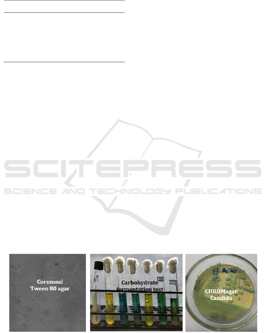
Table 1: Comparation between direct examination and
conventional methods
As for the carbohydrate fermentation test, for
example, positive result on dextrose and trehalose
means that the sample was Candida glabrata.
CAC revealed color of colony. All samples
showed one type of colony color, meaning one
Candida sp in every sample, and only 4 samples
shown two colony color (2 Candida sp). Light
green-colored colony is specific for Candida
albicans, while dark green colony is specific for
Candida dubliniensis. Purple-colored colony
characterized the colony of Candida glabrata.
These three conventional methods from 25
samples revealed that 14 sample were positive
forCandida albicans, and others were non-albicans
Candida sp. Five samples were positive for Candida
glabrata, 1 sample for Candida parapsilosis and 4
samples were positive for2 Candida sp (double
infections). From those 4 samples,1 sample were
positive for Candida albicans and Candida glabrata,
1 sample for Candida albicans and Candida famata,
and 2 samples were positive for Candida albicans
and Candida tropicalis. From the table 1 we can see
the comparation between microscopic and
conventional methods. Conventional methods
revealed species of the fungi while direct
microscopic examination can only show
pseudohypha and blastospore.
4 DISCUSSION
This was a descriptive cross-sectional study that
aimed to identify the causative agents of VVC. After
3 months sampling, 25 participants fulfilled the
inclusion criteria. The result showed that most
participants were in 15-24 age group. This is a
reproductive age group, with high level of estrogen.
Estrogen has been found to reduce the ability of
vaginal epithelial cells to inhibit the growth of
Candida albicans and also decreases immunoglobins
in vaginal secretions resulting in increased
vulnerability of women to vaginal Candidiasis.
Most patients were private employees. This group of
people tend to use vaginal hygiene products or
vaginal douche that contained antiseptic, and
prolonged misuse of antiseptic can cause VVC.
Most patient education were bachelor; they might
have better senses of personal vaginal hygiene than
patients with lower education group So it made them
went to hospital and checked their complaint and to
get a proper treatment from the hospital (Fidel et al.,
2000).
Most common duration of complaint of the
patients of VVC is 1 month until 9 months. From
this, we can deduct that there might be a recurrent
VVC that was ignored by the patientand causing
delayed proper treatment.
Clinical examination revealed edematous and
erythematous vulva and vagina on all 25 patients.
This means clinical sign of VVC is the first and
definite diagnosis of VVC withthe direct
microscopic examination was not the only parameter
to diagnose VVC (Sobel, 2008).
Figure 1: Conventional methods (SDA then Cornmeal Tween 80 agar, carbohydrate fermentation test, and CHROM agar
Candida).
Wet specimen & Gram
stain
Conventional methods
Microscopic
examination (wet
specimen and Gram
stain) can show fungal
morphology
(blastospore,
pseudohyphae, and
h
yp
hae
)
Conventional methods
can reveal species of the
fungi (Candida sp itself).
Profile of Candida Species in Vulvovaginal Candidiasis using Conventional Methods
283

Cornmeal Tween 80 agar showed the specific
morphology of every Candida. One sample with
budding yeast-like cell with no pseudohyfa indicates
the presence ofCandida glabrata. However, there
was also a sample that showed yeast cell only and,
no pseudohyfa.This could not conclude the presence
of Candida glabrata and additional examination
using other conventional method was needed to
identify the species (Golia et al., 2013; Suyoso,
2013).
The carbohydrate fermentation test shown
positive result based on the changing color of broth
to yellow and the gas formationin the Durham tube.
This test, however, is not reliable when used without
other additional method. For example, positive result
of dextrose and trehalose indicates presence of
Candida glabrata, while positive result of dextrose
and maltose, galactose and trehalose indicates
presence of Candida albicans. Therefore, a dubious
positive result of only dextrose and maltose could
not accurately indicate the presence of Candida
albicans.CAC will be useful for the additional
examination (Devi et al., 2014; Larone, 2011).
CAC revealed specific color for
eachcolony.There was a sample which color was
pink to purple, thus almost similar to Candida
glabrata. The carbohydrate fermentation test could
be used to differentiate dubious results from this
examination in order to get the final diagnosis (Faraz
et al., 2016; Suyoso, 2013; Vijaya et al., 2011).
5 CONCLUSION
There were increasing of non-albicans Candida sp
in the agents causing VVC. Non-albicans Candida
sp is an emerging threat fungi due to its antifungal
resistance. Identification of causative agent of VVC
down to the species level is important to give proper
treatment to the VVC patients. There are 3
conventional methods that can be used to identify
Candida sp to the species level. Cornmeal agar
showed the morphology of Candida sp.
Carbohydrate fermentation test revealed positive
result if there is brothcolor transformation to yellow
and Durham tube inside filled with gas. CHROM
agar Candida showed Candida sp by its colony
color. These 3 methods are still recommended for
identification of Candida sp to the species level and
should be performed simultaneously to support each
other's data to get the final definite species
identification result.
REFERENCES
Deorukhkar, S.C., Saini, S., Mathew, S., 2014. Non-
albicans Candida Infection: An Emerging Threat.
Interdisciplinary perspectives on infectious diseases,
615958. doi:10.1155/2014/615958
Sumitra Devi, L., Maheshwari, M., 2014. Speciation of
Candida Species Isolated From Clinical Specimens by
Using Chrom Agar and Conventional Methods.
International Journal of Scientific and Research
Publications 4, 2250–3153.
Faraz, A., Gaffar, U.B., Ansari, T., Sami, W. 2016.
Evaluation of diagnostic efficacy of chrom agar
candida for differentiation and identification of
common Candida species. Isra Med J 8(4): 224-6.
Farooqi, J.Q., Jabeen, K., Saeed, N., Iqbal, N., Malik, B.,
Lockhart, S.R., Zafar, A., Brandt, M.E., Hasan, R.,
2013. Invasive candidiasis in Pakistan: Clinical
characteristics, species distribution and antifungal
susceptibility. Journal of Medical Microbiology 62,
259–268. doi:10.1099/jmm.0.048785-0
Fidel, P.L., Cutright, J., Steele, C., 2000. Effects of
reproductive hormones on experimental vaginal
candidiasis. Infection and immunity 68, 651–657.
doi:10.1128/IAI.68.2.651-657.2000
Fidel, P.L., Vazquez, J.A., Sobel, J.D., 1999. Candida
glabrata: review of epidemiology, pathogenesis, and
clinical disease with comparison to C. albicans.
Clinical microbiology reviews 12, 80–96. doi:9880475
Golia, S., Reddy, K.M., Karjigi, K.S., Hittinahalli, V.
2013. Speciation of Candida using chromogenic and
cornmeal agar with determination of fluconazole
sensitivity. Al Ameen J Med Sci; 6(2): 163-6
Hedayati, M.T., Taheri, Z., Galinimoghadam, T., Aghili,
S.R., Cherati, J.Y., Mosayebi, E., 2015. Isolation of
different species of Candida in patients with
vulvovaginal candidiasis from sari, Iran. Jundishapur
Journal of Microbiology 8.
doi:10.5812/jjm.8(4)2015.15992
Kundu, R.V., Garg, A. 2012. Yeast infections: candidiasis,
tinea (pityriasis) versicolor, and malassezia
(pityrosporum) folliculitis. In: Goldsmith LA, Katz SI,
Gilchrest BA, Paller AS, Leffell DJ, Wolff K. editors.
Fitzpatrick dermatology in general medicine8th ed.
USA: McGraw Hill. p 2300-1
Larone, D.H. 2011. Medically important fungi. 5th ed.
New York: ASM Press. P. 117-36.
Naglik, J.R., Moyes, D.L., Wächtler, B., Hube, B., 2011.
Candida albicans interactions with epithelial cells and
mucosal immunity. Microbes and Infection.
doi:10.1016/j.micinf.2011.06.009
Mutiawati, V.K. Microbiologicalexamination for Candida
albicans. Jurnal Kedokteran Syiah Kuala 2016; 16 (1):
53-62.
Richter, S.S., Galask, R.P., Messer, S.A., Hollis, R.J.,
Diekema, D.J., Pfaller, M.A., 2005. Antifungal
susceptibilities of Candida species causing
vulvovaginitis and epidemiology of recurrent cases.
Journal of Clinical Microbiology 43, 2155–2162.
doi:10.1128/JCM.43.5.2155-2162.2005
RCD 2018 - The 23rd Regional Conference of Dermatology 2018
284

Sobel, J.D. Vulvovaginal candidiasis. In: Holmes, K.K.,
Sparling, P.F., Stamm, W.E., Piot, P., Wasserheit,
J.N., Corey, L. 2008. editors. Sexually transmitted
diseases. 4th ed. New York: McGraw Hill. p 823-38
Suyoso, S. Mucosal candidiasis. In: Bramono, K., Suyoso,
S., Indriatmi, W., Ramali, L.M., Widaty, S., Ervianti,
E. 2013. Editors. Dermatomikosissuperfisialis.
Jakarta: Badan penerbit FKUI. p.120-35.
Vijaya, D., Harsha, T.R., Nagaratnamma, T., 2011.
Candida speciation using chrom agar. Journal of
Clinical and Diagnostic Research 5, 755–757.
Profile of Candida Species in Vulvovaginal Candidiasis using Conventional Methods
285
