
The Reliability of Videomicroscopic Compared with Skin Scraping
Microscopic Examination in Detection of Sarcoptes Scabiei
Merlina Juhendy
1*
, Yulia Farida Yahya
1
, Inda Astri Aryani
1
, Legiran
2
1
Department of Dermatology and Venereology Faculty of Medicine Sriwijaya University/Dr. Moh. Hoesin
General Hospital Palembang, Jendral Sudirman Street Km 3.5, Palembang, South of Sumatera, Indonesia
2
Department of Anatomy, Faculty of Medicine Sriwijaya University, Dr. Mohammad Ali Street, Palembang,
South of Sumatera, Indonesia
Keyword: scabies, Sarcoptes scabiei, microscopic, videomicroscope, reliability
Abstract: Scabies is a contagious skin disease due to infestation of Sarcoptes scabiei var. hominis (S. scabiei). Skin
scraping microscopic examination is a standard diagnostic procedure, which is still considered as a “gold
standard” to confirm the diagnosis. This method is invasive, time consuming with risks associated
procedure. Videomicrosc1ope, a non medical digital microscope that can be used to visualize burrow, mites
and eggs of S. scabiei. The reliability of videomicroscope to detect mites has not been studied. This study is
to determine reliability of videomicroscope and skin scraping microscopic examination in detection of S.
scabiei in presumptive scabies patients.This is a diagnostic test study which was conducted from August to
October 2017 at the Orphanage Subulussalam, Orphanage Al-Wiam, Lembaga Wanita Peduli Sriwijaya,
Lembaga Pendidikan dan Sosial Pemuda Bersatu and Lembaga Perlindungan Anak Kota Palembang. A
total of 139 presumptive scabies patients who met the inclusion and exclusion criteria were recruited by
consecutive sampling. All subjects were examined with blinded method using videomicroscope and skin
scraping microscopic examination by researcher and other examiner. S. scabiei mites that was detected by
skin scraping examination was 49,6% of all subject study. Sensitivity and specificity of videomicroscope
examination compared to skin scraping microscopic examination were 89,8% and 82,9%. (PPV 83,8%;
NPV 89,2%; positive likelihood ratio 4,57; negative likelihood ratio 0,16; accuracy 86,3%, AUC 0,864).
Videomicroscope showed high sensitivity and specificity, which was useful as a rapid alternative diagnostic
method to detect S. scabiei in high risk population.
1 INTRODUCTION
Scabies is a skin disease due to infestation of
Sarcoptes scabiei var. hominis (S. scabiei) as an
obligate parasite on the epidermis (Burkhart and
Burkhart, 2012). Clinical manifestation of scabies are
skin lesions accompanied by pruritus due to allergic
reaction or inflammation to the mites and their
products, it can resemble to other diseases
(Chosidow, 2006). This can cause a misdiagnosis, an
inadequate therapy, also can increase the risk of
bacterial infection and morbidity, so further
examination is needed to confirm the diagnosis
(Heukelbach et al., 2013; Micali et al., 2016).
Standard diagnostic procedures to confirm the
diagnosis is microscopic examination of skin
scraping (SS). This examination method is invasive
and some literatures considered it as a “gold
standard” for definite diagnosis by the visualization
of the mites, eggs or scybala (Leung and Miller,
2011; PERDOSKI, 2017). Few disadvantadges of
microscopic examination by skin scraping are a pain
that cause discomfort especially in younger patients,
a risk of bleeding, a secondary bacterial infection, a
necessary to be repeated on a few locations and a
time consuming (Micali et al., 2016; Anderson and
Strowd, 2017).
Recently there is a new non invasive technique
such as a videomicroscope (VM), a digital
microscope with magnification until 1000x allowing
direct visualization burrow, mites and eggs of S.
scabiei for a definite diagnosis of scabies, faster and
more practical in its use, with affordable price
(Lacarrubba et al., 2015; Micali et al., 2015).
The objective of this study is to determine the
reliability of VM compared with SS microscopic
276
Juhendy, M., Yahya, Y., Aryani, I. and Legiran, .
The Reliability of Videomicroscopic Compared with Skin Scraping Microscopic Examination in Detection of Sarcoptes Scabiei.
DOI: 10.5220/0008155602760280
In Proceedings of the 23rd Regional Conference of Dermatology (RCD 2018), pages 276-280
ISBN: 978-989-758-494-7
Copyright
c
2021 by SCITEPRESS – Science and Technology Publications, Lda. All rights reserved

examination to detect S. scabiei in presumptive
scabies patients.
2 METHODS
This is a diagnostic test study with cross sectional
design from August to October 2017 at the
Orphanage Subulussalam, Orphanage Al-Wiam,
Lembaga Wanita Peduli Sriwijaya, Lembaga
Pendidikan dan Sosial Pemuda Bersatu and
Lembaga Perlindungan Anak Kota Palembang in
Palembang. The study was approved by the ethics
committee. A total of 139 patients with skin disease
who met the inclusion and exclusion criteria were
recruited by consecutive sampling. The inclusion
criteria was presumptive scabies patients, who
agreed to participate in the study. Exclusion criteria
was patient who had been treated with scabies
therapy within 4 weeks prior to study. Demographic
data were collected and all subjects were blinded
examined using VM and SS microscopic
examination by researcher and other examiner.
3 RESULTS
In this study majority of the subjects were male with
ratio of male is 52,5% and female is 47,5%. The
subjects of this study were divided into few groups
based on age which they were infants (3,6%),
children (31,7%), adolescents (61,2%) and adults
(3,6%). The education level of the subjects was
divided into 4 groups: non-school (12,2%), primary
school (28,8%), junior high (37,2%) and senior high
school (21,6%).
Table 1. Demographic and characteristic of the lesions
Characteristic Total (n) Percentage (%)
Gender
Male
Female
73
66
52,5
47,5
Age
Infants
Children
Adolescents
Adult
5
44
85
5
3,6
31,7
61,2
3,6
Education
Non school
Primary school
Junior high school
Senior high school
17
40
52
30
12,2
28,8
37,4
21,6
Sharing clothes/towels together
Sharing bed with others
Number of people in a bedroom
1 person
2-3 persons
4-5 persons
>5 persons
74
101
11
50
35
43
53,2
72,7
7,9
36
25,2
30,9
Location of the lesions
Interdigitalis manus
Radiocarpalisjoint
Dorsum manus
Antecubiti
Flexor extremitas superior
Abdominalis(umbilicus/periumbilicus)
Inguinalis/genitalia
Glutealis
125
102
106
9
44
28
44
29
89,9
73,4
76,3
6,5
31,7
20,1
31,7
20,9
Location where S. scabiei mites detected
5
7,2
D
orsum manus
F
lexor extremitas superio
r
5
7,2
Glutealis 2
2.9
I
nguinalis
/
g
enitalia 5
7,2
I
nterdigitalis manus 35
50,7
Abdominalis 4
5,7
R
adiocarpalis join
t
13
18,8
The Reliability of Videomicroscopic Compared with Skin Scraping Microscopic Examination in Detection of Sarcoptes Scabiei
277

Study subjects 53,2% were sharing
clothes/towels together with friends. A total of 36%
of the study subjects had 2-3 friends in one room
and 72,7% of the study subjects occupied the bed
with others. The most common lesions were
erythematous papules (99,3%) and most lesions
were found on the interdigital region (89,9%).
The total of S. scabiei detected using
microscopic skin scraping examination in this study
was 49,6% of all study subjects, with the most
commonly location found is on interdigital manus
(28,8%). Kappa value of VM examination between
two examiners was 0,82. The analysis of VM
examination results compared to SS microscopic
examination as a "gold standard" was obtained by
Sn, Sp VM value were 89,8% and 82,8% (PPV
83,8%, NPV 89,2%, PLR 5,25, NLR 0,12, 86,3% of
accuracy and AUC 0,864).
4 DISCUSSION
Scabies is a parasitic infestation of the skin caused
by S. scabiei, highly contagious through both direct
and indirect contact and affect all ages, races and
socioeconomic levels (Burkhart and Burkhart,
2012). Scabies is a global public health problem,
which is often overlooked, according to the WHO
including one "neglected tropical diseases". In poor
communities scabies potentially causing outbreaks
associated with its transmission method,
overcrowding, poor sanitation and low knowledge
(Heukelbach and Feldmeier, 2006). Non-specific
clinical features may lead to misdiagnosis and may
be a source of active transmission (Hewitt et al.,
2015). Late treatment and inadequate therapy,
increasing the risk of bacterial infections and
morbidity, so a diagnostic tool with high sensitivity
is needed (Micali et al., 2016). According to the
Japanese dermatological guidelines for diagnosis
and treatment 2017 the sensitivity of SS microscopic
examination varies between 10-70% (Ishii et al.,
2017). Dupuy et al. found that diagnosis of scabies
only on clinical grounds, because of the difficulty of
detecting S. scabiei mites, causing 27% of untreated
scabies patients, thus increasing the risk for an
outbreak of scabies. Videomicroscope is a non
medical digital microscope, commonly used for
botanical, entomology and microelectronics, that can
be used to make a definite diagnosis of scabies
(Micali et al., 2016; Anderson and Strowd, 2017).
The analysis of VM diagnostic test result compared
to SS microscopic examination as 'gold standard'
obtained by Sn and Sp VM were 89.9% and 82.9%
(PPV 83,8%; NPV 89,2%; PLR 4,57; NLR 0.16 and
86.3% accuracy, AUC 0.864). In this study of 139
patients (49.6%) presumptive scabies detected mites,
S. scabiei eggs in 69 patients. According to Micalli,
et al study 20 presumptive scabies patients were
examined using VM, VD was confirmed by SS
microscopic examination, S. scabiei mites were
detected in 15 patients, during follow-up 5 patients
remained negative. In the study, the VM diagnostic
test was not performed, and sample size population
were small (Lacarrubba et al., 2015; Micali et al.,
2015).
Burrow is scabies pathognomonic lesion, that
often can be difficult to find, especially in the
tropical climates, but VM can help to visualize it
(Walton and Currie, 2007; Burkhart and Burkhart,
2012).
Table 2. Diagnostic value results of VM compared with SS microscopic examination
No. Diagnostic test Value CI (95%)
1. Sensitivity 89,9% 87,5-90,5
2. Specificity 82,9% 81,5-84,3
3. Accurac
y
86,3% 84,9-87,7
4. Positive likelihood ratio 5,25 5,1-5,3
5.
N
egative likelihood ratio 0,13 0,11-0,15
6. Positive
p
redictive value 83,8% 82,4-85,2
7.
8.
Negative predictive value
A
rea under curve
89,2%
0,864
87,7-90,7
0,797-0,930
A good accuracy rate of VM (86.3%) also
reduces the risk of misdiagnosis so as to prevent
outbreaks of scabies especially in densely populated
and socioeconomic areas. VM examination is a non
invasive method, it can be well tolerated and
efficient in terms of time because it only takes ≤10
minutes to direct visualization of S. scabiei mites,
eggs. and burrows and may be used for the follow up
evaluation after therapy (Lacarrubba et al., 2010).
Further study is needed in multicenter with larger
sample size and more heterogeneous populations to
determine the reliability VM in the detection of S.
scabiei.
RCD 2018 - The 23rd Regional Conference of Dermatology 2018
278
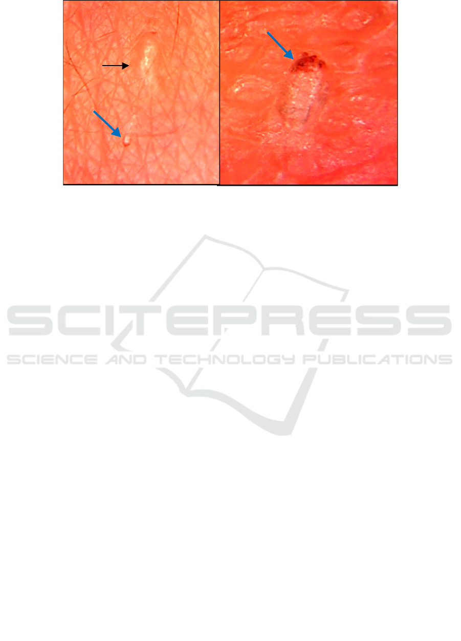
Figure 1. Videomicroscopic examination clearly evident the burrow (black arrow) and S. scabiei mite (blue arrow).
5 CONCLUSION
Videomicroscope has diagnostic value with high
accuracy and high level of accordance between the
examiners. The value of VM examination were:
sensitivity 89.9%, specificity 82.9%, PPV 83.8%,
89.2% NPV and 86.3% accuracy, AUC 0.864. Based
on this, VM diagnostic values are as good as SS
microscopic examination. Videomicroscope can be
used as a means of prevention of scabies in areas
with inaccessible and densely populated
communities.
ACKNOWLEDGEMENT
The authors would like to thank the Department of
Dermatology and Venereology, and Faculty of
Medicine of Sriwijaya University and all those who
assist in the effort of this research.
REFERENCES
Anderson, K.L., Strowd, L.C., 2017. Epidemiology,
Diagnosis, and Treatment of Scabies in a Dermatology
Office. The Journal of the American Board of
Family Medicine 30, 78–84.
doi:10.3122/jabfm.2017.01.160190
Burkhart, C.N., Burkhart, C.G. 2012. Scabies, other mites
and pediculosis. In: Goldsmith LA, Katz SI, Gilchrest
BA, Paller AS, Leffel DJ, Wolff K. eds. Fitzpatrick's
Dermatology in General Medicine 8
th
ed. Mc Graw-
Hill, New York Chicago pp. 2569-72
Chosidow, O., 2006. Scabies. New England Journal of
Medicine 354, 1718–1727.
doi:10.1056/NEJMcp052784
Dupuy, A., Dehen, L., Bourrat, E., Lacroix, C.,
Benderdouche, M., Dubertret, L., Morel, P., Feuilhade
de Chauvin, M., Petit, A., 2007. Accuracy of standard
dermoscopy for diagnosing scabies. Journal of the
American Academy of Dermatology 56, 53–62.
doi:10.1016/j.jaad.2006.07.025
Heukelbach, J., Feldmeier, H., 2006. Scabies. Lancet.
doi:10.1016/S0140-6736(06)68772-2
Heukelbach, J., Mazigo, H.D., Ugbomoiko, U.S., 2013.
Impact of scabies in resource-poor communities.
Current Opinion in Infectious Diseases.
doi:10.1097/QCO.0b013e32835e847b
Hewitt, K.A., Nalabanda, A., Cassell, J.A., 2015. Scabies
outbreaks in residential care homes: Factors associated
with late recognition, burden and impact. A mixed
methods study in England. Epidemiology and Infection
143, 1542–1551. doi:10.1017/S0950268814002143
Ishii, N., Asai, T., Asahina, A., Ishiko, A., Imamura, H.,
Kato, T., … Wada, Y., 2017. Guideline for the
diagnosis and treatment of scabies in Japan (third
edition): Executive Committee of Guideline for the
Diagnosis and Treatment of Scabies. Journal of
Dermatology,44(9), 991–1014.
https://doi.org/10.1111/1346-8138.13896
Lacarrubba F, Verzì AE, Micali G. 2015. A controlled,
dermatologist independently assessed, noninferiority
clinical trial of high resolution medically marketed
videodermatoscopy versus low cost nonmedical
videomicroscopy for the diagnosis of scabies. Journal
of the American Academy of Dermatology.
doi:10.1016/j.jaad.2015.02.510
Lacarrubba, F., D’Amico, V., Nasca, M.R., Dinotta, F.,
Micali, G., 2010. Use of dermatoscopy and
videodermatoscopy in therapeutic follow-up: A
review. International Journal of Dermatology.
doi:10.1111/j.1365-4632.2010.04581.x
The Reliability of Videomicroscopic Compared with Skin Scraping Microscopic Examination in Detection of Sarcoptes Scabiei
279

Leung, V., Miller, M., 2011. Detection of scabies: A
systematic review of diagnostic methods. The
Canadian journal of infectious diseases & medical
microbiology 22, 143–146.
Micali, G., Lacarrubba, F., Verzì, A.E., Chosidow, O.,
Schwartz, R.A., 2016. Scabies: Advances in
Noninvasive Diagnosis. PLoS Neglected Tropical
Diseases. doi:10.1371/journal.pntd.0004691
Micali, G., Lacarrubba, F., Verzì, A.E., Nasca, M.R.,
2015. Low-cost equipment for diagnosis and
management of endemic scabies outbreaks in
underserved populations. Clinical Infectious Diseases.
doi:10.1093/cid/ciu826
PERDOSKI. 2017. Panduan Layanan Klinis Dokter
Spesialis Dermatologi dan Venereologi. Jakarta. pp
80-83
Walton, S.F., Currie, B.J., 2007. Problems in diagnosing
scabies, a global disease in human and animal
populations. Clinical Microbiology Reviews.
doi:10.1128/CMR.00042-06
RCD 2018 - The 23rd Regional Conference of Dermatology 2018
280
