
The Effect of Advanced Adipose-derived Stem Cell Protein Extract to
Repairment of Collagen Deposition in Cultured Senescent Fibroblast
Erlina Pricilla Sitorus
1*
, Retno Dwi Utami
1
, Gita Hening Bunga
1
, Indah Julianto
1
, Harijono
Karionosentono
1
, Chandrani Khoirinaya
2
, Brian Wasita
3
1
Dermatology and Venereology Departement of Medical Faculty Sebelas Maret University, Surakarta
2
Dermama Biotechnology Laboratories, Surakarta
3
Pathology Anatomy Departement of Medical Faculty Sebelas Maret University, Surakarta
Keywords: Advanced adipose-derived stem cell protein extract, aging skin, collagen deposition
Abstract: Aging skin is progressive process in skin that stimulates by environment damage and influences the
appearance. There are two factors inducing aging, intrinsic i.e genetic and age, and extrinsic i.e sun expose,
air pollutant, smoking, alcoholic and malnutrition. Recently, cell based therapy has been widely reported as
the option for aging skin therapy. Advanced adipose-derived stem cell protein extract (AAPE) is a
conditioned medium cultured under a hypoxia of Adipose-derived stem cell (ADSC), which can stimulate
collagen synthesis and migration dermal fibroblast, thus induces wound healing and wrinkles repairment.
This study samples used cultured fibroblast from amnion, with donor from a 30 year old woman by
cesarean section on her first delivery. There were three samples cultured groups, normal fibroblast (control),
normal fibroblast with 30 minutes UVB exposure in basal media (study group 1), and normal fibroblast with
30 minutes UVB exposure in basal media added AAPE (study group 2). All groups were evaluated in 48
hours and checked the collagen density by using immunocytochemistry assay with collagen antibody-1. The
results were interpreted by Image-J software. Study group 2 showed significant increasing of collagen
density, with p value 0.001. Because the AAPE was derived from ADSC thus it has the same growth factors
as those of ADSC. AAPE had been proven in our study that it stimulated collagen deposition.
1 INTRODUCTION
Aging is a progressive degenerative process of all
organs in the body, including the skin. Aging in
human skin is caused by intrinsic factors such as
genetics and age and extrinsic factors like sun
exposure, air pollutant, smoking, alcoholic and
malnutrition (Yaar and Gilchrest, 2007; Ichihashi
and Ando, 2014), which disturb the function and
structures of epidermal cells and dermis, also the
extracellular matrix. Clinically, skin aging induced
by sun exposed or photoaging exhibits as wrinkles,
mottled pigmentation, rough skin, loss of skin tone,
dryness, sallowness, deep furrows, severe atrophy,
teleangiectasis, laxity, leathery appearance, solar
elastosis, actinic purpura, precancerous lesions, skin
cancer and melanoma (Pandel et al., 2013). The
histological features are epidermis and dermis
atrophy, elastosis in dermis, collagen changes and
elastin fibers fragments. Ultraviolet (UV) exposure
induces enhanced reactive oxygen species (ROS)
production so that the production of the components
and the oxidative destruction increase as well. This
destruction is a significant mechanism in aging
process (Stojiljković et al., 2014).
Cell based therapy has become a promising
therapy since it can induce repairment or cell
regeneration post tissue trauma or organ function
failure (Baer et al., 2016). One of cell based
therapies which show significant outcome is ADSC.
It is an adult stem cells which is also as
mesenchymal stem cell from human fat tissue. It has
ability to differentiate to be their derivates and to
secrete the various growth factors, which can repair
as well as replace the surrounding damaged cells
(Kim et al., 2011). The conditioned medium of
ADSC known as AAPE containing secreted growth
factors, which is beneficial for skin problem i.e face
wrinkles as well as wound repairment. AAPE can
induce collagen synthesis and fibroblast cell
migration into dermis, thus it can be used as
rejuvenation therapy and wound healing (Kim et al.,
2009; Zhou et al., 2016). Recently, the skin
regeneration has become cosmetical and
272
Sitorus, E., Utami, R., Bunga, G., Julianto, I., Kariosentono, H., Khoirinaya, C. and Wasita, B.
The Effect of Advanced Adipose-derived Stem Cell Protein Extract to Repairment of Collagen Deposition in Cultured Senescent Fibroblast.
DOI: 10.5220/0008155502720275
In Proceedings of the 23rd Regional Conference of Dermatology (RCD 2018), pages 272-275
ISBN: 978-989-758-494-7
Copyright
c
2021 by SCITEPRESS – Science and Technology Publications, Lda. All rights reserved

dermatologist concern as anti aging therapy due to
long term exposure to UV, for various non invasive
therapies to treat the skin aging symtomps (Moon et
al., 2012; Lee et al., 2014).
2 MATERIAL AND METHODS
2.1 Isolation and Culture of Fibroblast
The fibroblast cells were obtained from amnion
tissue from a 30 year old woman with history of her
first delivery by section cesarean as donor, with
informed consent and ethical clearance. After the
mechanical procedure, the tissue was cut in 0,5-1
cm
2
in size then placed to culture flask, immersed
with small amount of growth medium consisting of
high glucose Dulbecco’s Modified Eagle’s Medium
(DMEM) Gibco
®
. Then the part of these tissues was
incubated in 37°C and 5% CO
2
for 48 hours, until
the tissues were attached on the bottom of the well
plate. After 48 hours, when the sections had attached
the medium was exchange with new medium until
the entire of tissue section was submerged. The
medium was changed in 2 days until the fibroblast
growth with 80% confluences.
2.2 Induction of Cellular Senescence
The cultured fibroblast in DMEM was added with
10% FBS, 100 IU/ml Penicillin and 100 μg/ml
Streptomycin with 5% CO
2
in 37°C. After starvation
for 24 hours, the cells were washed with PBS and
exposed with UVB light dose 100 mJ/cm
2
in 30
minutes, using applied method by Kim et al. (2009).
After exposure, the PBS was aspirated and replaced
to complete growth medium.
2.3 Preparation of the Advanced
Adipose-derived Stem Cell Protein
Extract (AAPE)
ADSCs (4 x 10
5
cells) were cultured in DMEM
(Gibco
®
) serum-free medium. Conditioned medium
of ADSCs was collected after 72 h of culture,
centrifuged at 400 x g for 5 min and filtered using a
100 mm syringe filter (Kim et al., 2009)
2.4 Experiments
2.4.1 Treatment
Normal fibroblast (in DMEM) group as control
study (2 x 10
4
cells/400 μl). Second group were 2 x
10
4
cells/400 μl normal fibroblast + 30 minutes UVB
exposed in DMEM (basal media/without AAPE).
Third group were 2 x 10
4
cells/400 μl normal
fibroblast + 30 minutes UVB exposed in DMEM
added with AAPE 400 μl. Then all groups were
evaluated in 48 hours.
2.4.2 Immunocytochemistry with Collagen
Antibody-1
The coverslip was taken from the bottom of well
plate and pasted to object glass then was gave 1-3
drops of blocking reagen serum for 15 minutes.
Then dripped with blocking reagent 1-3 drops for 15
minutes, rinse with wash buffer then dried. After
that dripped with HSS-HRP in 30 minutes, rinse
with was buffer in every 2 minutes three times, then
dripped with 100-200 μL DAB until covered entire
tissues section for 20-30 minutes. Rinse in 10
minutes with was buffer for three times. Cover
stained tissue with a coverslip of an appropriate size.
Place slides vertically on a filter paper or towel to
drain excess mounting medium and allow them to
dry. Visualize tissue under a light microscope
(IHC/ICC Protocol Guide, 2014).
2.5 Measurement
2.5.1 Collagen Deposition
The slides which had been stained with collagen
antibody were examined under the microscope,
which one of it lenses used Optilab Olympus CX-
21
®
connected to computer installed with Image-J
software which enabled it to detect collagen
deposition and to score it with 0= negative; 1= low
positive; 2= positive; 3= high positive (Varghese et
al., 2014).
2.5.2 Results Analysis using Statistical Data
Statistical analysis using Kruskal Wallis, the
continued with Mann-Whitney assay, with
significant p value are < 0.05. The calculation of this
data using SPSS software.
2.6 Ethical Clearance
This study had received permission from Dr.
Moewardi Hospital’s Ethical Comissions after the
patient signed the informed consent.
The Effect of Advanced Adipose-derived Stem Cell Protein Extract to Repairment of Collagen Deposition in Cultured Senescent Fibroblast
273
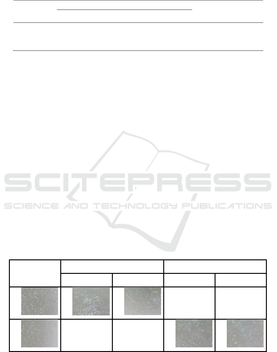
Table 1: Differentiation assay of collagen deposition in each group : control, AAPE (-) and AAPE (+)
Collagen
deposition
(fibroblast)
Groups
Total
(n=95)
p
Control
(
n=30
)
AAPE(-)
(
n=35
)
AAPE (+)
(
n=30
)
Negative 0 (0.0%) 0 (0.0%) 0 (0.0%) 0 (0.0%) 0.001
Low positive 9 (30.0%) 14 (40.0%) 0 (0.0%) 23 (24.2%)
Positive 21 (70.0%) 21 (60.0%) 30 (100.0%) 72 (75.8%)
3 RESULT
In the table 1, negative category of collagen
deposition was seen in each groups (0%). Low
positive score was observed in AAPE (-) or only
basal media groups (40%) and followed by control
group (30%), but not in AAPE (+) groups. The
increase of collagen deposition with the highest
proportion in AAPE (+) groups (100%) and the
lowest one in AAPE(-) groups (60%).
4 DISCUSSIONS
Changes in mechanical properties of the skin are
generally referred to extracellular aspects such as
alterations in polymerization and cross-linking of
collagen and elastin. In vivo study, the skin fibroblast
changes affect the separation of collagen fibers
leading to decreased collagen production (Schulze et
al., 2012).
AAPE are conditioned medium from
ADSC had specific ability to organize the protein and
secreted growth factors into extracellular
environment and had a relevant affect to various
organ and human body systems (Li et al., 2015).
Because it is derived from adipose mesenchymal
stem cell, this secretome has similar ability to its
source, such as promoting the collagen synthesis and
fibroblast migration in tissue repairment (Zhou et al.,
2016). Both ADSC and AAPE contain various
growth factors and they have ability to repaired and
renew the surrounding damaged cells. Our study
revealed there were significant differences in the
increase of collagen density among the control,
AAPE (-) and AAPE (+) groups. The highest of
collagen deposition was obtained in AAPE (+)
groups, as 100% of samples had shown positive
category in increasing of collagen deposition.
Kim et al. who studied the benefit of ADSC and
AAPE as anti aging through the activation of dermal
fibroblast by its secreted factors. Animal study
demonstrated that adding AAPE and ADSC to skin
fibroblast animal study exposed to UVB, resulting in
increased dermal layer thickness and collagen
amount, as well AAPE can reduced the apoptosis
death cell or UVB induced-apoptosis (Kim et al.,
2009).
Kim et al had studied highest concentration
of various cytokine concentration of AAPE proved
its ability to repair human dermal fibroblast, by
stimulating the wound healing in animal study, and
the last can play role in anti aging process.
Fibroblast +
UVB
exposed 30
minutes
AAPE (-)/ only basal media AAPE (+)
24 hours 48 hours 24 hours 48 hours
Figure 1. The differentiation of collagen deposition in treatment group, after UVB exposure in 30 minutes. After 30 minutes
UVB exposure, many damage fibroblasts were seen. After treatment, in AAPE groups showed repairmen of fibroblasts and
had more density of fibroblast than in basal media/ AAPE (-) groups.
RCD 2018 - The 23rd Regional Conference of Dermatology 2018
274

Analysis of cell cycle show that adding
conditioned medium of ADSC can prevent the
apoptotic cell process induced by reactive oxygen
species, by significant decreasing of sub-G1 phase
of dermal fibroblast cell (Kim et al., 2009).
5 CONCLUSIONS
From all samples (95 photos) there were significant
increasing of collagen deposition in control, AAPE
(-) and AAPE (+) groups, which the highes
deposition showed in AAPE (+) groups, with p
value 0.001. Therefore AAPE can repair the
damaged fibroblast by increasing the collagen
deposition.
ACKNOWLEDGEMENT
There is no conflict of interest while did this study
until we had all the results and did not sponsored by
any party.
REFERENCES
Baer, P.C., Overath, J.M., Urbschat, A., Schubert, R.G.H.
2016. Preconditioning of Human Adipose-derived
stromal/stem cells : Evaluation of Short-term
Preincubation regimens to enhance their regenerative
potential. J Stem Cell Res Ther;6(3):1–7.
Ichihashi, M., Ando, H., 2014. The maximal cumulative
solar UVB dose allowed to maintain healthy and
young skin and prevent premature photoaging.
Experimental dermatology. doi:10.1111/exd.12393
IHC / ICC Protocol Guide.
Kim, J.-H., Jung, M., Kim, H.-S., Kim, Y.-M., Choi, E.-
H., 2011. Adipose-derived stem cells as a new
therapeutic modality for ageing skin. Experimental
Dermatology 20, 383–387. doi:10.1111/j.1600-
0625.2010.01221.x
Kim, W.-S., Park, B.-S., Sung, J.-H., 2009. Protective role
of adipose-derived stem cells and their soluble factors
in photoaging. Archives of Dermatological Research
301, 329–336. doi:10.1007/s00403-009-0951-9
Lee, H.J., Lee, E.G., Kang, S., Sung, J.H., Chung, H.M.,
Kim, D.H., 2014. Efficacy of microneedling plus
human stem cell conditioned medium for skin
rejuvenation: A randomized, controlled, blinded split-
face study. Annals of Dermatology 26, 584–591.
doi:10.5021/ad.2014.26.5.584
Li, Y., Lei, D., Swindell, W.R., Xia, W., Weng, S., Fu, J.,
Worthen, C.A., Okubo, T., Johnston, A., Gudjonsson,
J.E., Voorhees, J.J., Fisher, G.J., 2015. Age-
Associated Increase in Skin Fibroblast-Derived
Prostaglandin E2Contributes to Reduced Collagen
Levels in Elderly Human Skin. Journal of
Investigative Dermatology 135, 2181–2188.
doi:10.1038/jid.2015.157
Moon, K.M., Park, Y.-H., Lee, J.S., Chae, Y.-B., Kim, M.-
M., Kim, D.-S., Kim, B.-W., Nam, S.-W., Lee, J.-H.,
2012. The Effect of Secretory Factors of Adipose-
Derived Stem Cells on Human Keratinocytes.
International Journal of Molecular Sciences 13,
1239–1257. doi:10.3390/ijms13011239
Pandel, R., Poljšak, B., Godic, A., Dahmane, R., 2013.
Skin Photoaging and the Role of Antioxidants in Its
Prevention. ISRN Dermatology 2013, 1–11.
doi:10.1155/2013/930164
Schulze, C., Wetzel, F., Kueper, T., Malsen, A., Muhr, G.,
Jaspers, S., Blatt, T., Wittern, K.P., Wenck, H., Käs,
J.A., 2012. Stiffening of human skin fibroblasts with
age. Clinics in Plastic Surgery.
doi:10.1016/j.cps.2011.09.008
Stojiljković, D., Pavlović, D., Arsić, I.,
Stojiljković@bullet, D., 2014. Oxidative Stress, Skin
Aging and Antioxidant Therapy. Scientific Journal of
the Faculty of Medicine in Niš 31, 207–217.
doi:10.2478/afmnai-2014-0026
Varghese, F., Bukhari, A.B., Malhotra, R., De, A., 2014.
IHC profiler: An open source plugin for the
quantitative evaluation and automated scoring of
immunohistochemistry images of human tissue
samples. PLoS ONE 9.
doi:10.1371/journal.pone.0096801
Yaar, M., Gilchrest, B.A., 2007. Photoageing: Mechanism,
prevention and therapy. British Journal of
Dermatology. doi:10.1111/j.1365-2133.2007.08108.x
Zhou, B.R., Zhang, T., Bin Jameel, A.A., Xu, Yang, Xu,
Yan, Guo, S.L., Wang, Y., Permatasari, F., Luo, D.,
2016. The efficacy of conditioned media of adipose-
derived stem cells combined with ablative carbon
dioxide fractional resurfacing for atrophic acne scars
and skin rejuvenation. Journal of Cosmetic and Laser
Therapy 18, 138–148.
doi:10.3109/14764172.2015.1114638
The Effect of Advanced Adipose-derived Stem Cell Protein Extract to Repairment of Collagen Deposition in Cultured Senescent Fibroblast
275
