
The Morphology of Contaminant Organism in Kappaphycus
alvarezii Tissue Culture
Ulfatus Zahroh
1
, Apri Arisandi
2
1
Biotechnology of fisheries and Marine Science, Universitas Airlangga
2
Departement of Marine Science, Universitas Trunojoyo Madura
Keywords: Kappaphycus alvarezii, Tissue culture, Cell morphology.
Abstract: The main problem in increasing the production of seaweed cultivation of Kappaphycus alvarezii is the
availability of quality seeds. One of the causes is because the seeds are susceptible to infectious diseases.
Tissue culture is one of the techniques to produce Specific Pathogen and Epiphyte Free /SPE
Nevertheless, the presence or absence of contamination needs to be analyzed to determine the cause of
contamination,, morphology of contaminating organisms, and changes of morphological cell of tissue
culture which can be used to prevent contamination during the next tissue culture and cultivation at the sea.
Based on the results, it could be revealed that the occurring contamination caused by bacteria and fungi as
well as caused by the less sterill culture process. Thallus morphology affected by the disease has slower
growth. There were also black spots, cotton-like substance as contaminated fungi (Saprolegnia sp and
phytopthora), and the fading of green and slimy pigment as bacteria contamination. In addition, the
morpology of ill seaweed cells has smaller cells and shrinked tissue compared to the healthy ones with their
bigger cells and no shrinkage.
1 INTRODUCTION
The species of seaweed widely cultivated in Madura
is Euchema Cottonii which is also known as
Kappaphycus alvarezii. It is the most important
seaweed and the largest on production volume in
Indonesia. This type contains Karaginan which is
useful as a Gelling agent, solidified agent, anda
fertilizer (Suryaningrum, 1998). The main problem
in increasing production of seaweed (Kappaphycus
Alvarezii) cultivation is the availability of good
seeds. One of the cause is that the seeds are
susceptible to the disease. Epiphytic attack caused
the decrease on seeds quality;, hence, the resistance
of seaweeds toward the disease is an indicator of
seaweed cultivation accomplishment. A widely
applied plain and low-cost technologyof seaweed
cultivation is not supported by the availability of
seeds which are unrestrained from disease and
epiphytic (specific pathogen and epiphyte free / spef).
Tissue culture is one of techniques to produce
quality seeds stock. Research that has been carried by
Parenrengi, et. a. in 2007; Hurtado and Biter in 2007;
Hurtado, et. al. 2009; and Yunque, et.al in 2010
revealed that seaweed K. alvarezii can be cultivated
by using tissue culture. However, those research did
not specifically examine occuring contamination and
identify the contaminant species. One of the
problems occurring in tissue culture is the
contamination. The condition of in vitro favored by
Eksplan contains sucrose and nutrient in high
concentration, high moisture, and suitable
temperature. These situations are also preferred by
microorganisms that grow more rapidly than eksplan.
A source of contaminating organisms can come from
unsterile environment, or they have existed in the cell
when eksplan of Kappaphycus alvarezii will be
cultivated through tissue culture. Therefore, it needs
to be controlled. The first step is by identifying the
types of contaminated microorganisms and by
acknowledging those symptoms.
2 LITERATURE REVIEW
2.1 Biology kappaphycus alvarezii
Kappaphycus alvarezii is a seaweed of class
Rhodophyceae. Based on the identification of the
karaginan fraction produced by Kappaphycus
alvarezii, kappa karaginan type, it is taxonomically
changed its name from Eucheuma alvarezii become
Zahroh, U. and Arisandi, A.
The Morphology of Contaminant Organism in Kappaphycus alvarezii Tissue Culture.
DOI: 10.5220/0007547605870594
In Proceedings of the 2nd International Conference Postgraduate School (ICPS 2018), pages 587-594
ISBN: 978-989-758-348-3
Copyright
c
2018 by SCITEPRESS – Science and Technology Publications, Lda. All rights reserved
587
Kappaphycus alvarezii (Patadjai 2007). The name
"alvarezii" given to Kappaphycus alvarezii comes
from the name of the Vicente (Vic) Alvarez. Vic is a
pioneer in cottonii cultivation method (Patadjai
2007). The world of seaweed trade is more familiar
with the name Eucheuma cottonii or cottonii only.
Meanwhile, seawater disease is defined as disruption
of structures and normal functions such as changes in
growth rate and appearance (color and shape) that
can affect productivity levels. Culture or tissue
culture in vitro is a method to isolate parts of the
plants grown on sterile artificial media, in sterile
culture bottles, and in aseptic conditions. Thus the
parts can proliferate and regenerate into a complete
plant. Tissue culture is a series of activities
undertaken to make plant parts (roots, buds, plant
growing tissues) grow into the whole (perfect)
condition in vitro (in glass) plants (Indrianto 2002).
The basic theories of tissue culture are: a. Cells of
a multicellular organism wherever it is located is
actually the same as a zygote cell because it
originates from one cell (omne cellula ex cellula). b.
The Cell Totipotency Theory by Schwann and
Schleiden (1898) states that the cell has Totipotency
nature that every living plant cell is equipped with
genetic information and complete physiological
devices to grow and develop into whole plants if the
conditions are appropriate. This theory believes that
every part of the plant can breed because all parts of
the plant are made up of living tissues. According on
Thorpe, (1981) , there are three main principles in
tissue culture:
a. Isolation of plant parts of whole plants (organs,
roots, leaves, stems);
b. Maintainance of the plant’s parts in the
appropriate environment and conditions of the
culture;
c. Maintenance under aseptic conditions.
3 METHOD
The implementation of tissue culture was carried out
in the Tissue Culture Laboratory Agroecotechnology
and research on contaminant organisms was
conducted at Marine Science Laboratory of
Trunojoyo University Madura. Tools used for tissue
culture process included Bottle Culture, Filter Paper,
Funnel, Glass Beaker 250 ml and 500 ml, Autoclave,
Laminar Flow, Microscope, Glass preparation +
cover glass and other tools.
Seaweed materials used were Kappaphycus
alvarezii cultivated by farmer groups in Aengdake
Village, Bluto District, Sumenep Regency. Conway
was utilized for tissue culture media and the seawater
was also sterilized.
The selected explant source had the following
criteria: (a) had many branches, dense and spiky
leaves, (b) no spots found and peeling (c) had specific
bright color (d) was live less 35 days, (e) weighed
between 50-100 grams per rumpon and was not
exposed to ice-ice disease. Tool sterilization used
70% alcohol and autoclave; while the material for
eksplan sterilization utilized 0,5% betadine and 25
ppt sterile sea water. The stages of tissue culture
implementation to explant observation was
conducted by methods from Hurtado and Biter
(2007) as mentioned below:
a. Preparation of tools and materials
1. Preparing the tool
▪ Washing the appliance with running water.
▪ Sterilizing equipment to be used with alcohol,
wet sterilization using autoclave with
temperature of 1210C at 1.5 atm pressure for 20
minutes
2. Preparing materials
▪ Filtering sea water into culture bottle ± 100ml
▪ Saving seawater that had been in autoclave in
the sterile room
▪ Filtering the media conway and sterilizing it
into an autoclave with a temperature of 121
0
C at
a pressure of 1.5 atm for 15 minutes.
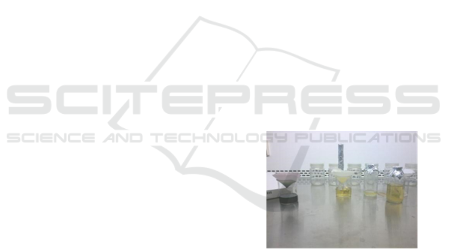
Figure 3.1 Media Conway filter
.
b. Planting stage culture
Bottles containing media, as well as other
planting tools, were sprayed first with 70% alcohol.
Cut where Thallus was taken and put it into a
disinfectant solution I (1% betadine solution where
concentration 1 ml per 100ml seawater). Ten minutes
later, rinsed with sterile distilled water for subsequent
inclusion in a solution of disinfectant II (0.5%
betadine solution where 0.5 ml per 100ml seawater)
for 10 minutes. Then rinsed with sterile aquades for
3 times, aiming for no remnants of disinfectant
material stick on eksplan then drained in petridish.
ICPS 2018 - 2nd International Conference Postgraduate School
588

Implant planting was done in laminar air flow.
Eksplan should be planted in culture bottle
containing media; there was an eksplan planted on
each bottle by using tweezers clamp. This activity
was carried out in a laminar flow, placed in a room
temperature of 25 oC. The media was of 25 ppt
salinity, pH 7.5 and the explants were cultured in a
culture bottle amounted to 120 units. Then the bottle
was closed, removed, and arranged on a culture rack
in accordance with the placement plan, given 10
watts fluorescent lighting.
The location of the lamp was 30 cm on the top of
the shelf away from the culture bottle. The lighting
was programmed for 24 hours continuously.
c. Subculture stage
Every 5 days, eksplants grew in subculture to a
new culture bottle containing tissue culture media
with the same concentration and parameters with
previous culture media.
d. Maintenance stage
Maintenance is conducted on the culture room by
maintaining cleanliness and room temperature.
Culture bottles containing media and explants were
sprayed with 70% alcohol every day and
contaminated plants and media were immediately
removed from the room and observed.
e. Stage of data collection
Observation was carried out every day to see the
contamination of bacteria or fungi. Contaminated
media and explants were removed from the room and
their cells as well as their contaminants (Hayashi et
al. 2008) were observed.
The data taken include:
a. Percentage of contamination
The percentage of contamination is calculated
using the formula previously performed by
Amiluddin (2007) as listed below:
C (%) = A x 100%
T
description:
C: percentage of infected seaweed (%)
A: number of explants or infected seaweed
T: number of explants or bonded seaweed
points observed
b. Morphology cell
Cell of morphology was observed using the
microscope 100L Olympus with 100 times
magnification. It was photographed on size
1600 x 1200 with ISO 100 (Yulianto 1993).
c. Contaminant agent
The presence of epiphytic diseases and epiphytes
that infect the explants (seaweed) was observed
based on signs of morphological abnormalities.
Observed morphological abnormalities were
identified by looking at and comparing with the
images contained in the literature. The
morphology of contaminant organisms was
observed using the Olympus 100L microscope
with magnification 100 times. It was
photographed at the size of 1600 x 1200 with ISO
100 (Yulianto 1993).
d. Survival rate
The rate of seedling survival explants of K.
alvarezii. Based on Amiluddin (2007) was
calculated using the following formula:
S
SR (%) =
S
+
M
x 100
description: SR = Survival Rate (%)
S = Number of living seaweed
M = Number of dead seaweed
4 RESULTS
Epifit conditions on control treatment
Seaweed affected epiphytes in the sea
is characterized by the presence of
algae filament attached to the epidermis of the thallus
of appaphycus alvarezii as seen in Figure 4.1. Based
on the identification in the type and characteristics of
filament algae in accordance with Largo (2002), the
species identified are polysiphonia sp which is an
epiphytic competitor algae.
(a) (b)
(c)
Figure 4.1 Polysiphonia sp. on Kappaphycus alvarezii. a)
The state of Thallus the contaminated. b) morphology of
Polysiphonic attached to the epidermis thallus in a
magnification microscope 100x. c) Polysiphonia according
on Largo (2002).
The Morphology of Contaminant Organism in Kappaphycus alvarezii Tissue Culture
589
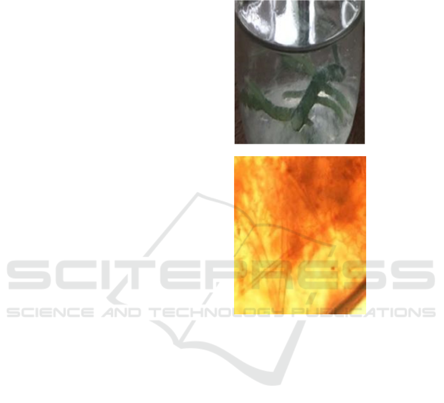
The existence of competitor algae can result in
disruption of the thallus in obtaining nutrients and
light. If the components needed in metabolism are
reduced; then over time, the seaweed Kappaphycus
alvarezii can become thin, flabby, pale and can cause
death.
The observation showed the presence of
abnormalities in tissue are characterized by: a
dangling lump in the epidermis, morphological
observations through a microscope found that
Polysiponia had afilament dangling and the base
through the part of the cell wall of Kappaphycus
alvarezii. According to Darmayati et. al. (2001),
Polyshiponia sp. is type of Rhodophyta-rhinoceros
and is a competitor algae in seaweed that can cause
disturbance in seaweed photosynthesis resulting
from the covering of thallus surface by
Polyshiponia.
Epiphytic organisms in tissue culture
Organisms that contaminate seaweeds resulting
from tissue culture which consists of fungi and
bacteria.
Contamination of fungus
Contaminated fungi consists of Saprolegnia sp.
and Phythopthora sp.,mushrooms of Oomycota class
that can only grow in environment with high
moisture or aqueous.
a. Saprolegnia sp.
The contaminated seaweed Saprolegnia sp. is
characterized by the whitening of thallus, mucus
covered by dirt like white flour, the peeled outer skin
or epidermis to reveal deep cell or medullaryin of the
tissue thallus, and the presence of white hifa on the
surface of the media.
Morphology Saprolegnia sp. as contained in Figure
4.3.
Saprolegnia sp. has a characteristic feature that
can grow at a temperature range of 0-35° C, with an
optimum growth interval of 15-30°C. It is a fungus
group of Oomycota also called a water fungus that
can live in aqueous / high humidity environment.
Saprolegnia sp. commonly attacks the injured part,
and may subsequently spread to other healthy tissues.
According to Wilfred et al. (1965) in Ningsih (2011),
the fungus of the family Saprolegniaceae can live in
freshwater and saltwater. Zoospora groups of these
fungi search for fertile substrate, then settle down and
start producing hypha. Mycelia grow over the
wounded tissue or the site of infection, and then
spread to normal tissue around the site of infection.
The fungal enzyme secreted by the fungus destroys
the surrounding tissue, kills cells, and progresses
mycelia. It is very dense and sticking out into the
water, making it look likes cotton.
(a)
(b)
Figure 4.3. Morphology of saprolegnia sp. a) Thallus
affected by fungi b) Endogenous of Saprolegnia sp. in
thallus tissue.
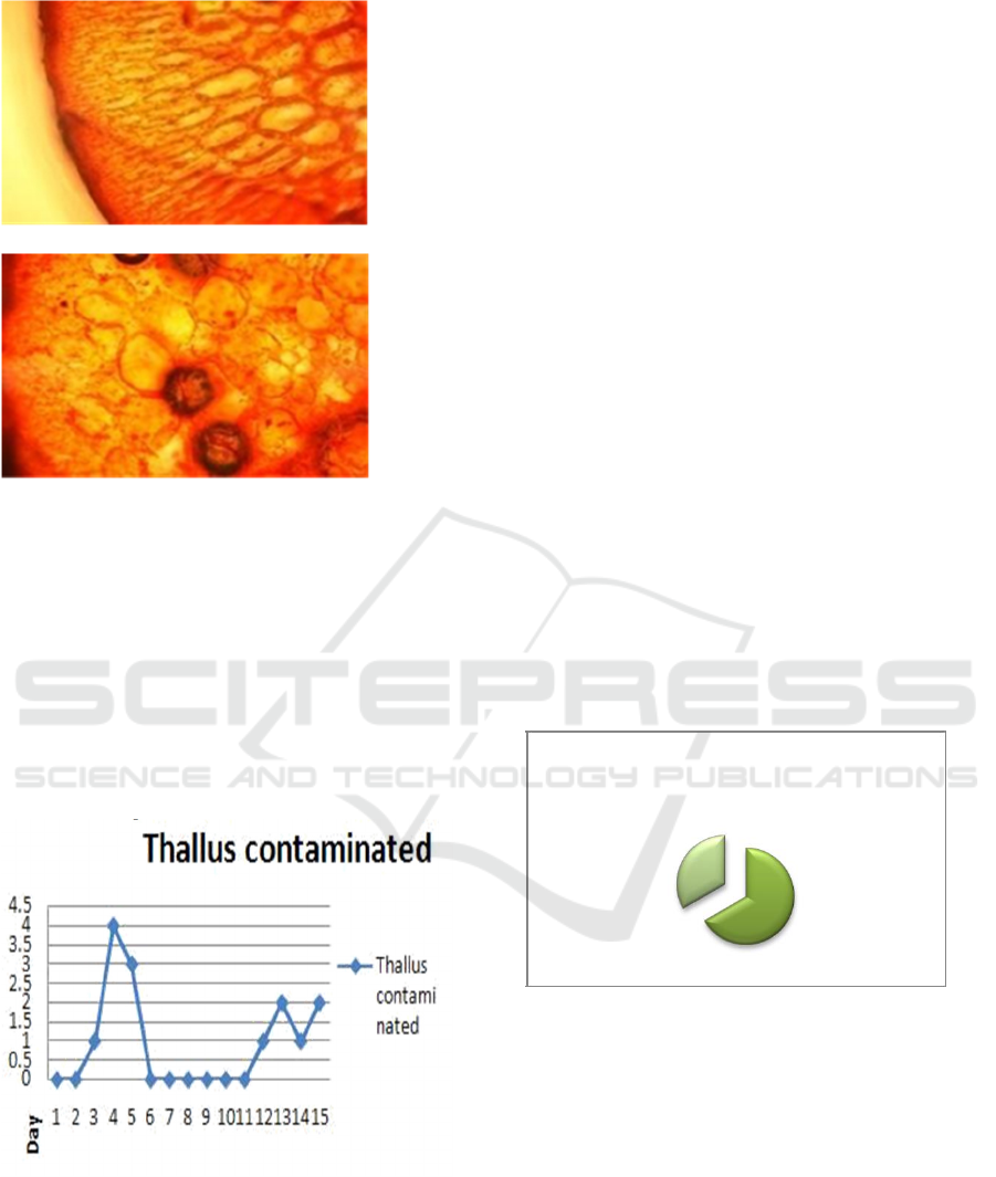
b. Phytophthora sp
Contaminated Thallus is pale green and has
specks of black spots. The morphological
observations of the tissues show the presence regions
of dark and rounded as shown in Figure 4.4 below:
ICPS 2018 - 2nd International Conference Postgraduate School
590
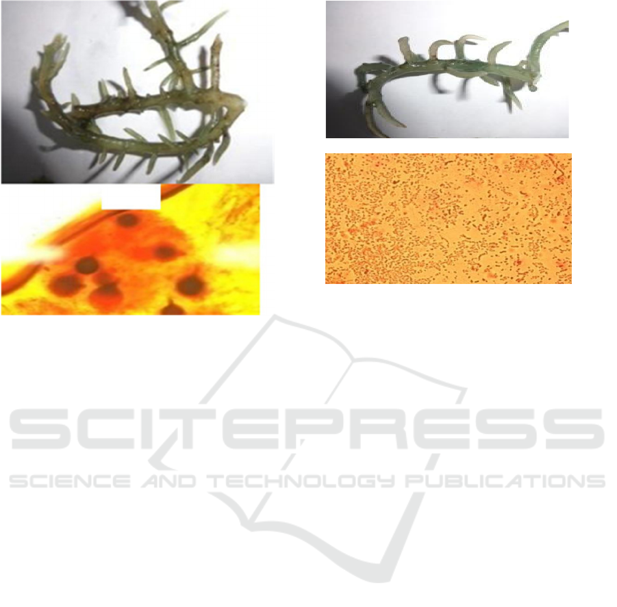
Figure 4.4 Contamination Phytophthora sp in thallus a)
Contaminated Morphology thallus, b) Morphology of
contaminated tissue Phytophthora sp at 100x
magnification microscope.
Diagnosis done in laboratories by taking mycelia
wasplaced on the surface of the glass slide and given
a little water for further observation under the
microscope. Mycelia cause saprolegniasis to have
branching with aseptate hypha structure.
The morphology of Phytophthora sp.,
sporangium, is oval to slightly rounded or pear-
shaped, whieh its spore has a whip feather (flagella)
that can move in water.
The pathogens may form in round
chlamydospores (Directorate of Holticulture
Protection 2011).
Contamination of bacteria
Contamination due to bacteria characterized by
exsplants showed symptoms of wilting and stingy
media smells. The observation of the thallus tissue
shows the presence of a ruptured cell due to a
bacterial infection. Observation of bacterial
morphology is done by first breeding bacteria
attached to wall of thallus in media agar and given
gram staining, then identified through electron
microscope. After doing bacterial culture in media
agar and identification under microscope, the result
showed that morphology of bacteria is single coccus,
paired, chained, and in the form of gram negative.
Thus, it indicated that the bacterium was
Streptococcuss sp.
The morphology is as shown in Figure 4.5 below:
(a)
(b)
Figure 4.5 Contamination on thallus by bacteria a)
morphology Thallus b) bacterial on a 100x magnification
through microscope.
Based on Figure 4.5, it was found that the
morphology and color of bacteria originating from
the culture room on the control are relatively the
same as the result of identified contaminant in the
contaminated explants. So, the contaminant
organism is indicated to have been present in the
source of the explants.
Changes in cell morphology
Cells observed in healthy thallus tissue appear more
clearly with scattered prevalence of staining (safranin)
throughout the surface of seaweed tissue. The vividly
dispersed safranin indicates that the liquid cell is
absorbed evenly by every space within the cell because
the state of the cell
wall is intact and no cytoplasm is
broken while the contaminated thallus tissue has an
incomplete cell shape and looks uneven due to the
damage on cell components or rupture of the
cytoplasm.
According to Juwono and Juniarto (2002) and
Lakitan 2011 in Arisandi (2011), the cell wall of the
plant has a main function as a protector of the cell
framework. When the cell wall is damaged by the
disease, it can lead to changes on shape and size of
the cell. Damage to the cell wall can interfere with
the absorption of nutrients into cells; it will also
disrupt metabolism and inhibit cell division. The
worsening damage causes the cell wall to burst,
leaving the fluid out and causing the cell to become
irregular and shrink (plasmolysis) subsequently as a
beginning to death (Musa and Wei 2008 in Arisandi
2011). The difference between healthy and
contaminated cells can be seen in Figure 4.6
The Morphology of Contaminant Organism in Kappaphycus alvarezii Tissue Culture
591
(a)
(b)
Figure 4.6. The difference between healthy and
contaminated cells in a 100x magnification. a) Healthy
cells, b) cells are contaminated
.
Contamination percentage
The percentage of contamination is the ratio of
thallus of infected seaweed to cultured seaweed,
calculated in percent. Based on the calculation of the
contamination, percentage on culture tissue activity
is 11.7% and the percentage of healthy seaweed is
88.3% with the amount of contamination per day is
as shown in Figure 4.7 below
Figure 4.7 Number of Contaminated Culture Bottles.
In the first week, contaminated grass sea culture
were in among 8 bottles; where the 6 bottles were
characterized by bleaching thallus, mucus covered by
dirt such as flour white, and the outer skin or
epidermis flaked. While in two other bottles, there
were black spots and brown line on the wall of the
thallus. Contamination in the first week was occurred
on day 3, day 4, and day 5; with the highest number
occurred on day 4. In the next week, the
characteristics of contamination were among in 6
bottles. Three bottles illustrated bleaching thallus
and three other bottles obtained black spots and
brown line on the wall of the thallus. Semangun
(2001) said that between the infection and the
seemingly occasional symptom, there is a long period
of time yet usually the symptoms of the disease will
appear after infection.
Surival rate
In the tissue culture activities conducted, the
survival rate was approximately 62.5%, which the
number of dead seaweed were as many as 45 bottles
and alive ones were 75 bottles as shown in Figure 4.8.
This indicates that the success rate of culture is
proven through the number of alive seaweed was
higher than the dead ones. Based on Perkasa (2011),
this survival rate is one of the determinants of success
in tissue culture activities. If the number of living
seaweed on the harvest is high and the number of
death is low, then the value of survival will be high.
Otherwise, if the number of the death is higher than
the other, the survival rate might be lower or below
as in the diagram:
Survival Rate
Figure 4.8 Survival rate level of tissue culture.
5 CONCLUSIONS
Based on the results obtained, it could be concluded
as follows:
1. There were two species of contaminants in
seawater tissue culture of Kappaphycus alvarezii:
fungus Saprolegnia sp., phythophthora
sp. and bacterium Streptococcus sp.
2. Fungal contamination was indicated that it is
already exist in prospective explant of seaweed to be
cultured, because the type of fungus is a water fungus
Not Survive
31,3 %
Survive
62,50%
ICPS 2018 - 2nd International Conference Postgraduate School
592

or Oomycota that can only grow in environments
with high moisture or watery levels.
3. Contaminated cells were smaller in size and
looked more shrunken than cells in tissue. A healthy
thallus looks bigger and has more sturdy cell walls
in support of its cell shape.
REFERENCES
Amiluddin, NM, 2007. Study Growth and Composition
Carrageenan Seaweed Disease Affected Kappaphycus
alvarezii Ice-ice in the Sea of Pari Island Thousand
Islands. Thesis. IPB. Bogor. 78 p.
Anggadiredja J, S Irawati, and Kusmiyati. 1996. Potential
and Benefits of Indonesian Seaweed in the
Pharmaceutical Sector. National Seminar on Seaweed
Industry. Jakarta. p. 49-62
Atmadja WS, A Kadi, Sulistijo and
Rachmaniar.1996. Introduction of Indonesian Seaweed
Types. Puslitbang Oseanologi -
LIPI, Jakarta.
Atmadja WS and Sulistijo. 1977. Efforts to Utilize
Euchema spinosum (L) J. AGRADH Sea Algae
Cuttings Seeds in Thousand Islands to be Cultivated.
In: Jakarta Bay, Resource Oceanological Properties
and Problems. Editor: M. Hutomo, K Romimohtarto
and Burhanudin. LON LIPI, Jakarta: pp. 433-449.
BBPP Lembang. 2008. About Network Culture,
(Online), http://www.bbpp-lembang.info/
Accessed July 14, 2013. Gadjah
Mada. Yogyakarta.
Darmayati, Y., A. Hatmanti, N. Farida and Surahman.
2001. Study of pests and diseases. Final report on
Research on Development of Seaweed Superior Seeds,
Management of Water Quality and Pests and Diseases.
Deep Sea Resources Research, Development and
Utilization Project. Oceanography Research Center-
LIPI Jakarta. 7 things.
Ministry of Maritime Affairs and Fisheries. 2003. Profile
of Indonesian Seaweed. Draft.
Directorate General of Aquaculture. Jakarta. 111 p.
Directorate General of Fisheries. 2004. Seaweed Pests and
Diseases.
Doty M. 1985. Euchema alvarezii sp.nov (Gigartinales,
Rhodophyta) from Malaysia. Inside: Abbot IA, Norris
JN (editors). Taxonomy of Economic Seaweeds.
California Sea Grant College Program. P 37-45.
Haryono Semangun. 2011. Plant disease. Gadjah Mada
University press: Yogyakarta.
Hayashi, L., Yokoya, N.S., Kikuchi, D.M., and Oliveira,
E.C., 2008. Callus Induction and Micropropagation
Improved by Colchicine and Phytoregulators in
Kappaphycus alvarezii (Rhodophyta, Solieriaceae). J.
Appl. Phycol., 20: 653-659.
Hurtado A.Q., and Biter A.B., 2007. Plantlet Regeneration
of Kappaphycus alvarezii var. younger siblings by
tissue culture. Journal of Applied Phycology, 19: 783-
786
Hurtado, A.Q., Yunque, D.A., Tibubos, K., and Critchley,
A.T., 2009. Use of Acadian Marine Plant Extract
Powder from Ascophyllum nodosum in Tissue Culture
of Kappaphycus alvarezii.Journal of Applied
Phycology, 21:633–639
Hutabarat J. 1995. Workshop on Marine Cultivation: Bio
Hydrography Conditio Evaluation in Determining
the Location of Aquaculture, Jepara.
Indriani H and E Sumiarsih. 1999. Aquaculture,
Management and Marketing of Seaweed. PT. Penebar
Swadaya, Depok. IPB (1997).
Indrianto. 2002. In Vitro Selection of Andalas Plant
(Morus macroura Miq.) Tolerant Drought Stress Using
Polyethylene Glycol (PEG). Proceedings of the 21st
Western Conference BKS-PTN Seminar and Meeting.
Kresno Yulianto. 2004. Phenomenon of controlling factors
causing losses in caraginophyte cultivation in
Indonesia. OseanaISSN Journal 0216-1877 LIPI, 29:
Largo, B.D., K. Fukami and T. Nishijima. 1995.
Occasional pathogenic bacteria promoting ice-ice
disease in the carrageenan-producing red algae
Kappaphycus alvarezii and Eucheuma denticulatum
(Solieriaceae, Gigartinales, Rhodophyta). Journal of
Applied Phycology 7: 545 – 554
Lobban CS, PJ Harison. 1994. Seaweed Ecology and
Physiology. Cambridge Univ. Press
New York.
Lobban, C. S and Harrison, P. J. 1995. Seaweed Ecology
and Physiology. Cambridges University Press. 366 pp.
Nasution MH 2005. Pathogenicity of Several
Bacterial Isolates Against Seaweed Kappaphycus
alvarezii from Pari Island, Thousand Islands. Faculty
of Biology, National University of Jakarta. Jakarta.
Neish, I.C. 1989. Alkali Treatment of Carragenan Bearing
Seaweeds Past, Present and Future. FMC Corporation,
Marine Colloid Div. 11 pp.
Nontji 1993. Laut Nusantara. Penerbit Djambatan: Jakarta
Patadjai Hospital. 1993. Effect of TSP Fertilizer on the
Growth and Quality of Seaweed Gracilaria gigas Harv.
Thesis Study Program on Aquatic Sciences. IPB
Postgraduate Program. Bogor.
Fish quarantine center of the marine and fisheries
department. 2007. Standard method of bacterial HPIK
examination: Directorate General of aquaculture.
Fish quarantine center of the marine and fisheries
department. 2007. HPIK class of HPIK examination
standard method: Directorate General of aquaculture.
Fish quarantine center of the marine and fisheries
department. 2007. Standard method of HPIK parasitic
examination: Directorate General of aquaculture.
Schwann dan Schleiden.1898. Plant Propagation by Tissue
Culture 3rd Edition. Springer Publisher: Netherlands
Soegiarto A., WS Atmadja, Sulistijo and
H. Mubarak. 1978. Seaweed (Algae):
Benefits, Potential and Cultivation Efforts. LON-LIPI,
Jakarta.
Sulistiyo. 1988. Root Induction from Leaf Leaf Explants of
Three Patchouli Varieties (Pogos temon cablin Benth)
in MS Media Containing Paclobutrazol In Vitro Zuriat.
19 (1): 16-31
The Morphology of Contaminant Organism in Kappaphycus alvarezii Tissue Culture
593

Suryaningrum TD. 1988. Study of Quality Properties of
Seaweed Commodities Cultivation of Eucheuma
cottonii and Eucheuma spinosum. IPB Thesis. Bogor.
Thorpe.1981, Review: In Vitro Selection and
45Somaclonal Variation for Biotic and Abiotic stress
Tolerance.Biodiversitas. 7 (3): 297-301
Widianti. 2003. Introduction to Tissue Culture Techniques
and Vegetative-Modern Plant Propagation Guidelines.
Yogyakarta: Kanisius.
Wetherell, D. F. 1982. Introduction to Plant Propagation in
Vitro. Koensoemardiyah S. SU, translator; Semarang:
IKIP Semarang Press. Translation of: Introduction to
In Vitro Propagation
Yusuf M.I., 2004. Production, Growth and Content of
Seaweed Carrageenan Kappaphycus alvarezii (Doty)
Doty (1988) which was cultivated with Different Water
Media Systems and Thallus Seeds. Dissertation: Ppps.
Universitas Hasanudin. Makassar. Pp.13-15.
ICPS 2018 - 2nd International Conference Postgraduate School
594
