
Effect of Macrophage Addition and Incubation Time to Interferon
Gamma (IFN-) Levels on Tuberculosis Granulomas In Vitro Models
Yusuf Eko Nugroho
1,2
, Agung Dwi Wahyu Widodo
3
and Jusak Nugraha
4
1
Post Graduate Student of Master of Immunology, Faculty of Pasca Sarjana, Universitas Airlangga,Indonesia
2
Department of Stem Cell Institute of Tropical Disease, Universitas Airlangga, Indonesia
3
Department of Microbiology Clinic, Faculty of Medicine, Dr. Soetomo Hospital, Indonesia
4
Department of Patology Clinic, Faculty of Medicine, Dr.Soetomo Hospital, Indonesia
Keywords: granuloma, macrophage, IFN-.
Abstract: Tuberculosis (TB) is an infectious disease caused by stem bacteria Mycobacterium tuberculosis (Mtb)
which can cause latent infection. Mtb enters through aerosolization which then infects and activates
macrophages and dendritic cells present in the lungs. The active dendritic cells of coumadin present an
antigen which has been processed in peptide form into the lymphocyte cell which later becomes granuloma.
Granulomas are compounds of tissue composed of infected macrophages and multinucleated giant cells,
known by aggregates of new monocytes or macrophages, and neutrophils and lymphocytes. Macrophages
have a duty to kill Mtb germs. Different types of macrophage phenotype in granulomas with various
functions, including anti-mycobacterial effector action, produce cytokines ie IFN-γ. In humans, IFN-,
released by activated Th1 cells, is the major lymphokine to activate the search process and antimicrobial
activity that eliminates mycobacteria. This study aims to determine the relationship between IFN- and In-
vitro method using PBMC infected with Mtb germ with the addition of Macrophage with concentration
1,2,3x105 with incubation time 1,2,3,4 and 5 days to determine the level of IFN - using ELISA test. With
use with day variables of macrophages 1x10⁵, 2x10⁵, and 3x10⁵ analized with One-Way ANOVA obtained
values p = P = 0.7201 The value is greater than 0.05 (p> 0.05). There is no meaningful difference.
1 INTRODUCTION
Tuberculosis (TB) is an infectious disease caused by
stem bacteria Mycobacterium tuberculosis (Mtb).
TB is a disease with the highest rates of morbidity
and mortality especially in developing countries and
is also a problem of chronic infection in the world
(Santoso et al., 2017). In addition to causing active
disease, Mtb can cause latent infection. Latent
infection results in one third of the world's
population carrying asymptomatic infections that
can produce 8 million new TB cases and 2 million
deaths annually (WHO, 2011; Birkness et al., 2007).
Mtb enters through aerosolization which then
infects and activates macrophages and dendritic cells
present in the lungs. The active dendritic cells of
kemuadin present treated antigens in peptide form to
CD4 T cells. The activated lymphocytes and
infected macrophages, in the inflammatory response
of cytokines and chemokines migrate to the infection
section and form an arrangement called granuloma,
where Mtb is in the dorman phase (Kapoor et al.,
2013).
Granulomas are tissue compounds consisting of
infected macrophages and multinucleated giant cells,
surrounded by aggregations of new monocytes or
macrophages, and neutrophils and lymphocytes
(Parasa, 2014). Macrophages have an important role
of granuloma. Macrophages serve as early cells of
granuloma formers. Macrophages have a duty to kill
Mtb germs. Different types of macrophage
phenotype in granulomas with various functions,
including anti-mycobacterial effector mechanisms,
produce proinflammatory and anti-inflammatory
cytokines, chemokine secretions and proteins
associated with tissue remodeling. These cells play a
major role in infection control in granulomas (Flynn
et al, 2011).
IFN-γ is a major cytokine involved in the
immune response to mycobacteria, and its primary
function is the activation of macrophages, enabling
them to use their microbicide role function (Khan et
392
Nugroho, Y., Widodo, A. and Nugraha, J.
Effect of Macrophage Addition and Incubation Time to Interferon Gamma (IFN-g) Levels on Tuberculosis Granulomas Invitro Models.
DOI: 10.5220/0007543603920397
In Proceedings of the 2nd International Conference Postgraduate School (ICPS 2018), pages 392-397
ISBN: 978-989-758-348-3
Copyright
c
2018 by SCITEPRESS – Science and Technology Publications, Lda. All rights reserved

al., 2016). IFN-γ along with IL-12, IL-6, and TNF-α
stimulate the production of oxidative explosions,
thus mediating the function of tuberculostatic
macrophages, as well as stimulating immune cell
migration to the site of infection, contributing to
granuloma formation, which controls disease
progression (Khan et al., 2016).
Based on this background, researchers wanted to
know the role of adding macrophages to IFN-y
levels in granuloma models using ELISA.
2 MATERIALS AND METHODES
2.1 RPMI
The growth media used in this study was the
Roswell Park Memorial Institute (RPMI) 1640
which was obtained already in the form of ready-to-
use solution. RPMI 1640 media is a medium used
for cell and tissue culture, usually used for the
growth of human lymphoid cells. This medium
contains a large amount of phosphate and is
formulated for use in air with a 5% CO
2
atmosphere.
RPMI 1640 uses the bicarbonate buffer system so
that it enables the growth of several types of cells,
especially T lymphocytes, hybridomas. There are
several series of RPMI most often used is RPMI
1640.
2.2 PBMCs
Peripheral Blood Mononuclear Cells = PBMCs are
cells made from human blood which are then
processed for the PBMC cell capture. Sample
criteria are adult blood, the blood used should be
new blood taken can not be blood that has been
stored for too long, blood comes from healthy
people and does not suffer from tuberculosis
infection, there is no specific provision for sex either
male or female. Suggestions from the blood
researcher used should come from one person only,
because the immune response of each individual is
different so if coming from more than one person in
worry affects the outcome. The number of PBMC
used in this study was 10
6
in each well.
2.3 Mycobacterium Tuberculosis
This study used bacterial isolates Mycobacterium
tuberculosis H37Rv obtained from the Laboratory of
Microbiology, Institute of Tropical
Disease,Airlangga University Surabaya with
concentration 10
5
in each well. Comparison of
PBMC and bacteria concentrations used in this study
was MOI 1: 0.1.
2.4 Macrophage Issolation
Taken buffy-coat about 60 ml. Then prepare 50 ml
conical tubes with histopags each 15 ml. Prepared
one tube every 10 ml of buffy-coat. Histopags are
used at room temperature. Plate at 10 cm culture
dishes (10 ml / dish) incubated at 1-2 hours at 37ºC
5% CO2. Observe macrophage cells under a
microscope and then make doses 1,2 and 3x10
5
.
2.5 Procedures
Divided into 4 groups. Group I was given PBMC
and Mycobacterium tuberculosis bacteria on RPMI
media as control. Group II was given PBMC,
Mycobacterium tuberculosis and 1x10
5
macrophages on RPMI media. Group III was given
PBMC, Mycobacterium tuberculosis and 2x10
5
macrophages on RPMI media. Group IV was given
PBMC, Mycobacterium tuberculosis and 3x10
5
macrophages on RPMI media. Prepared well plates
that already contain RPMI media. Enter 1x10
5
PBMC cells into all wells. Inoculated with 1x10⁵
M. tuberculosis strain H37Rv bacterial isolates into
all wells. Added macrophages as much as 1x10
5
,
2x10
5
and 3x10
5
cells into group II, III and IV.
Plate was incubated at 37°C with 5% CO2
condition. Observed on days 1,2, 3, 4 and 5. Every
day 100uL of supernatant was taken to test IL-10
levels using the ELISA test at 450nm wavelength.
3 RESULTS
3.1 Direct Granuloma Observation
The method used for direct observation is performed
directly under an inverted microscope using
specimens of living cell cultures in the plate / well
with the above lighting system followed by the lens
system on the microscope base. The direct
observational images present in this journal against
the control group (no macrophage), macrophage
addition of 1,2 and 3x10
5
and then incubate for 1
until 5 days in 37
o
C.
Effect of Macrophage Addition and Incubation Time to Interferon Gamma (IFN-g) Levels on Tuberculosis Granulomas Invitro Models
393
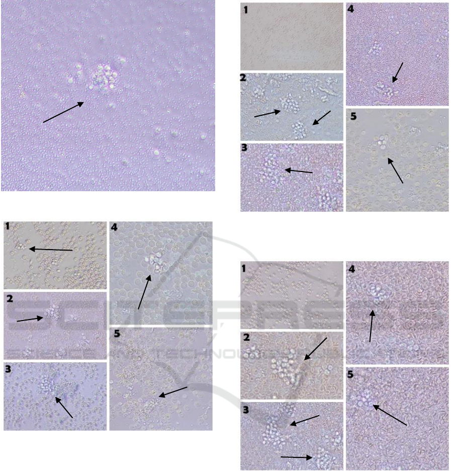
Figure 1: PBMC infected M.Tb without macrophage
Figure 2: Direct observation of the group with the addition
of 2x10
5
macrophage (400x magnification
Figure 3: Direct observation of the group with the addition
of 2x10
5
macrophage (400x magnification)
Figure 4: Direct observation of the group with the addition
of 3x10
5
macrophage (400x magnification)
Day three (each dose) is the culmination of
granuloma formation. It is clearly visible from the
solid structure, with the many number of cells. Cell
aggregation appears larger than the first and second
days. Immune cell cells on the third day begin to
respond to Mtb for further elimination.
ICPS 2018 - 2nd International Conference Postgraduate School
394
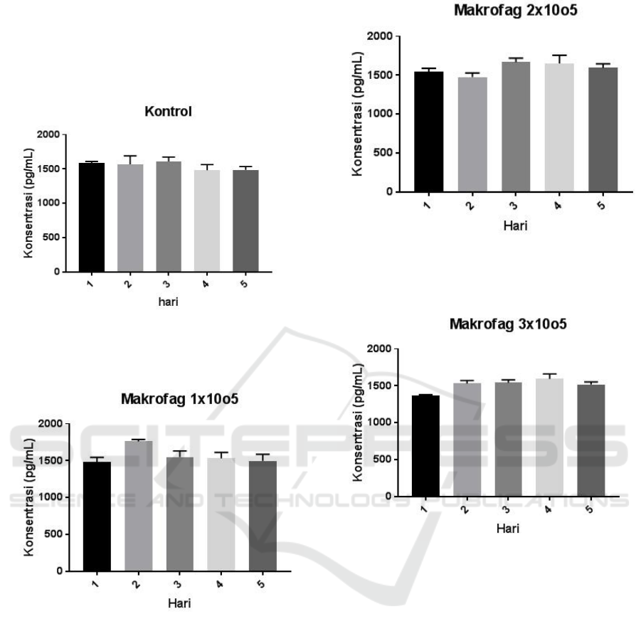
3.2 Examination the levels of IFN-
Samples in the form of supernatant were then examined by
IFN-g using ELISA method and got the average result
from each treatment either control or sample.
Figure 5: IFN- secretion control without the addition of
macrophages by day variation (pg / mL)
Figure 6: IFN-y secretion with the addition of 1x10
5
macrophages based on day variation (pg / mL)
Figure 7: IFN-y secretion with the addition of 2x10
5
macrophages based on day variation (pg / mL)
Figure 8: IFN-y secretion with the addition of 3x10
5
macrophages based on day variation (pg / mL)
IFN-y examination results with ELISA method of 4
groups ie without the addition of macrophages with the
addition of macrophages 1x10
5
, 2x10
5
, 3x10
5
showed high
levels. The highest level occurred on day 2 at
concentrations of 3x10⁵. On the 3rd day showed an
increase in levels which then tend to fall on days 4 and 5.
Effect of Macrophage Addition and Incubation Time to Interferon Gamma (IFN-g) Levels on Tuberculosis Granulomas Invitro Models
395
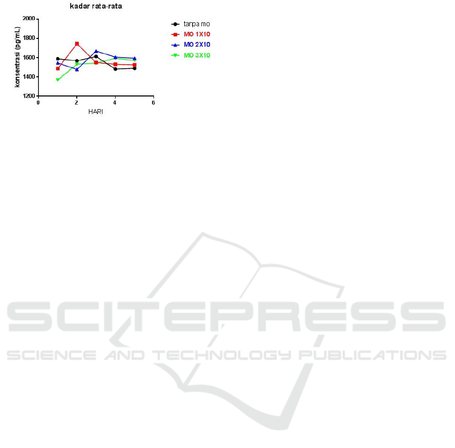
Figure 9: average IFN-y levels
The test results using One-Way ANOVA which
aims to determine the significance of the price of the
proportion (p). In groups without macrophages with
a group of 1x10⁵, 2x10⁵, and 3x10⁵ macrophages, p
= 0.451 was obtained. the value is greater than 0.05
(p> 0.05) thus indicating no significant difference.
3.3 Discussion
The formation of granulomas is a dynamic process
that begins immediately after infection and
continues to develop over time. Typically,
granulomas can be divided into three distinct phases:
(1) "congenital granuloma," a loose aggregate
consisting of macrophages and recruited neutrophils;
(2) "immune granuloma" is formed after the
emergence of antigen-specific T cells; and (3)
"chronic granulomas," resulting from different
morphological changes in granuloma structures
(Shaler et al., 2013).
After innate activation, APC cells are recruited to
the lungs and transport mycobacteria to mediastinal
lymph nodes. APC activates antigen-specific T cells.
Because of the nature of M.tb infection, the majority
of bacilli and antigen are in the endosome, and most
efficiently loaded into the major histocompatibility
complex (MHC) class II. Class II MHC loading
facilitates priming of the interferon gamma TH1
(IFN-γ) which is T cell discretion, which rapidly
returns the lung. While the dominant subset of T
cells is CD4 +, the cross presentation also allows
strong induction of CD8 + T cells, collectively
resulting in a polarized type 1 adaptive immune
response
Macrophages are important effector cells in
immunity against intracellular bacteria. In infection,
macrophages (MO) recognize mycobacteria with
Toll Like Receptor (TLR) involvement (mainly
TLR1 / 2 and TLR2 / 6) followed by phagocytosis
and mycobacterial growth control. In addition,
macrophages and dendritic cells also secrete
cytokines such as IL-12 and IL23 to induce IFN-
produksi production by T and NK cells, which, in
turn, increase phagocytosis, fagolososomes fusion,
oxidative bursts (Khan et al., 2016).
The addition of macrophages with different
doses does not affect the levels of IFN-y. this is
because IFN-y levels tend to be produced by T cells,
especially Th1 to stimulate macrophages more
actively in phagocytosis mtb. In this case the T cell
in generating IFN-y is independent.
ACKNOWLEDGEMENTS
The authors would like to thank the technicians of
the Stem cell Research Centre and Tuberculosis and
Leprosi Laboratory of Tropical Diseases (ITD) of
Airlangga University and all those who have assisted
in the completion of this research..
REFERENCES
Birkness A K, Jeannette G, Suraj B S, Ralph A T,
Kathryn L K, Jeanine B, Frederick D Q.
2007. An In Vitro Model Of The Leukocyte
Interactions Associated With Granuloma
Formation In Mycobacterium Tuberculosis
Infection. Immunology and Cell Biology vol.
85, 160–168.
Cowley Siobhán C. and Karen L. Elkins (2003).
CD4 T Cells Mediate IFN Independent
Control of Mycobacterium tuberculosis
Infection Both In Vitro and In Vivo .The
Journal of Immunology. 171:4689-4699.
Flynn JR, Chan J, Lin PL. 2011. Macrophages And
Control Of Granulomatous Inflammation In
Tuberculosis. Mucosal Immunol. Vol. : 4(3) ;
271-278.
Kapoor N, Santosh P, Tatiana D S, Chirajyoti D,
William L W, Pappachan E K. 2013. Human
Granuloma In Vitro Model, for TB Dormancy
And Resuscitation. PLOS ONE vol. 8 : issue
1.
Parasa V R, Muhammad J R, Anh T N H, Mattias S,
Susanna B, Maria L. 2014. Modeling
Mycobacterium tuberculosis Early
Granuloma Formation In Experimental
Human Lung Tissue. Disease Models &
Mechanisms vol. 7 : 281-286.
Santoso G A, Hidayat S, Dwi YNH. 2017. Pengaruh
Infeksi Mycobacterium Tuberculosis Strain
ICPS 2018 - 2nd International Conference Postgraduate School
396

H37rv Terhadap Ekspresi Tnf-Α Pada
Jaringan Otak. MNJ , Vol.03, No.01.
WHO.(2011). GlobalTuberculosisCon- trol.
Geneva:WHO Press,World Health
Organization.
Effect of Macrophage Addition and Incubation Time to Interferon Gamma (IFN-g) Levels on Tuberculosis Granulomas Invitro Models
397
