
Laten Blood Detection Test Sensitifity of Lizard (Varanus salvator)
Using Leuco Malachite Green (LMG) with Different Dilution
Farah Aidah Nurreza
1
, Bilqisthi Ari Putra
1
, Nadia Yohana
1
and Djoko Legowo
2
1
Forensic Science, Postgraduate School Airlangga University, Surabaya, Indonesia 60115
2
Veterinary Pathology Department, Faculty of Veterinary Medicine, Airlangga University, Surabaya, Indonesia 60115
Keywords: Forensic, Forensic veterinary, Lizard, Blood Pattern staining, Leuco Malachite Green, LMG
Abstract: This study was undertaken to investigate reaction sensitivity blood detection Leuco Malachite Green (LMG)
of Varanus salvator’s blood. Forensic veterinary may be requested in both criminal and civil cases. Blood
tests at crime scene can provide useful information for the investigation process. Indonesia is home to several
varanid species. Varanus salvator known as water lizard. This animal is an endemic lizard species of
Indonesia. Leuco Malachite Green is one of latent blood staining method that commonly used to detect blood
pattern on the crime scene or tools. The aim of research is to compare the sensitivity of LMG reaction to each
different blood varanus salvator dilution (10
1
, 10
2
, 10
3
, 10
4
, 10
5
). 35 total samples were used in this study.
The staining results show different sensitivity to different dilutions. The higher the dilution rate becomes less
visible.
1 INTRODUCTION
Blood tests at crime scene can provide useful
information for the investigation process (Idries et al,
2011). Blood is the most common and perhaps the
most important form of evidence in today's criminal
justice world (Tobe et al, 2007). An investigator can
interpret certain basic patterns at the scene, such as
drag marks, smears, or blood trails. It is important to
keep in mind that an injured animal may be mobile
and may shake his head or body, causing spatter
(Merck, 2007). Mature reptile erythrocytes are
generally larger than bird erythrocytes or mammals.
The reptile erythrocytes are ellipsoid cells with an
oval or rounded middle core. The color of the
cytoplasm is more pink uniform with the staining of
Wright's Stain (Bijanti et al, 2010).
Presumptive tests for blood utilize a variety of
chemicals to identify the presence of blood through a
reaction with the haemoglobin molecule (Spalding,
2006). They are described as presumptive because
there are substances other than haemoglobin which
may cause a false positive reaction and in forensic
settings further testing is required to confirm the
result. These are rapid tests that are used to identify
whether an unknown substance is likely blood and to
identify areas of a crime scene that should be
investigated in more detail. The benefit of utilizing
these tests is the rapidity of results and the ease of
interpretation (Colotelo, 2009). Leuco Malachite
Green (LMG) is widely used for presumptive testing
in casework at crime scene investigations and in the
laboratories. A presumptive test will indicate if a
biological substance such as blood is present in a stain
found (Andersson, 2017). Hemoglobin makes up the
greater share of the solid content of the red blood cell
(Dessauer, 1970). Heme-reacting chemicals react
with the heme group in haemoglobin present in blood.
These chemicals, also known as peroxidase reagents,
are colourless dyes that are oxidised to form a
coloured product. The reaction between LMG and
blood results in a green colour (Farrugia et al, 2010).
Research on blood detection in animals that
have similarities with reptiles has already been done
on fish blood using fluorescein, Bluestar©,
phenolphthalein, Hemastix®. The result conclude
that fluorescein was found to be the best (i.e., low rate
of false positives, detected highest proportion of true
positives). Based on this information, fluorescein was
investigated further to refine its application in
fisheries research. Using fluorescein, injuries could
be detected up to 5 hours after the injury occurs and
once fluorescein is applied, there is significantly less
detectable fluorescein after one hour (Colotelo,
2009). The research on reptil’s blood using LMG to
Nureza, F., Putra, B., Yohana, N. and Legowo, D.
Laten Blood Detection Test Sensitifity of Lizard (Varanus salvator) Using Leuco Malachite Green (LMG) with Diffrent Dilution.
DOI: 10.5220/0007542903610364
In Proceedings of the 2nd International Conference Postgraduate School (ICPS 2018), pages 361-364
ISBN: 978-989-758-348-3
Copyright
c
2018 by SCITEPRESS – Science and Technology Publications, Lda. All rights reserved
361
see the quality and reaction results has never been
done before.
2 MATERIALS AND METHODS
Materials:
LMG, H
2
0
2
, Filter paper, Pipette, Reptile’s blood,
container, Aquades, EDTA.
Forensic Photography:
Photographs are taken in close-up
(photomacrography) of the labeled bloodstain to give
an interpretation of the using a Digital Single-lens
Reflex (DSLR) Camera and 50mm fixed lens
Sample:
Lizard’s blood (Varanus salvator) was taken and lied
in EDTA tube. Lizard’s blood is placed on the ice box
and brought to pathology laboratory, Faculty of
Veterinary Medicine, Airlangga University.
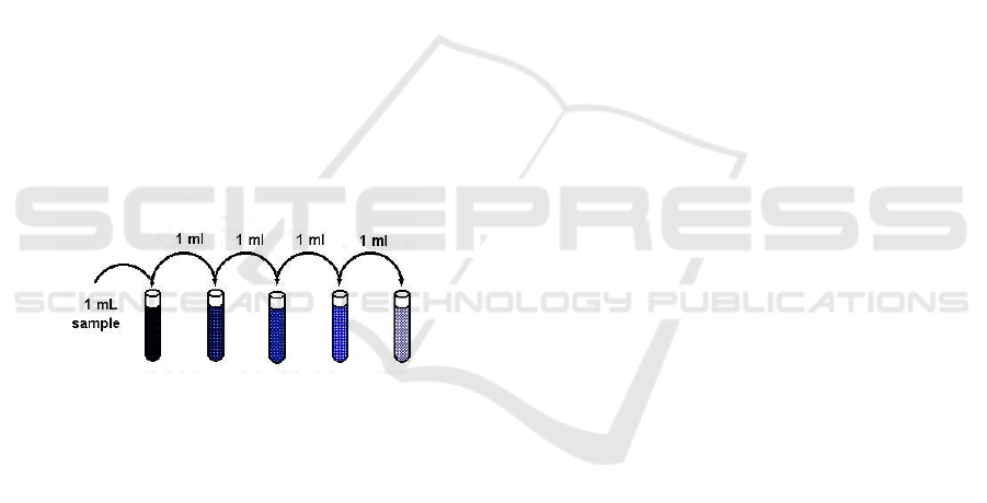
Blood Dilution:
The blood was diluted with aquadest with dilution 10
-
1
, 10
-2
, 10
-3
, 10
-4
and 10
-5
.
Figure 1: blood dilution scheme.
Reagen Test and Interpretation
Adapted from
Andersson (2017), The LMG solution
was prepared by adding 150 ml of aquadest and 100
ml of concentrated acetic acid to 1 g of Leuco
Malachite Green into a brown chemical bottle. The
solution was stirred until all the Leuco Malachite
Green was dissolved and the solution was filtrated.
The prepared LMG solution was afterwards
transferred from the brown chemical bottle with LMG
solution into a smaller separate tube. A 30 % H
2
0
2
(MERCK®) was diluted with deionised water to a 10
% H
2
0
2
solution and the H
2
0
2
was then transferred to
a smaller separate tube. A volume of 25 μl of LMG
solution and 25 μl of H
2
0
2
solution was used
throughout this study. When not in use, all solutions
where kept in a refrigerator.
Methods:
Leuco Malachite Green (LMG) and H
2
O
2
were
prepared according to Manufacturer guideline and
Forensic Laboratory of Indonesian’s Police
Department as well. All reagents were used according
to the manufacturer’s guidelines. Positive controls
were taken by applying the reagent to a bloodstained
piece of filter paper. Negative controls were
performed by applying the reagents to a fresh piece of
filter paper with no trace of blood.
Sensitivity Testing Autoclaved bottles and
distilled H
2
O were used. Water was measured using a
graduated cylinder and blood was added using a
Gilson pipette. Based on Webb et al (2006), differing
low concentrations of blood were achieved by making
a stock solution of blood and distilled water.
Solutions of 10
-1
, 10
-2
, 10
-3
, 10
-4
and 10
-5
were
prepared. A set of 35 3cmx3cm pieces of filter paper
were placed in each of the diluted blood solutions for
each of the presumptive reagents tested. The pieces of
filter paper were then removed and allowed to dry for
10 minutes. Each of the pieces of filter paper was then
tested with its corresponding reagent to see whether
the blood present was detectable. The time taken for
the reagent to register a positive result was
determined and recorded. Tests were considered
negative if reagents failed to react within 4min of
exposure to the blood-stained filter paper.
ICPS 2018 - 2nd International Conference Postgraduate School
362
3 RESULT AND DISCUSSION
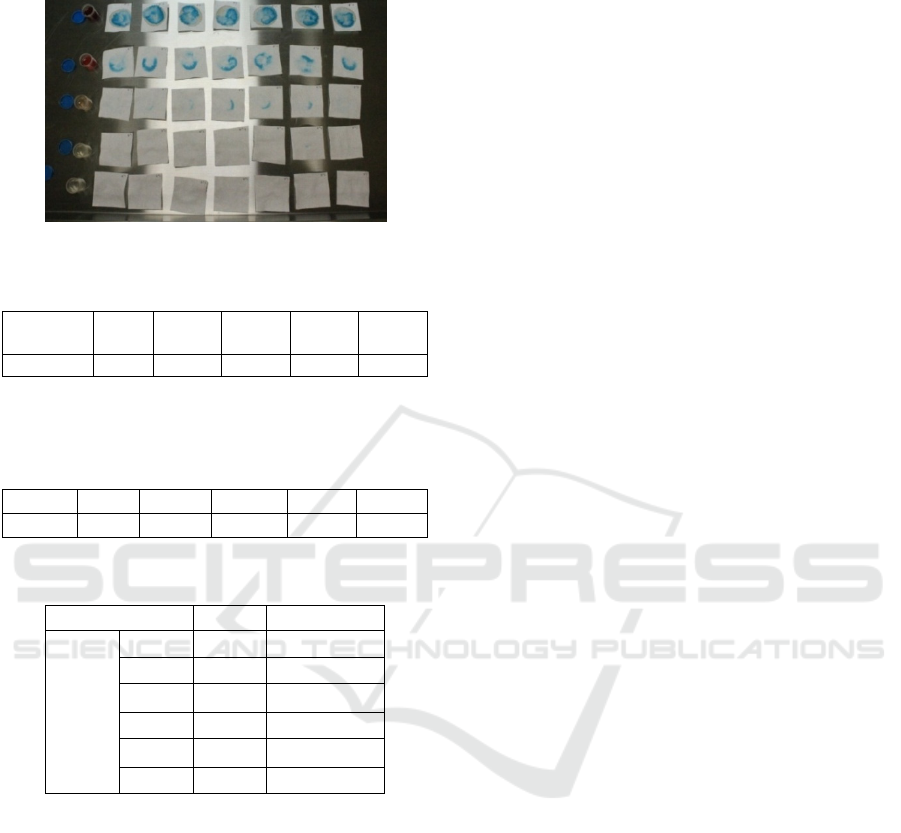
Figure 2: LMG reaction toward lizard’s blood dilution.
Sensitivity
Blood
Dilution
10
-1
10
-2
10
-3
10
-4
10
-5
Lizar
d
+ + + + -
The table above shows that LMG in lizard’s blood can
still react to dilution 10
-4
.
Descriptive
N
Mean SD Min. Max.
Lizar
d
35 1.743 1.6688 .0 5.0
Quality
Kruskal-Wallis Test
Dilution N Mean Rank
Lizard 10
-1
7 32.00
10
-2
7 25.00
10
-3
7 17.71
10
-4
7 8.29
10
-5
7 7.00
Total 35
All the data was analyzed by Kruskal-Wallis test with
significancy p<0.05 for all groups.
The result of this study shows that different
dilution resulted on different interpretation of lizard’s
blood. Dilution of 10
-1
, 10
-2
,10
-3
and 10
-4
obtained
positif (+) result and 10
-5
obtained negative (-) result.
Hemoglobin concentration can affect the quality
of the LMG test reaction. At the higher dilutions it
causes a decrease in hemoglobin concentration.
Based on Gul and Tosunoglu (2011), the amount of
hemoglobin lizard ranges from 8-13 g/dL (Mean:
10.54 g/dL). It is estimated that hemoglobin can no
longer be detected by LMG at dilutions of more than
10
-4
, but can be re-examined in the narrower range
between 10
-4
to 10
-5
.
4 CONCLUSIONS
The conclusions of this study show that the level of
dilution affects the level of sensitivity of LMG test to
blood Varanus salvator. Sensitivity LMG in lizard’s
blood can still react to dilution 10
-4
.
ACKNOWLEDGEMENTS
The author would like to thank Bilqisthi Ari Putra,
DVM, Djoko Legowo, DVM and Nadia Yohana,
DVM as my partner team for this research.
REFERENCES
Andersson, A., 2017. An Evaluation of Two Presumptive
Blood Tests and Three Methods to Visualise Blood.
Linköping University.
Bijanti, R., Yuliani, M.G.A., Wahjuni, R.S and Utomo,
R.D., 2010. Patologi Klinik Veteriner. Airlangga
University Press. Surabaya.
Colotelo, A.H., 2009. Evaluation and Application of
Presumptive Tests for Blood for Fish Epithelial Injury
Detection. Carleton University Ottawa, Ontario.
Dessauer H. C., 1970. Blood chemistry of reptiles:
physiological and evolutionary aspects. In Biology
ofthe Reptilia (Edited by Gans C. and Parsons T. S.),
Vol. C, pp. l-72. Academic Press, London.
Farrugia, K.J., NicDaéid, N., Savage, K.A., Bandey, H and
Ciuksza, T., 2010. Chemical enhancement of footwear
impressions in blood on fabric — part 2: peroxidase
reagents. Elsevier.
Gul, H. And Tosunoglu, M., 2011. Hematological
Reference Intervals of Four Agamid Lizard Species
from Turkey. Herpetozoa Journal.
Idries, A.M and Tjiptomartono, A.L., 2011. Penerapan
Ilmu Kedokteran Forensik Dalam Proses Penyidikan.
Jakarta: CV Sagung Seto.
Marck, M.D., 2007. Veterinary Forensics Animal Cruelty
Investigations, Blackwell Publishing. USA, 1
st
edition.
Spalding, R.P., 2006. The identification and
characterization of blood and bloodstains. In: James
SH, Nordby JJ (eds) Forensic science: an introduction
to scientific and investigative techniques, 2nd edn. CRC
Press, Boca Raton, FL, p 237–260
Tobe, S.S., Watson, N., and Dae’id, N.N., 2007. Evaluation
of Six Presumptive Tests for Blood, Their Specificity,
Sensitivity, and Effect on High Molecular-Weight DNA.
Journal Forensic Science. Vol. 52; p: 102-109.
Laten Blood Detection Test Sensitifity of Lizard (Varanus salvator) Using Leuco Malachite Green (LMG) with Diffrent Dilution
363

Webb, J.L., Creamer, J.I. and Quickenden, T.I., 2006. A
comparison of the presumptive luminol test for blood
with four non-chemiluminescent forensic techniques.
University of Western Australia
ICPS 2018 - 2nd International Conference Postgraduate School
364
