
Type 1 Leprosy Reaction in Multibacillary (MB) Leprosy Patient
That Have Not Received MDT-MB Therapy
Donna Partogi
1
, Cut Putri Hazlianda
1
, Dina Arwina Dalimunthe
1
1
Department of Dermatovenereology, Universitas Sumatera Utara, Jalan Dr. Mansyur Kampus USU 20155, Medan,
Keywords: Leprosy, type 1 leprosy reaction, multibacillary leprosy.
Abstract: Leprosy reaction is a variety of symptoms and signs of acute inflammation of leprosy reactions, which can be
considered as part of journey of leprosy. In the type 1 reaction that plays a role is cellular immunity. A 31-
year-old man complaint of reddish skin thickness on the face, body, back, hands, and feet experiences since
2 weeks ago. On dermatological examination, erythematous plaque is found in the facial region, thoracic,
abdominal and posterior trunk, erythematous macules of the superior extremity and the inferior extremity, the
right side of the sinus. On examination of the peripheral nerve, left and right N. Auricularis Magnus are found
enlarged, N. Ulnaris, N. Popliteal lateralis and N. posterior tibialis tenderness are present. Bacterial
examination (BTA) +1. Diagnosis in this patient was multibacillary leprosy who had type 1 reactions that had
not received MDT-MB therapy. the patient was given Prednison 40 mg/day and was reduced gradually every
2 weeks as much as 5-10 mg. leprosy reactions can occur becfor, during, and after treatment. Various factors
that are considered to cause leprosy reactions. In this case, stress was the trigger factor.
1 INTRODUCTION
Leprosy is one of chronic infection disease, caused by
Mycobacterium leprae, mainly affect peripheral
nerve but can also affect skin, mucosa and other tissue
organ, except central nerve system (Bryceson and
Pfaltzgraff, 1990).
The prevalence of leprosy worldwide is estimated
less than 1 case per 10.000 populations. Nevertheless,
leprosy is still one of health problems in Indonesia.
There are still many provinces and districts in
Indonesia that have not achieved leprosy elimination
that was targeted in 2000 (Lewis et al, 2012; Hernani
et al 2004).
Diagnosis of leprosy is based on cardinal signs
that consist of anesthetic skin lesion, thickened
peripheral nerve with impaired nerve function, and
positive acid-fast bacilli (AFB) from slit skin smears
(Hernani et al, 2004; Amirudin et al, 2003).
According to WHO classification in 1981 that was
modified in 1988, leprosy can be classified into
Paucibacillary (PB) and Multibacillary (MB)
(Amirudin et al, 2003; Noordeen 1994; Kosasih et al,
2008). This classification was based on clinical
features and AFB from slit skin smear examination
(Amirudin et al, 2003).
Leprosy treatment in Indonesia was based on
WHO classification, using Multi Drug Therapy
(MDT). MDT-PB consists of Rifampicin and
Dapsone, while MDT-MB consists of Rifampicin,
Dapsone and Clofazimine (Hernani et al, 2004).
Leprosy reaction is a group of acute inflammatory
sign and symptoms on leprosy skin lesions that were
considered as part of leprosy. There are two types
leprosy reaction. In type 1 reaction, cellular immunity
takes the main role, whereas there are shifting toward
tuberculoid pole (reversal/upgrading) or lepromatose
(downgrading). The clinical manifestations of type 1
reaction are skin lesion become more erythematous,
thickened peripheral nerve with tenderness and
function disorder, with minimal systemic
manifestations (Hernani et al, 2004). Type 2 reaction,
also known as erythema nodosum leprosum, is type
III reaction according to Coomb and Gell with
clinical manifestations such as bright red, painful and
tender nodules, on normal looking skin. It can be
found in skin or subcutaneous tissue on any body part,
especially on the face, hands, and limbs with systemic
symptoms (Bryceson and Pfaltzgraff, 1990;
Martodihardjo and Susanto, 2003).
The principle treatment of leprosy reaction consist
of antireaction medication, rest or immobilization,
analgetic or sedative to treat the pain and continue
Partogi, D., Hazlianda, C. and Dalimunthe, D.
Type 1 Leprosy Reaction in Multibacillary (MB) Leprosy Patient That Have Not Received MDT-MB Therapy.
DOI: 10.5220/0010097108710875
In Proceedings of the International Conference of Science, Technology, Engineering, Environmental and Ramification Researches (ICOSTEERR 2018) - Research in Industry 4.0, pages
871-875
ISBN: 978-989-758-449-7
Copyright
c
2020 by SCITEPRESS – Science and Technology Publications, Lda. All rights reserved
871
antileprosy medication (Martodihardjo and Susanto,
2003).
We report a case of multibacillary leprosy with
type 1 reaction in 31 years old male patient.
2 CASE
A male, age 31 years old, Bataknese, entrepreneur,
came to Dermatology and Venereology Policlinic H.
Adam Malik General Hospital on March 2
nd
2009
with reddish thickened skin without itchiness on his
face, body and back with reddish patch, also without
itchiness on his hand and leg since 2 weeks ago. At
first, the reddish patches were seen in his body and it
got bigger. There is history of white patches that were
not itchy on his back since 2 years ago. His body felt
unwell and he also had family problem. He never
went to seek medical advice for his condition. There
was no similar family history.
Physical examination showed his general
condition was good with good nutritional status.
Dermatological examination showed erythematous
plaques on facial, thorax, abdomen, and trunk
posterior region, erythema maculae on both superior
and inferior extremities. Examination on peripheral
nerve showed thickened auricularis magnus nerve,
there were no thickened and tenderness on ulnar
nerve, lateral popliteal nerve, and tibial posterior
nerve. There were anesthesia found in the lesion and
both inferior extremities. There were no anomalies in
motoric nerve function test.
The differential diagnoses for this patient are
multibacillary leprosy with type 1 reaction that have
not received MDT-MB, paucibacillary leprosy with
type 1 rection that have not received MDT-MB, and
urticaria. The temporary diagnosis for this patient is
multibacillary leprosy with type 1 reaction that has
not received MDT-MB.
The patient was then referred to dr. Pirngadi
General Hospital Medan to have AFB examination
and get appropriate treatment.
Bacteriological examination (AFB) from right
earlobe showed (+) 1, left earlobe showed 1(+), and
back (+) 1. The patient refused to undergo biopsy
examination. Laboratorium examination of blood and
urine sample is within normal range.
Working diagnosis for this patient is
multibacillary leprosy with type 1 reaction that has
not received MDT-MB.
The patient was advised to rest and was given
MDT-MB, which consist of Rifampicin 600
mg/month, Clofazimine 300 mg/month, and continue
with Clofazimine 50 mg/day and Dapsone 100
mg/day. For his reaction, the patient was given
Prednisone 40 mg/day (1x8 tablet/day) and the
dosage was planned to be reduced 5-10 mg every 2
weeks, Paracetamol 3x500 mg.
After 2 weeks, erythematous plaques are become
less thick and reduced; there were no new
erythematous maculae and plaques. There was no
fever. From peripheral nerve examination, we found
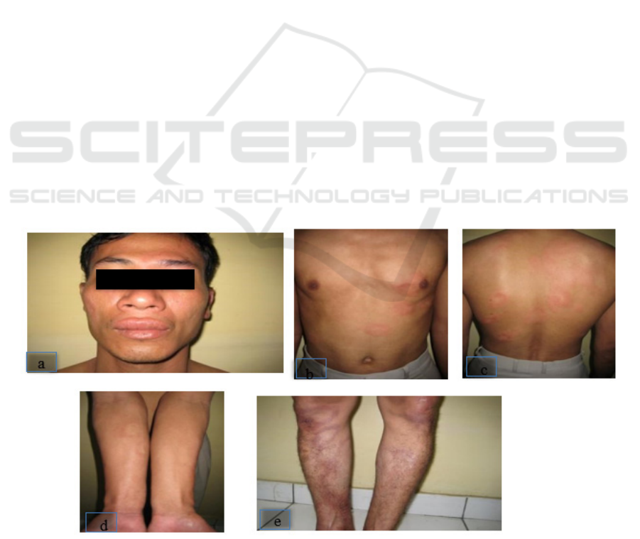
Figure 1. a-e First visit of the patient. (a,b,c) erythematous plaques on facialis, thorax, abdomen and posterior trunk; (d,e)
erythematous maculae on superior and inferior extremities.
ICOSTEERR 2018 - International Conference of Science, Technology, Engineering, Environmental and Ramification Researches
872
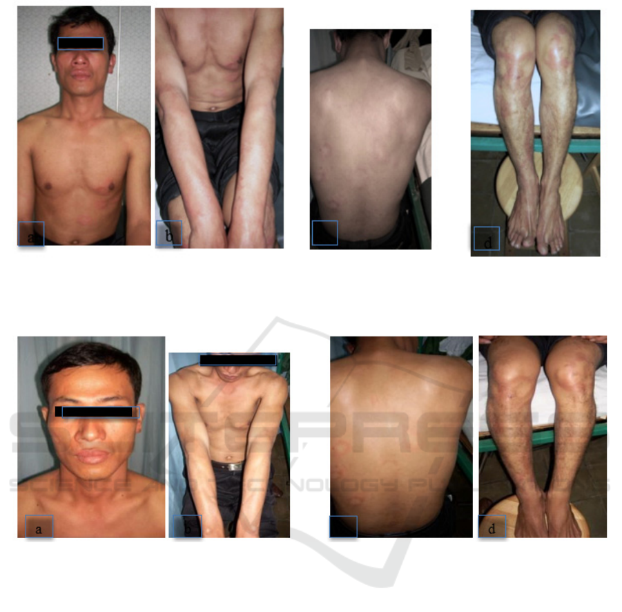
Figure 2. (a) First Control, (b) erythematous plaques were reduced and less thick on facial, thorax, abdomen, and posterior
trunks region, (c) erythematous maculae were also reduced on superior extremities (d) erythematous maculae were reduced
in the inferior extremities.
Figure 3. (a,b,c) Erythematous plaques were less thick on facial, thorax, abdomen, posterior trunk; previous erythematous
maculae were not seen on superior extremities (d) erythematous maculae became less on inferior extremities.
thickened both auricularis magnus nerve, tenderness
in ulnar nerve was not found anymore, but it still can
be found on popliteal lateral nerve. Sensory function
test showed anesthesia is decreased in both inferior
extremities. Motoric nerve function test showed no
anomalies.
Prednisone dose was reduced to 30 mg/day (1x6
tablet/day). We still continue MDT-MB treatment
until 12-18 months.
After 2 weeks, in the next control, the
erythematous plaque and erythematous maculae had
reduced, and there were no new lesion were found.
The peripheral nerve examination found that there
were no thickened peripheral nerve on both
auricularis magnus, no tenderness on ulnar and lateral
popliteal, but tenderness can still be found on
posterior tibial nerve. Sensory nerve function showed
that less anesthesia on both inferior extremities. There
were no anomalies in motoric nerve function.
The prednisone dosage was lowered to 20 mg/day
(1x4 tablet/day). We continue the MDT-MB
treatment and hopefully it will be finished in 12-18
months.
The prognosis of this patient is quo ad vitam ad
bonam, quo ad functionam ad bonam, quo ad
sanationam dubia.
3 DISCUSSION
Diagnosis of leprosy can be concluded based on
cardinal signs, such as numb skin lesion, thickened
peripheral nerve lesion with tenderness and positive
Type 1 Leprosy Reaction in Multibacillary (MB) Leprosy Patient That Have Not Received MDT-MB Therapy
873

AFB examination (Hernani et al, 2004; Amirudin et
al, 2003).
From anamnesis, there are erythematous plaque
without itchiness on his face, body, and back with
erythematous patch without itchiness on his hand and
legs since 2 weeks ago. At first, the reddish patch was
seen on his body and slowly spreading. The patient
also felt feverish. The patient also said that he had
family problem and never seek medical advice for his
skin condition. Leprosy reaction is a group of acute
inflammatory sign and symptoms on leprosy skin
lesions that were considered as part of leprosy
(Martodihardjo and Susanto, 2003). Leprosy reaction
can happen to leprosy patient before, during, and after
treatment (Bryceson and Pfaltzgraff, 1990; Hernani et
al, 2004). Various factors that contributed to this
condition is physical stress caused by pregnancy or
after labor, after vaccination, infection, anaemia,
malnutrition, fatigueness, and psychological stress
that caused by shame, also drug that enhance
immunity (Hernani et al 2004, Rea and Modlin,
2008). In this case, leprosy reaction probably caused
by stress.
Dermatological examination showed
erythematous plaques on facial, thorax, abdomen, and
posterior trunk; erythematous macules on both
inferior and superior extremities. Peripheral nerve
examination showed thickened both auricularis
magnus and tenderness also shown in ulnar, lateral
popliteal, and posterior tibial nerve. Sensory nerve
function test showed anesthesia on skin lesion and
both inferior extremities. Clinical manifestation of
type 1 leprosy reaction is erythematous and
edematous skin lesion that sometimes with ulceration
and followed by tenderness and nerve disorder with
minimal systemic manifestation such as fever,
malaise, and joint pain
(Bryceson and Pfaltzgraff,
1990; Hernani et al, 2004).
Bacteriological examination (AFB) on right
earlobe is (+) 1, on left earlobe (+) 1, and back (+) 1.
This examination support MB leprosy diagnosis.
According to WHO classification in 1988, positive
AFB examination is classified as MB leprosy
(Kosasih et al, 2008).
The differential diagnoses for this patient are
multibacillary leprosy with type 1 reaction that have
not received MDT-MB, paucibacillary leprosy with
type 1 rection that have not received MDT-PB, and
urticaria. The diagnosis of paucibacillary leprosy with
leprosy reaction that have not received MDT-PB can
be removed because we found AFB (+)1 (Hernani et
al, 2004). Differential diagnosis of urticaria can be
removed based on clinical manifestation. Usually in
urticaria, the skin lesions suddenly appear and
disappear gradually. In urticaria, we will not found
AFB and sensory disorder (Aisah S, 2008).
For his treatment, the patient was given MDTMB
that consist of Rifampicin 600 mg/month,
Clofazimine 300 mg/month followed by Clofazimine
50 mg/day and Dapsone 100 mg/day with prednisone
40 mg/day (1 x 8 tablet/day, taken every morning)
with reduced dosage every 2 weeks and paracetamol
3x500 mg. The principle treatment of leprosy reaction
consist of antireaction medication, rest or
immobilization, analgetic or sedative to treat the pain
and continue antileprosy medication (Kosasih et al,
2008).
Prednisone should be started at high dose,
which is 40-80 mg/day depending on the reaction
degree of severity and taken in the morning. The
dosage is decrease gradually, 5-10 mg every 2 weeks
until reaching 5 mg. If there are no clinical
improvement, the dosage should be increase and
reevaluate (Bryceson and Pfaltzgraff, 1990; Hernani
et al, 2004).
Generally, the prognosis of this patient is good,
but there are possibilty of recurrence. After finishing
antileprosy medication for 12 weeks and avoid factors
that caused the reaction, it is hoped that the patient is
going to recover from reaction.
Nevertheless, recurrence can happen if the patient
is exposed to predispose factor (Rea and Modlin,
2008; James, 2006).
4 CONCLUSIONS
Type 1 leprosy reaction can occur before, during and
after completed MDT therapy. In this case, the type 1
leprosy reaction occurred before MDT therapy and
the trigger factor was stress.
ACKNOWLEDGEMENTS
Author wishing to acknowledge financial assistance
from Universitas Sumatera Utara.
REFERENCES
Aisah S., 2008. Urtikaria in Djuanda A., Hamzah M., Aisah
S. (eds.) Ilmu Penyakit Kulit dan Kelamin, 5
th
edition.
Jakarta: Balai Penerbit FK UI. pp.169-76.
Amirudin M.D., Hakim Z., Darwis E., 2003. Diagnosis
Penyakit Kusta in Daili E.S., Menaldi S.L., Ismiarto
S.P., Nilasari H. (eds.) Kusta, 2
nd
edition. Jakarta : Balai
Penerbit FK UI. pp.12–6.
ICOSTEERR 2018 - International Conference of Science, Technology, Engineering, Environmental and Ramification Researches
874

Bryceson A., Pfaltzgraff R.E. 1990. Leprosy. 3
rd
edition.
London: Churchill Livingstone. pp.1, 115-6,122,129.
Hernani, Damari A., Hartati F., et al. 2004. Buku Nasional
Pemberantasan Penyakit Kusta. Jakarta : Departemen
Kesehatan RI. pp.11,42,64-8,70-1.
James W.D. 2006. Hansen’s diseases in James WD (ed).
Andrew’s diseases of the skin, clinical dermatology 10
th
edition. Canada: Saunders company. pp.343-52.
Kosasih A., Wisnu I.M., Daili E.S., Menaldi S.L. 2008.
Kusta. in Djuanda A., Hamzah M., Aisah S. (eds.) Ilmu
Penyakit Kulit dan Kelamin, 5
th
edition. Jakarta: Balai
Penerbit FK UI. pp.73-88.
Lewis F.S., Conologue T., Harrap E. Leprosy [online].
Available at : www.emedicine.com (Accessed: 1 Juni
2012).
Martodihardjo S., Susanto R.S.D. 2003. Reaksi kusta dan
penanganannya. in Daili E.S., Menaldi S.L., Ismiarto
S.P., Nilasari H. (eds.) Kusta, 2
nd
edition. Jakarta : Balai
Penerbit FK UI. pp. 75-82. Noordeen SK. 1994. The
Epidemiology of Leprosy. in Hasting R.C. (ed).
Leprosy, 2
nd
edition. London: Churchill livingstone;. p.
308.
Rea TH, Modlin RL. 2008. Leprosy, in: Wolff K,
Goldsmith LA, Katz SI, dkk, editor. Fitzpatrick’s
Dermatology in General Medicine, 7
th
edition. New
York: Mc Graw Hill. pp.1786-96.
Type 1 Leprosy Reaction in Multibacillary (MB) Leprosy Patient That Have Not Received MDT-MB Therapy
875
