
Description of Liver Histology of Mice (Mus musculus L) after Giving
Nano Herbal Haramonting (Rhodomyrtus tomentosa)
Putri Cahaya Situmorang
1
and Syafruddin Ilyas
1
1
Department of Biology, Faculty of Mathematics and Natural Sciences,
Universitas Sumatera Utara, Medan, Indonesia, 20155
Keywords: Nanoherbal, Rhodomyrtus tomentosa, Liver.
Abstract: The objective of this study was to determine the description of liver histology of male mice (Mus musculus
L.) after giving Nano Herbal Haramonting (Rhodomyrtus tomentosa). This research use the Completely
Randomized Design (CRD), which consists of six groups of CMC 0.5% and Nano Herbal Haramonting
(R.tomentosa) at dosage of (100; 141,42; 200; 282,82; 400)mg/20g body weight. The livers were made into
preparations by using the paraffin method and Hematoxylin Erlich-Eosin staining (HE). The value of each
cell with Manja Roenigk's histophatolgy model. Nano-R.tomentosa administration was not significantly
different in body weight of mice (P>0,05), but It was significantly different to weigh of liver (P<0,05). The
results of histological liver showed that there is a significant difference (P<0,05) between control group and
treatment groups with damaged cells liver of male mice Mus musculus L.) after giving of Nano Herbal
Haramonting (R.tomentosa).
1 INTRODUCTION
Nanoherbal is the herb that uses nanoscale technology
(wave power) and use quality herbal raw materials.
The use of herbal remedies has been practiced for
thousands of years and a part of several countries such
as Indonesia, China and India. Nanotechnology
commonly refers to structures that are up to several
100 nm in size which can be increased up to 1000 nm.
Nanotechnology and nano science studies have
develops briskly during the past years in a large range
of product results. It gives opportunities for the
development of materials, including those for medical
applications, where conventional techniques may
reach their limits (Ratnam et al, 2006). Herbal
medicines have less adverse effects as compared with
modern medicines due to poor solubility, poor
permeability, low bioavailability, instability in
biological milieu and extensive first pass metabolism
by developing new formulation as nano herbal
medicines like nanoparticles, dendrimers, Nano
crystals, Quantum dots, Nanosperes, Nanocapsules,
herbal market get good feedback (Rinku et al, 2018)
One study using nano herbal is Nano curcumin. In
this study Nano-curcumin significantly inhibited the
growth of MCF-7 breast cancer cell line and resulted
in synergism cytotoxicity effects (Parisa et al, 2018)
Haramonting (R.tomentosa) is an ornamental,
evergreen shrub grows up to four meters. This plant
species is native to southern and southeastern Asia
(Awinita et al, 2005). The potential of R. tomentosa
as a new source of health-promoting compounds such
as dietary fibers, essential fatty acids, and phenolic
compounds. A total of 19 phenolic compounds were
tentatively characterized, including stilbenes and
ellagitannins as major components, followed by
anthocyanins, flavonols, and gallic acid. Piceatannol,
a promising health-promoting stilbene component,
was the major phenolic compound found in R.
tomentosa fruits (Lai et al, 2013).
Hepatic sistem is the major organ system involved in
the metabolism, detoxification and excretion of
various endogenous and exogenously. Liver damage
is always asssociated with necrosis, Strengthening of
inbuilt protective mechanism or exogenous
administration of antioxidant may be useful in
protecting the liver (Pramodh et al, 2008). In vitro
antioxidant activity of the different extracts of R.
tomentosa has been reported by different methods
(Geetha et al, 2010). Natural antioxidants are known
to exert beneficial effects in hepatitis induced by
antitubercular agents (Limsuwan et al, 2009).
982
Situmorang, P. and Ilyas, S.
Description of Liver Histology of Mice (Mus musculus L) after Giving Nano Herbal Haramonting (Rhodomyrtus tomentosa).
DOI: 10.5220/0010083409820986
In Proceedings of the International Conference of Science, Technology, Engineering, Environmental and Ramification Researches (ICOSTEERR 2018) - Research in Industry 4.0, pages
982-986
ISBN: 978-989-758-449-7
Copyright
c
2020 by SCITEPRESS – Science and Technology Publications, Lda. All rights reserved
Oxidative stress induced damage to hepatocytes has
been found to have a key role in antitubercular drugs
induced hepatitis (Shakun and Shmnan’ko, 1986). R.
tomentosa extract demonstrated its free radical
scavenging effects in concentration dependent
manner and the results suggest that R. tomentosa
extract can serve as a potent antioxidant (Sodhi,
1998). So in this study, we examined the effect of
Nano- R.tomentosa to liver.
2 METHODS
2.1 Materials
The material used is male mice (Mus musculus L)
strain DDW, Haramonting leaves (R.tomentosa)
obtained from plantation residents in Tapanuli North
Sumatera, Feed the mice no. PB 551, staining
Hematoxylin and Eosin. This research use the
Completely Randomized Design (CRD) using 30
male mice with an average weight of 20-25g age 12-
18 weeks. Male mice are kept in Animal Cages
Biological Laboratory, Faculty Mathematics and
Natural Science, Universitas Sumatera Utara.
Experimental animal handling is done ethically
(Ethical Clearance).
2.2 Making of Nano- R.tomentosa
R.tomentosa leaves are washed, then dried in
accordance with the requirements of water content
using high energy milling (HEM), then Simplicia as
the destructive medium is inserted into the jar
container, Inserting balls with larger diameter size
then continued by inserting small balls and the last
sample. The total volume of the balls and the samples.
entered does not exceed 2/3 of the volume of the jar.
The usual Ball to Powder Ratio (BPR) is 20:1, 10:1,
8:1. Example BPR 20:1 means 1 gram of sample then
milled with 20 grams of ball weight. Jar that has been
filled with the ball, samples are closed tightly then
mounted on the jar inside the HEM tool, then HEM is
turned on for 2 hours.
2.3 Treatment and Observation of
Hystology
The treatment consisted of 6 groups consisting
of 5 male mice ie CMC 0.5% and Nano Herbal
Haramonting (R.tomentosa) at dosage of (100;
141,42; 200; 282,82; 400)mg/20g bw in 14 days, then
killed by the method of disclasio cervicalis to take
the liver. Preparation of histologist by paraffin
method and Hematoxylin Erlich-Eosin staining (HE)
to observe the damage to liver cells due to the
administration of Nano-R.tomentosa. Mixture is
observed with magnification 400x. In each field of
view counted 20 cells at random and in the value of
each cell with Manja Roenigk's histophatolgy model
(1: Normal, 2: Parenchymatous Degeneration, 3:
Hydrophic Degeneration 4: Necrosis) then the data
processed with SPSS 22 program with Kruskal Wallis
test.
3 RESULT AND DISCUSSION
The result will be discussed in 3 subsection i.e
average weight of body, weight of liver and damage
of liver cells.
3.1 Weight of Body
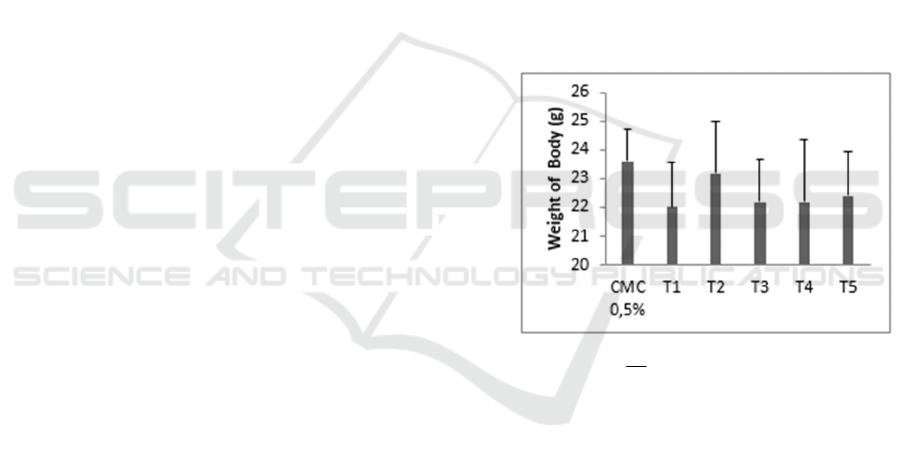
Figure 1. Average of weight of male mice after giving
Nano-R.tomentosa. (
X
± SD).
The results of average body weight of mice
CMC 0,5% group has the highest average weight
while the lowest weight is in T1 (Figure 1). Nano-
R.tomentosa administration was not significantly
different in body weight of mice (P>0,05). Nano-
R.tomentosa does not affect the weight of male mice,
Allegedly due to outside variables that can not be
controlled such as the psychological condition of the
mice that is affected by the surrounding environment,
repeated treatment, different appetite and fights
between mice.
3.2 Weight of Liver
The result of statistic test P<0,05 showed that Nano-
R.tomentosa administration at each treatment was
significantly different in weight of liver except in
Control group with T1, T3,T4 and T5, T1 group with
T4 and T5, T2 group with T3, T3 group with T4
Description of Liver Histology of Mice (Mus musculus L) after Giving Nano Herbal Haramonting (Rhodomyrtus tomentosa)
983
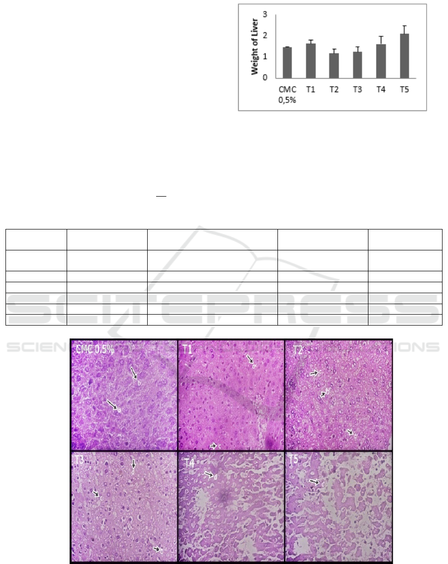
(P>0.05) . T5 group has the highest average liver
weight while the lowest liver weight is in T2 (figure
2). T1 higher than T2, and also T1 higher than control
is assumed because the outside variable is difficult to
control such as weight of body, different appetite and
activity of mice.The liver is the largest organ in the
body that is involved in the body's metabolism,
neutralize toxins and toxic substances through the
detoxification process by cell kupfer in the body. So
that the weight of the liver can be increased or
decreased due to the entry of excessive toxic
substances in the body.
3.3 Damage of Liver Cells
Table 1. Average Hepatocyte Normal and degree of damage (Parenchymatous Degeneration, Hydrophic Degeneration and
necrosis) after giving Nano-R.tomentosa (
X
± SD).
Figure 3. Histology of Liver, a: Normal, b: Parenchymatous Degeneration, c: Hydrophic Degeneration, d: Necrosis (400x).
The results P<0,05 showed cell damage due to
nano-R.tomentosa administration is significantly
different from each treatment except Control group
with T1 (P>0.05). Description of liver damage is
different for each treatment (figure 3). Damage with
Parenchymatous degeneration occurs lowest in CMC
Treatmen
t
Normal Parenchymatous
Degeneration
Hydrophic
Degeneration
Necrosis
CMC
0.5%
14.40 ± 1.66 4.56 ± 1.47 4.20 ± 3.00 7.52 ± 4.05
T1 13.60 ±1.50 4.88 ± 1.42 5.16 ± 2.81 9.12 ± 4.09
T2 12.30 ±1.14 5.52 ± 1.94 12.5 ± 2.48 10.24 ± 4.01
T3 6.76 ±1.51 6.80 ± 2.00 12.5 ± 4.30 22.72 ± 8.54
T4 4.84 ±1.52 7.76 ± 2.60 11.5 ± 3.32 29.76 ± 8.01
T5 3.40 ±1.35 6.80 ± 2.71 9.48 ± 3.84 40.16 ±8.06
Figure 2. Weight of liver after giving N
ICOSTEERR 2018 - International Conference of Science, Technology, Engineering, Environmental and Ramification Researches
984
0
20
40
60
80
CMC
0.5%
T1 T2 T3 T4 T5
Percentage of cells
damage
0,5% (Control) and highes in T4 (table 1) due the
mice are also given aquades so that the cells can
regenerate. cells in liver organ with parecimatous
degeneration may improve, cells undergoing necrosis
over time will be replaced with new liver cells due
cell regeneration process in liver organ (Geetha et al,
2012). Parenchymatous degeneration is the mildest
degenerate levelIn parenchymatous degeneration
cells, granules are found in the cytoplasm due the
precipitate that causes the cytoplasm to become turbid
and swelling of the cells. Hydrophic Degeneration is
low in control, and increases in T2 and T3 (table 1).
This degeneration is more severe damage, there are
vacuoles containing water and cytoplasm that do not
contain fat and glycogen. This change is generally a
result of metabolic disorders such as hypoxia or
chemical poisoning. This degeneration is also
reversible although it may be irreversible if the cause
of the injury persists. The process of Necrosis
increases from Controls to P5 and Normal cells
decreases from control to treatment level (table
1)(figure 3). At each dose level, the toxic ingredients
in the liver are getting out of process, causing
parenchymatous degeneration, hydrophic
degeneration and necrosis in liver. The target of a
toxic substance in the body is the molecular structure
of transport of bile acids, membranes, intracellular
fats, proteins and nucleic acids. As a result the target
molecule becomes a non-functioning unit and may
activate secondary pathways such as apoptosis,
necrosis, autofagocytes and mitochondrial disorders
and other immunological reactions (Kandena et al,
2011).
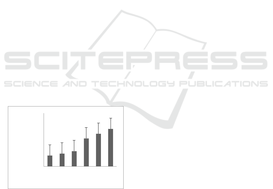
Figure 4. Percentage of cells damage.
The degree of damage is known that the
percentage of liver damage from control to treatment
with high doses continue to be damaged (figure 4).
R.tomentosa leaves contain secondary metabolites of
phenol, flavonoids, saponins, tannins, steroids and
triterpenoids. Phenols, flavonoids, tannins, steroids
and triterpenoids that have an antioxidant effect.
Saponins can cause haemolysis by affecting the lipid
bilayer in the protein membrane of the red blood cells
causing the formation of pores in the red blood cell
membrane (Kaplowitz, 2002)(Baumann et al, 2000).
Damage of liver cells is thought to be caused by
saponins, and tannins in nano-R.tomentosa who
accumulated and irritant or toxic. Sentrolobuler
damage in liver due tannin and saponin compound
administration in research that spans short time,
usually seen cell swelling, necrosis to cause death in
mice. So the liver loses its function by no longer able
to change the compounds that are very toxic to be less
toxic.
4 CONCLUSIONS
Cell damage due to Nano-R.tomentosa administration
is significantly different from each treatment
(P<0.05) and Giving Nano herbal Haramonting
(Rhodomyrtus tomentosa) with excessive doses can
cause a decrease in hepatic weight and damage to
liver cells, So the liver loses its function especially the
detoxification function of toxic.
ACKNOWLEDGEMENTS
We are grateful to Directorate of research and
community service, Directorate general of research
and development, Ministry of research, Technology,
and Higher Education in accordance with research
and community service funding agreement for
budgeting year 2018 (Fund of the research
postgraduate team I) to funding our research.
REFERENCES
Awinita, T., Wright, and J., A., Goolsby, 2005. “Herbivores
in Thailand on Rhodomyrtus tomentosa
(Myrtaceae), an invasive weed in Florida,”
Florida Entomologist, vol. 88, no. 1, pp. 104–
105.
Baumann, E., Stoya, G., Volkner, A., Richter, W., Lemke,
C., et al. 2000. Hemolysis of human erytrocytes
with saponin affects the membrane structure.
Acta histochemia.
Geetha, K.,M., Sridhar, C., Murugan, V., 2010. Antioxidant
and Gastroprotective activities of Rhodomyrtus
tomentosa (Ait.) Hassk. Int J Pharm Tech Res.
2:283‑91.
Geetha, K.,M., Patil, V., Murugan, V.. 2012.
Hepatoprotective activity of aqueous alcoholic
(70%) extract of Rhodomyrtus tomentosa (Aiton)
Description of Liver Histology of Mice (Mus musculus L) after Giving Nano Herbal Haramonting (Rhodomyrtus tomentosa)
985

Hassk against antitubercular drugs induced
hepatic damage. Int J Green Pharm. 6:295-8.
Kandena, I.,M dan Winaya, I., B., O., 2011. Kadar Perasan
Kunyit yang Efektf Memperbaiki Kerusakan Hati
Mencit yang Dipicu Karbon Tetrachlorida.
Jurnal Veteriner. 1:34-49.
Kaplowitz, N., 2002. Biochemical and cellular mechanisms
of toxic liver injury. Semin. Liver. Dis. 22: 137-
144.
Lai, T.,N., Herent, M.,F., Quetin-Leclercq, J., Nguyen,
T.,B., Rogez, H., Larondelle, Y., et al. 2013,
Piceatannol, a potent bioactive stilbene, as major
phenolic component in Rhodomyrtus tomentosa.
Food Chem. 138(2-3):1421-30.
Limsuwan, S., Trip, E.,N., Kouwen, T.,R., Piersma, S.,
Hiranrat, A., Mahabusarakam W., et al, 2009,
Rhodomyrtone: A new candidate as natural
antibacterial drug from Rhodomyrtus tomentosa.
Phytomedicine, 16(6), 645–651.
Parisa, Z., AmirReza, H., Malihe, B., Maryam, B., Faezeh,
G., 2018. Evaluation of Cytotoxicity Effects of
Combination Nano-Curcumin and Berberine in
Breast Cancer Cell Line. Iranian Journal of
Toxicology. 12(4).
Pramodh, K., Deval, R.,G., Lakshmayya, Ramchandra,
S.,S., 2008, Antioxidant and hepatoprotective
activity of tubers of Momordica tuberosa cogn.
Againts CCl4 i duced liver in rats. Indian J Exp
Biol. 46:510-513.
Ratnam, D.,V., Ankola, D.,D., Bhardwaj, V., Sahana,
D.,K., Kumar, M.,N., 2006, Role of antioxidants
in prophylaxis and therapy: a pharmaceutical
prospective, J control release. International
journal of molecular sciences. 113:189- 207.
Rinku, Y., Pati, Shubhangi, A., Patil, Niranjan, D., Chivate,
Yogesh, N., Patil, 2018, Herbal Drug
Nanoparticles: Advancements in Herbal
Treatment. Research J. Pharm. and Tech. 11(1)
Shakun, N.,P., Shmnan’ko, V.,V., 1986.Antioxidant
effectiveness in isoniazid induced lesions of liver.
Farmakol Toksikol. 49:86‑9
Sodhi, C.,P., Rana, S., Mehta, S., Yaiphe,i K., Mehta, S.,K.,
1998. Oxidative hepatic injury of isoniazid ‑
rifampicin in young rats subjected to protein
energy malnutrition. Drug Chem Toxicol. 21: 305
‑17.
ICOSTEERR 2018 - International Conference of Science, Technology, Engineering, Environmental and Ramification Researches
986
