
Histopathologic Feature of Breast Cancer Patients in Young Women
M. S. Sitorus
1
, L. I. Laksmi
2
1
Department of Anatomy, Universitas Sumatera Utara
2
Department Of Pathology Anatomy, Universitas Sumatera Utara
Keywords: Breast Cancer, Histopathologic Feature, Young Woman.
Abstract. : Breast malignancy is still the most common cancer in the worldwide that attacks women. Causing a high
mortality rate that is about 15% of all deaths in women. The incidence of breast cancer cases in young age
under the 40 years of age is low but they have particular attention because of its unique behavior. Breast
cancer biology in young women is more aggressive than women of premenopausal age. This is related to
many factors associated with poor prognosis, including high proliferation index, grade three and four disease
and negative Estrogen Receptor expression. This study aims to explore further histopathologic feature,
immunohistochemical expression, and subtype of breast cancer biology, in women under 40 years of age. It
was found that the most common type, of breast cancer histopathology was invasive breast cancer (88%). The
most histopathologic grading was grade I which is 74%. Based on the ER immunohistochemical staining
found that 60% gives a negative expression, only 40% gives positive. A total of 68% gave a positive PR
expression, 68% Her-2 positive one, 30% Her-2 positive three. As many as 56% positive Ki-67. All cases are
Her-2 positive and have a moderate to poor prognosis.
1 INTRODUCTION
Breast cancer is the most common malignancy in
women. Each year affects 1.5 million women, with a
high number of deaths. By 2015, 570,000 women die
caused by breast cancer - about 15% of all deaths in
women (WHO, 2018). The incidence of breast cancer
is more frequent in developing countries while the
death is higher in less developed countries
(Ghoncheh, 2016). In Indonesia, this cancer is also
the most cancer with a number of cases 48,998 in
2014, with a mortality rate of 21.4% (WHO, 2014).
Now, it is so rapidly progressing concerning
therapeutic and screening of breast cancer to reduce
mortality at all ages. However, breast cancer in young
age although rarely encountered has its uniqueness
compared with old age sufferers. However breast
cancer in young age woman has an aggressive
behaviour (Lee HB, 2014). Around 7% of breast
cancers are found in young women under 40 years
old. The survival rate is worse than that of older
people (Anders CK , 2009). This paper aims to obtain
a histopathologic feature, immunohistochemical
expression, and subtype of breast cancer biology, in
young women under 40 years old.
2 METHODOLOGY
The sample of the study was female breast cancer
patients aged under 40. Patient has performed surgery
and histopathology examination of the network in
Installation of Pathology Anatomy of Adam Malik
Hospital Medan. Anatomy Pathology examination
results indicate the presence of breast cancer. The
number of samples is 50 people with a distribution
showed in table 1.
Table 1 : Distribution of breast cancer patients by age.
A
g
e
(y
ears
)
Number
(p
erson
)
Percenta
g
e
(
%
)
35-40 45 90
30-34 3 6
25-30 2 4
<24 - -
50 100
Some breast cancer patients aged 35-40 people were
found as much as 90%. Only two persons (4%) aged
25-30 years, which are one person age 29 years and
one person aged 26 years. There were no breast
cancer patients under 24 years of age.
578
Sitorus, M. and Laksmi, L.
Histopathologic Feature of Breast Cancer Patients in Young Women.
DOI: 10.5220/0010079205780581
In Proceedings of the International Conference of Science, Technology, Engineering, Environmental and Ramification Researches (ICOSTEERR 2018) - Research in Industry 4.0, pages
578-581
ISBN: 978-989-758-449-7
Copyright
c
2020 by SCITEPRESS – Science and Technology Publications, Lda. All rights reserved
3 RESULTS
3.1 Histopathology Feature
A tumor had removed and sent to Installation of
Pathology Anatomy. The tissue is processed in the
Installation of Pathology Anatomy RS Adam-Malik
Medan to become a slide. Then stained with
Hematoxylin-Eosin to be observed with a light
microscope. The type and grading histopathologic
breast cancer are established. The most frequently
type of histopathologic breast malignancy is invasive
breast cancer with no specific type (44 cases) (88%),
five patients are invasive lobular cancer and one clear
cell carcinoma patient as shown in table 2.
Table 2: Breast malignancy types.
Breast cancer
malignancy types
Number Percentage
(%)
Invasive breast
carcinoma
44 88
Invasive lobular
carcinoma
5 10
Clear cell
carcinoma
1 2
50 100
The Bloom & Richardson classification
determines histopathologic grading. Classification is
determined by tubular formation, cell atypia, and
mitosis. The histopathologic Grading was found in
Table 3.
Table 3: Grading breast cancer.
Grading breast
cancer
Number Percentage
(%)
Grade
I
12 24
Grade I
I
37 74
Grade III 1 2
50 100
The most histopathologic grading was grade I of
74%, at least grade III of 2%.
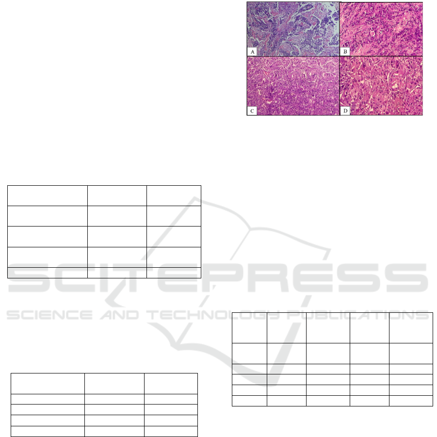
Figure 1: Microscopic view of invasive ductal carcinoma of
breast. A. Grade III (HE, 50x), B. Grade III infiltratif
pattern (HE,50x), C.Solid pattern (HE,50x), D(HE,100x).
3.2 Immunohistochemical (ihc)
Expression
Then all histopathologic tissue blocks were
immunohistochemically stained (ihk) with Estrogen
Receptor (ER), Progesterone Receptor (PR), Her-2,
and Ki-67 antibodies. The immunohistochemical
examination is assessed on a scoring system.
Immunohistochemical examination are shown in
Table 4.
Table 4: Immunohistochemical expression of breast cancer.
Ihc
expres
sion
ER(%) PR(%) Her-
2(%)
Ki-
67(%)
Negati
ve
30(60) 34(68) 1(2) 22(44)
+20
(
40
)
16
(
32
)
34
(
68
)
28
(
56
)
++ 0 0 0 0
+++ 0 0 15
(
30
)
0
50(100) 50(100) 50(100) 50(100)
Based on the ER ihc expression found that 60%
gives a negative expression, only 40% gives a
positive. A total of 68% gave a positive PR
expression, 68% Her-2 positive one, 30% Her-2
positive three. As many as 56% positive Ki-67.
Histopathologic Feature of Breast Cancer Patients in Young Women
579

Figure 2: ER expression negative (A), PR +(B), Her-2
+1(C), Her-2 2+(D), Her-2 3+(E), Ki6 +(F).
3.3 Histopathology Subtypes
Through ihc examination, a subtype of breast cancer
can be grouped based on a classification of St. Gallen.
There were five intrinsic subtypes of breast cancer,
Luminal-A, Luminal-B, Her-2 overexpression,
Basal-like and Normal-like as in table 5.
Table 5: Classification of molecular subtypes of breast
cancer (Dai X, 2015).
Intrinsic Subtype Ihc
Luminal A [ER+
|
PR+] HER2-KI67-
Luminal B [ER+|PR+] HER2-KI67+
[ER+
|
PR+] HER2+KI67+
HER2
Overexpression
[ER-PR-] HER2+
Basal
(Triple neg)
[ER-PR-] HER2-, basal
marker+
Normal-like [ER+|PR+] HER2-KI67-
In this study, it was found that all cases were Her-
2 positive, and had a moderate to poor prognosis
(table 6). No Luminal-A subtype was found.
Table 6. Molecular subtypes of breast cancer.
Intrinsic
Subtype
Number Percentage (%)
Luminal A 0 0
Luminal B 21 52
HER2
Overexpression
28 56
Basal
(Triple
neg)
0 0
Normal-like 1 2
Jumlah 50 100
4 DISCUSSION
The risk of breast cancer will increase with age. Also,
will decrease in young age. So the amount of breast
cancer patients in young age women is lower. In
United States, an estimated 11,000 women diagnosed
with invasive breast cancer are under 40 years old.
That is 4.7% -4.9% of all breast cancer patients (Lee
HB, 2014). The less number of breast cancer in young
age is associated with pathogenesis and breast cancer
risk factors. There are two risk factors: internal and
external factors. The internal factors are age,
hormonal status (age of menarche, menopause,
breastfeeding) and genetics. External factors are the
form of low-fiber diet, carcinogenic food, radiation
exposure, smoking, hormonal therapy also play an
important role in the pathogenesis of breast
malignancy (Cable AM , 2015).
In this study, the most common type of breast
cancer was invasive breast cancer in 44 cases (88%).
While invasive lobular carcinoma were 5 cases
(10%). When viewed from the literature, this type of
cancer is indeed the most cases of 60-75% (Eliyatkin
N, 2015, Malhotra GK, 2010).
Breast malignancy at a young age, although
slightly but has unique biological characteristics. This
makes the researchers interested in exploring more
about this. Breast cancer biology in young age women
is more aggressive than women of premenopausal
age. This is related to many factors associated with
poor prognosis, including high proliferation index,
grade three and four disease and negative ER (Lee
HB, 2014).
In this study, no luminal-A subtype had a good
prognosis. In fact, 98% of cases show positive Her-2
and 56% of cancer with Her-2 overexpression. The
presence of Her-2 overexpression has been stated to
be an adverse prognostic factor of breast cancer. This
situation is found in 20-25% of cases that can be
detected with ihc. Her-2 is a member of the epidermal
growth factor receptor functioning in the stimulation
of cell growth, survival and differentiation (Punglisi
F, 2015).
The existence of these different subtypes of breast
cancer biology, as well as the basis for determining
prognosis, may also determine appropriate treatment
measures. In breast cancer that expressed positive ER
will be treated with antihormonal, Her-2 positive will
be treated with anti-HER-2. Targeting therapy can
increase the life expectancy rate and the death rate
decreases.
Another worse prognostic factor is triple negative
breast cancer (ER-/PR-/Her-2-). In some papers, it is
said that this subtype more often found at a young
ICOSTEERR 2018 - International Conference of Science, Technology, Engineering, Environmental and Ramification Researches
580

age. This will make two bad things together. The risk
of death would be 2.7-fold if a triple negative was
encountered at a young age. There was no triple
negative subtype in this study(Bano R et., 2015).
5 CONCLUSION
Breast cancer at a young age woman is a rare case.
However, cancer cases in young age will show a
worse prognosis. The grading, ihc appearance and
subtype biology of breast cancer are worse compared
with cancer found in older age.
REFERENCES
Anders CK, Johnson R, Litton J, Phillips M, Bleyer A.
2009. Breast Cancer Before Age 40 Years. Semin
Oncol; 36(3): 237–249.
Bano R, Salim M, Abid M, Zaidi A, Khan AI. 2016.
Prognosis Of Breast Cancer In Very Young Age (Less
Than 30 Years). Journal Of Cancer & Allied
Specialties;2(1). Available at:
http://journals.sfu.ca/jcas/index.php/jcas/article/view/5
3/html.
Dai X, Li. T, Bai Z, Yang Y, Liu X, Zhan J, Shi B. 2015.
Breast cancer intrinsic subtype classification, clinical
use, and future trends. Am J Cancer Res.;5(10):2929-
2943.
Eliyatkin N, Yalcin E, Zengel B, Aktas S, Vardar E. 2015.
Molecular Classification of Breast Carcinoma: From
Traditional, Old-Fashioned Way to A New Age, and A
New Way J Breast Health; 11: 59-66.
Ghoncheh M, Pournamdar Z, Salehiniya H. 2016.
Incidence and Mortality and Epidemiology of Breast
Cancer in the World. Asian Pac J Cancer Prev,17,
Cancer Control in Western Asia Special Issue, 43-46.
Kabel AM, Baali FH. 2015. Breast Cancer: Insights into
Risk Factors, Pathogenesis, Diagnosis, and
Management. Journal of Cancer Research and
Treatment; 3(2):28-33.
Lee HB, Han W. 2014. Unique Features of Young Age
Breast Cancer and Its Management. J Breast Cancer
17(4): 301-307.
Malhotra GK, Zhao X, Band H, Band V. 2010.
Histological, molecular and functional subtypes of
breast cancers. Cancer Biol Ther; 10(10): 955–960.
Puglisi F, Fontanella , Amoroso V, Bianchi GV, Bisagni G,
Falci C, Fontana A, Generali D, Gianni L, Grassadonia
A, Moscetti L,Portarena I, Rossi E, Marchetti P. 2016.
Current Challenges in HER2-positive breast cancer.
Critical Reviews in Oncology/Hematology 98:11–221.
World Health Organization-Cancer-Breast Cancer, 2018.
www.who.int>diagnosis-screening>bre.
World Health Organization – Cancer country profile, 2014.
Indonesia. Available at www.who.int>country-
profiles>idn_en.
Histopathologic Feature of Breast Cancer Patients in Young Women
581
