
Differences in Radiopacity Value of RMGIC, GIC and Composite
Resin Materials with Secondary Caries using Conventional and
Digital Radiography
Cek Dara Manja
1
, Kholidina Imanda Harahap
2
1
Department of Dentomaxillofacial Radiology, Faculty of Dentistry, Universitas Sumatera Utara, Medan, Indonesia
2
Department of Dental Material and Technology, Faculty of Dentistry, Universitas Sumatera Utara, Medan, Indonesia
Keywords: Radiopacity, Materials, Secondary Caries, Conventional Radiography, Digital Radiography
Abstract: Radiopacity is an important feature of the restorative material because the ability of the dentist differs in
interpreting a lesion or caries on a radiograph. The purpose of this study is to determine the differences in
radiopacity value of RMGIC, GIC and bulkfill composite resin materials with secondary caries as well as to
evaluate the radiographic technique used to obtain the radiograph. This type of research is descriptive
analytics by using comparative group design. The samples in this study were dental radiographs filled with
RMGIC, GIC and bulkfill composite resin materials produced from conventional and digital radiographic
sampling. Then conventional and digital radiographs are measured using Image J software to distinguish the
respective radiopacity of restoration materials and secondary caries. Using RMGIC average restorative
materials on conventional radiographs of 191,226 ± 17,908 and on digital radiographs of 187.490 ± 11.734.
Using the average GIC restoration material on conventional radiographs of 191,063 ± 52,527 and on digital
radiographs of 186,809 ± 15,663. Using a bulkfill resin composite resin on average radiopacity on a
conventional radiograph of 177.960 ± 39.147 and on a digital radiograph of 192.293 ± 11.704. The mean
secondary caries radiodencity on conventional radiographs was 195,651 ± 10,191 and the digital radiograph
was 104,293 ± 15,114. Furthermore, the data were analyzed by using T test with significance value p <0,05.
There was no significant difference in radiopacity value of RMGIC, GIC and bulkfill composite resin
materials on secondary caries using conventional radiography. There are significant differences in the
radiopacity value of RMGIC, GIC and bulkfill composite resin materials on secondary caries using digital
radiography.
1 INTRODUCTION
Radiopacity is a physical property of restorative
materials that have no specific standard for use in
dental restorations I and II (ISO, 2000).
Manufacturers of ingredients add radiopacity
ingredients to the dental resins. Adequate radiation
will make the material distinguishable from enamel
and dentin tissue on the radiograph so as to facilitate
the dentist in diagnosing secondary caries.
Radiopacity of resin material is related to the
percentage of barium, strontium and zirconia content
in volume or weight (Power JM, 2006).
The thickness of the material also affects the
radiopacity of the radiograph. By considering the
system used, there is a significant relationship
between the type of material used and the diagnosis.
(Pedrosa, 2011).
The presence of restorative materials may affect
the diagnosis of a carious lesion on a radiograph.
Enforcement of secondary caries diagnoses is a
challenge for dentists because they are often fooled
by low-radiation-grade restorations. On radiography,
to be able to diagnose the presence of secondary
caries, several factors can affect such a close distance
between lesions with restoration, size and orientation
of the lesion and geometry and projection (Nair MK,
2001).
By using a charge coupled divice (CCD) and with
a phosphor plate, the secondary caries image looks
similar, but when contrast and brightness are
increased it is superior to the image obtained with the
phosphor plate without additional( Nair MK, 2001).
The aim of this study was to evaluate conventional
and digital radiographs in assessing the radiopacity
of GIC restoration materials and bulkfill composite
568
Manja, C. and Harahap, K.
Differences in Radiopacity Value of Rmgic, Gic and Composite Resin Materials with Secondary Caries using Conventional and Digital Radiography.
DOI: 10.5220/0010078505680571
In Proceedings of the International Conference of Science, Technology, Engineering, Environmental and Ramification Researches (ICOSTEERR 2018) - Research in Industry 4.0, pages
568-571
ISBN: 978-989-758-449-7
Copyright
c
2020 by SCITEPRESS – Science and Technology Publications, Lda. All rights reserved
resins to distinguish them from secondary caries
features.
2 METHODOLOGY
This type of research is descriptive analytics by using
comparative group design. The study was conducted
in dental practice, Pramitha clinic
laboratory and dental radiology clinic hospitals
teeth and mouth University of Sumatera Utara. The
samples in this study were dental radiographs that
had been restored with RMGIC, GIC and bulkfill
composite resin materials and obtained from
conventional and digital radiography systems.
Inclusion criteria were a) conventional and digital
radiographs with details and contrast of teeth clearly
visible from the occlusal surface to the root tip, b) for
secondary caries, visible radiolucent images under
the fillings. Exclusion criteria are conventional and
digital radiographs that are blurred and experience
cone cutting. A sample size of 18 divided into six
groups, each group consisting of three radiographs,
namely:
1. Group of conventional dental radiographs
restored with RMGIC.
2. Group of conventional dental radiographs
restored with GIC.
3. Group of conventional dental radiographs
restored with bulkfill composite resins.
4. Group of digital dental radiographs restored with
RMGIC.
5. Group of digital dental radiographs restored with
GIC.
6. Group of digital dental radiographs restored with
bulkfill composite resins.
Assessment of the radiopacity of conventional
radiograph groups using the indirect method of
conventional radiographs scanned and digital
photographs was obtained. The radiopacity
assessment of the digital radiograph group uses a
direct method where an optical density value is
directly obtained by using a direct photo analysis.
Assessment of radiopacity of GIC, bulkfill
composite resin and RMGIC restoration Material
and secondary caries radiodensity using Image J
software by:
Choosing a radiograph that will be analyzed, then
giving a sign to the restoration area and secondary
caries found on the tooth.
1. Open the Analyze menu and select the Histogram
menu.
2. The mean value and standard deviation will come
out computerized.
3. Calculate the average value for the entire
radiograph based on the restoration material and
the radiographic technique used.
Furthermore, a comparison of each restoration
material with secondary caries was used using T test
analysis to see significant differences.
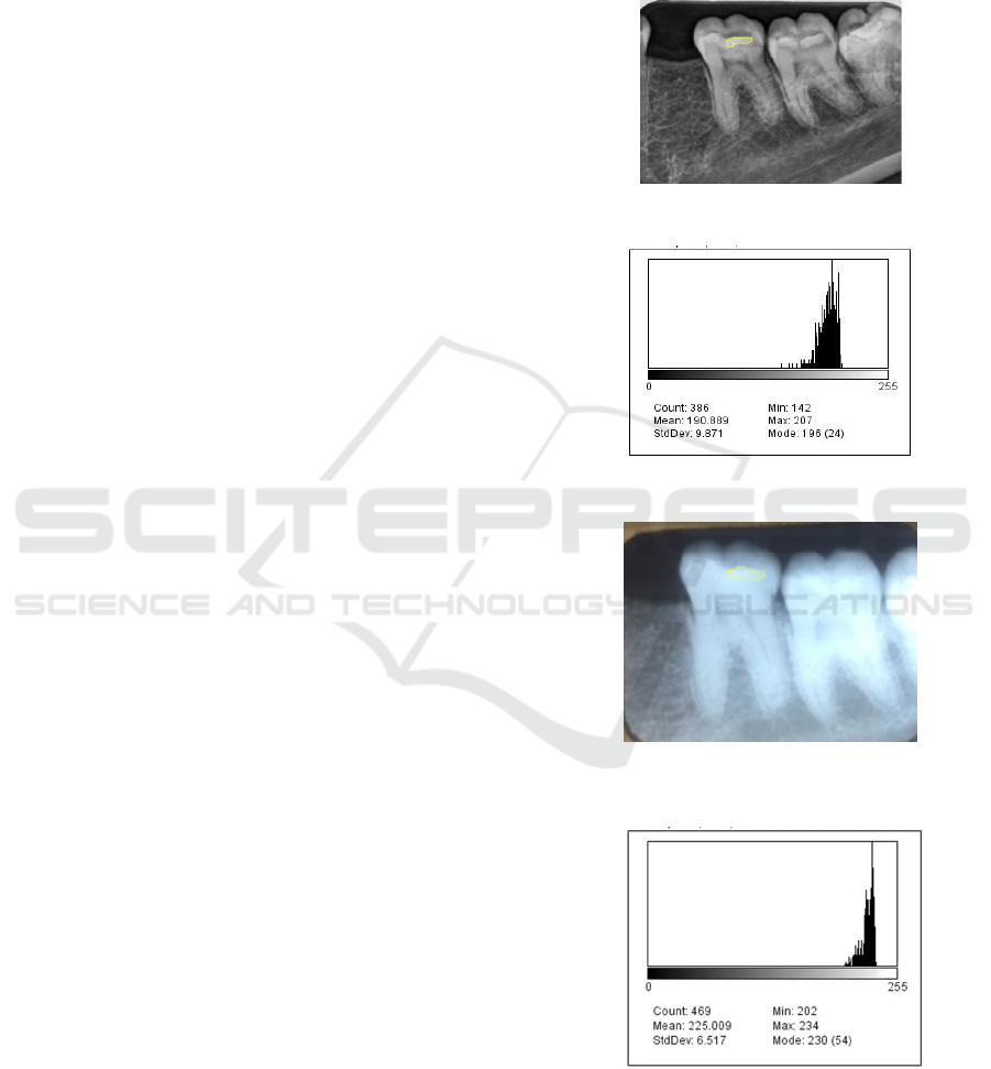
Figure 1. Digital radiograph with Class I restoration on 36.
Figure 2. Histogram analysis using Image J software.
Figure 3. Conventional radiograph with Class I restoration
on 36.
Figure 4. Histogram analysis using Image J software.
Differences in Radiopacity Value of Rmgic, Gic and Composite Resin Materials with Secondary Caries using Conventional and Digital
Radiography
569
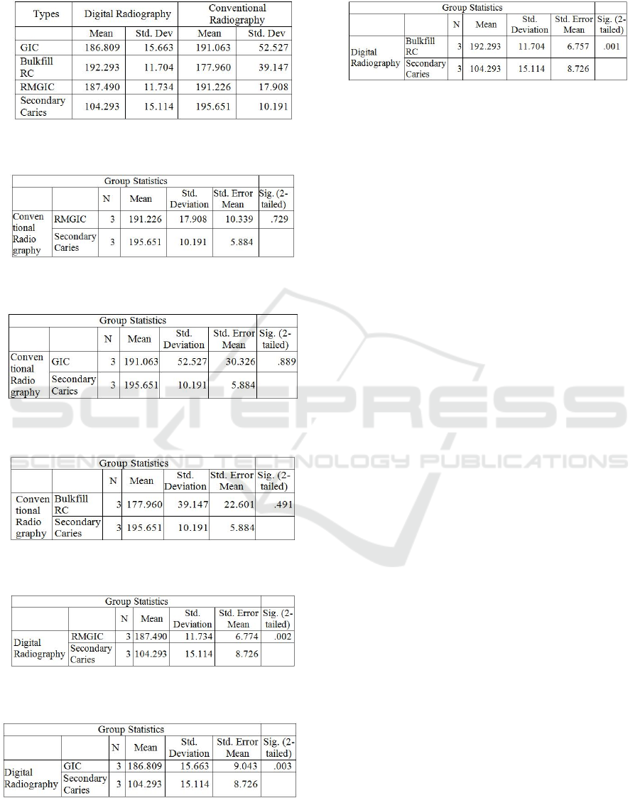
Table 1: The average radiopacity value of restoration
material and radiodensity of secondary caries.
Table 2: Comparison of RMGIC and secondary caries with
conventional radiography.
Table 3: Comparison of GIC and secondary caries with
conventional radiography.
Table 4: Comparison of bulkfill composite resins and
secondary caries with conventional radiography.
Table 5: Comparison of RMGIC and secondary caries with
digital radiography.
Table 6: Comparison of GIC and secondary caries with
digital radiography.
Table 7: Comparison of bulkfill composite resins and
secondary caries with digital radiography.
3 DISCUSSION
Radiopacity of dentistry is very important to
distinguish the dental curing material with the tooth
tissue and its surroundings. Radiopacity is a property
needed for dental materials, including restorative
materials, cavities, core enhancers, adhesives,
adhesives for root canal fillers, temporary crowns,
bridges and ceramics (Anusavice KJ, 2013). Material
radiopacity will increase with increasing particles
containing high atomic number elements (Powers
JM, 2006).
Radiopacity of dentistry material is defined as the
value of optical density of a material (Candeiro,
2012). Factors that can influence the radiopacity of
dental materials are the thickness and chemical
composition of dental materials (Pekkan, 2016).
Another factor is the setting of exposure to light, X-
ray beam angulation, the distance of the film to the
light source and the exposure method used.
Radiopacity of the restoration material used does not
have sufficient radiopacity on the radiograph
including some glass ionomer cement, so the dentist
must know about it (Tsuge, 2009). Restorative
materials vary in radiographic appearance depending
on the thickness, density, atomic number and x-ray
energy rays used to make the radiograph (Eric, 2013).
The results showed that the RMGIC, GIC and
bulkfill composite resin materials using conventional
radiography had different radiopaquality values with
secondary but not significant caries. On conventional
radiographs it is difficult to distinguish the image of
restoration materials with secondary caries. Diagnosis
of secondary carious lesions seen using imaging is
influenced by the type of restoration material
(Antonijevic, 2014).
This may be due to factors such as variations in
the film positioning technique and X-ray rays can
greatly affect the picture of the carious lesion, the
lighting factor may produce marks that affect the
overall contrast of the radiograph thus affecting the
shape or size of the carious lesions on the radiograph
and the exact position of the carious lesion for
example buccal / lingual or caries expansion into
buko-lingual (Eric, 2013).
ICOSTEERR 2018 - International Conference of Science, Technology, Engineering, Environmental and Ramification Researches
570

Another thing that can affect is the distance
between the caries lesion and the pulp horn where
these two shadows can be close together or even
visually interconnected but may not be in the same
plane. The presence of a carious lesion and the
density of the enamel top layer may obscure the
decalcification zone. The presence of secondary
caries and existing patches may coat thoroughly the
existing carious lesions causing errors in interpreting.
The imaging system affects the image of the
restoration. Restoration material with radiopacity is
greater than enamel, will be beneficial for true-
negative diagnosis (Antonijevic, 2014). The
radiopacity value of the restoration material which is
between the enamel and dentin values, or lower than
dentin, tends to create confusion in the test and is
susceptible to false positive diagnosis of secondary
carious lesions (Pedrosa RF, 2011).
The results showed that the RMGIC, GIC and
bulkfill composite resin materials using digital
radiography had significantly different
radiopaquality values with secondary caries. This is
probably because digital radiographs use detectors
that can show significant changes in how we acquire,
store, retrieve, and display images (White and
Pharaoh, 2009).
Digital detectors have the characteristics of
contrast resolution that is the ability to distinguish
radiographic image density and space resolution ie
the capacity to distinguish in detail (Gu, 2006). The
sensitivity of the detector has the ability to respond
to a small amount of radiation. The International
Organization for Standardization classifies the
sensitivity of intraoral films based on speed (ISO,
2000).
The usefulness of digital receptor sensitivity is
influenced by a number of factors including detector
efficiency, pixel size and noise system (White and
Pharaoh, 2009).
4 CONCLUSIONS
The conclusion of this study is that there is no
significant difference in radiopacity value of
RMGIC, GIC and bulkfill composite resin materials
on secondary caries using conventional radiography.
There are significant differences in radiopacity value
of RMGIC, GIC and bulkfill composite resin
materials on secondary caries using digital
radiography. It is better to conduct further research
using different restorative materials in the posterior
and anterior tooth regions.
ACKNOWLEDGEMENTS
This research was funded by the University of North
Sumatra in accordance with the TALENTA Research
Contract of the University of North Sumatra 2018
Fiscal Year Number: 2590 / UN5.1.R / PPM / 2018
dated March 16, 2018
REFERENCES
Antonijevic, D., Ilic, D., Medic, V., Dodic, S., Obradovic-
Djuricic, K., Rakocevic, Z., 2014. Evaluation of
Conventional and Digital Radiography Capacities for
Distinguishing Dental Materials on Radiograms
Depending on The Present Radiopacifying Agent.
(Vojnosanit Pregl) pp 1006-1012.
Anusavice, KJ., Shen, C., Rawls, HR., 2013. Philip’s
Science of Dental Materials ( Saunders Missoury) pp
275-300.
Candeiro, GTM., Correia, FC., Duarte, MAH., Ribeiro-
Siqueira, DC., Gavini, G., 2012. Evaluation of
Radiopasity, pH, Release of Calcium Ions and Flow of
a Bioceramic Root Canal Sealer (JOE) pp 842-45.
Eric Whaites., 2013. Essentials of Dental Radiography and
Radiology 3th edition (Churchill Livingstone: London)
pp 505-530
Gu, S., Rasimick, BJ., Deutsch, AS., Musikant, BL., 2006.
Radiopacity of Dental Materials Using a Digital X-ray
System (Dent Mater) pp 765–70.
ISO, 4049., 2000. Polymer-Based Filling, Restorative and
Luting Materials (Geneva, Switzerland: International
Organization for Standardization)
Nair, MK., Ludlow, JB., May, KN., Nair, UP., Johnson,
MP., Close, JM., 2001. Diagnostic Accuracy of
Intraoral Film ang Direct Digital Images for Detection
of Simulated Recurrent Decay. Oper Dent : 26: 223-30.
Pekkan, G., 2016. Radiopacity of Dental Materials: An
Overview (Avicenna J Dent Res) e36847.
Pedrosa, RF., Brasiliero, IV., dos Anjos Pontual, ML., dos
Anjos Pontual, A., da Silveira, MMF., 2011. Influence
of Materials Radiopacity in the Radiographic
Diagnosis of Secondary Caries: Evaluation in Film and
Two Digital Systems. Dentomaxillofacial Radiology pp
344-350
Powers, JM., Sakaguchi, RL., 2006. Craig’s Restorative
Dental Materials. 12th ed. (Missoury:Mosby) pp 194-
7.
Tsuge, T., 2009. Radiopacity of conventional, resin-
modified glass ionomer,and resin-based luting
materials. J Oral Sci. pp 223–30.
Van Noort, R., 2007. Introduction to dental materials. 3th
ed. (Elsevier: Philadelphia) pp 217-22.
White., Pharaoh., 2009. Oral Radiology, Principles and
Interpretation. 6th edition. ( Mosby Elsevier) pp 335-
450
Differences in Radiopacity Value of Rmgic, Gic and Composite Resin Materials with Secondary Caries using Conventional and Digital
Radiography
571
