
The Effect of Noise Exposure on Signal to Noise Ratio Changes of
Distortion Product Otoacoustic Emissions (DPOAE) Examination in
Rattus Norvegicus
Tengku Siti Hajar Haryuna
1
* and Susi Mulyana
1
1
Department of Otorhinolaryngology-Head and Neck Surgery, Faculty of Medicine, Universitas
Sumatera Utara, Jl. dr. Mansur Kampus USU Indonesia Medan 20155
Keyword: Noise-exposure, Outer hair cell, Otoacoustic emissions, Signal to noise ratio.
Abstract: Distortion Product Otoacoustic Emission (DPOAE) was used to assess outer hair cells. The aim of this study
is to assess the damage to outer hair cells of the noise model Rattus norvegicus by assessing the Signal to
noise ratio (SNR). Twenty-seven Rattus norvegicus were grouped into 3: controls; 2-hours 100 dB noise-
exposure; and 2-hours 110 dB noise-exposure. DPOAE was assessed 3 times in the beginning before
treatment, 2 hours after the second day of exposure (day 2), and after 3 days of no exposure (day 5). The
results showed that the SNR of DPOAE after the noise exposure decreases at all frequencies compared to
before the treatment. A significant decrease in SNR especially at 2000Hz - 4000Hz frequencies. These results
suggest that DPOAE may be used as a sensitive procedure at various frequencies for early diagnosis and
screening of NIHL
.
1 INTRODUCTION
Noise is an important issue in occupational health;
excessive exposure to noise has the social and
psychological impacts caused by noise-induced
hearing loss (NIHL) (Nassiri et al., 2016). The
bilateral sensorineural hearing loss may cause
irreversible and progressive cochlear damage (Souza
Alcaraz et al., 2012;Jahani et al., 2016) In NIHL, a
notch at 3000, 4000, or 6000 Hz is a common finding
in audiometric examination (Derekoy et al., 2004;
Lee Preel &Miller, 2016).
Physiological changes from noise exposure can be
assessed on cochlear cells, where the outer hair cells
are more susceptible to damage than inner hair cells.
Degenerative outer hair cell is the most sensitive
initial process against sound (Balatsouras, 2004).
Otoacoustic emission measures the microscopic
biochemical activity of healthy outer hair cell. OAE
provides bicochlear mechanical stimulation that flow
from the tympanum to the external ear through the
external acoustic meatus (Nassiri et al.,
2016;Moussavi-Najarkola et al., 2012).
The DPOAE measures the bicochlear intensified
emissions recording of a different frequency (f1 and
f2) (Moussavi-Najarkola et al., 2012). The DPOAE
examination uses two simultaneous pure sound
stimuli, of different frequencies and intensities, called
f1 and f2 (f1 < f2) (Janssen et al., 2006;Prieve &
Fitzgeral, 2015).
Studies on animal noise-model have used high-
frequency DPOAE of 1-8 kHz and assess the Ldp
(level distortion product). In this study, the group and
intensity of noise exposure were different from prior
studies which use a low-frequency DPOAE of 1-5
kHz, which is expected to be able to assess the
damage to cochlear outer hair cell of noise model
Rattus norvegicus by Signal to Noise Ratio (SNR)
from DPOAE.
2 METHOD
Twenty-seven Rattus norvegicus aged 2-3 months
with 200-300 gr of weight. All Rattus norvegicus
were examined by the veterinarian and declared
542
Haryuna, T. and Mulyana, S.
The Effect of Noise Exposure on Signal to Noise Ratio Changes of Distor tion Product Otoacoustic Emissions (DPOAE) Examination in Rattus Norvegicus.
DOI: 10.5220/0010077505420546
In Proceedings of the International Conference of Science, Technology, Engineering, Environmental and Ramification Researches (ICOSTEERR 2018) - Research in Industry 4.0, pages
542-546
ISBN: 978-989-758-449-7
Copyright
c
2020 by SCITEPRESS – Science and Technology Publications, Lda. All rights reserved
healthy. The animals are placed in polycarbonate
cage at 25°C with humidity ranging from 40%-60%.
In its care, they get sufficient light source and free
access to water and food until the end of the study.
All procedures are carried out in accordance to
Animal Research Ethicss Committees/AREC Faculty
of Science University of North Sumatera
(No.560/KEPH-FMIPA/2017).
The study group was divided into 3 groups (n =
9), Group I (control); Group 2 (2-hours 100 dB noise-
exposure); and Group 3 (2-hours 110 dB noise-
exposure). The noise was exposed to Rattus
norvegicus in a noise-box of 64.5 x 45 x 40 cm made
by the Faculty of Electro University of North
Sumatra. The source of sound provided by Compact
Disc (CD) containing sound recordings, CD player,
and amplifier which generate the noise with a
frequency of 1-10 kHz and an intensity of 100 dB and
110 dB as measured by a sound level meter.
The Distribution Product Otoacoustic Emission
(DPOAE) examination (Elios Elito Otodia, Echodia
Ltd., London, UK) were performed on all animal
under anesthetic drugs with ketamine 50 mg/kg body
weight and Xylazine 10 mg/kg body weight
intraperitoneal in a soundproof room. The probe was
modified from a plastic pipe adjusted to the size and
placed in the ear canal. DPOAE was assessed 3 times:
early before treatment; 2 hours after the second day
of noise exposure and after 72 hours of the noise-free
period. Acoustic emission results that can be
evaluated at frequencies of 1.5, 2, 3, 4, 5 kHz. All
study samples must have normal SNR (equal to or
more than 6) before noise exposure.
To assess the SNR differences between groups
and between frequency, the data were averaged
between right and left SNR then analyzed with
independent K Kruskal Wallis followed by Mann
Whitney Test, significancy demonstrated as P < 0.05.
3 RESULTS AND DISCUSSIONS
Table 1 shows the mean and standard deviation of
SNR on DPOAE at the frequency of 1500-5000 Hz in
the control group. Table 2 and Table 3 show the mean
and standard deviation of SNR DPOAE at 100 dB and
110 dB, respectively; there was a decrease of SNR
value on the second and fifth day in both groups of
100 dB and 110 dB.
Table 1. The mean and standard deviation of SNR in control
group.
Frequency
(Hz)
Signal to noise ratio (dB)
Day 0 Day 2 Day 5
1500 18,3±9,71 16,7±7,95 9,2 ±8,53
2000 4,1 ± 1,28 8,1 ± 4,81 6,1 ±2,02
3000 9,7 ± 1,88 8,6 ± 3,2 9,7 ±4,11
4000 10,4±3,97 15,8 ±3,25 11,1±2,80
5000 12,1±5,72 11,7 ±7,91 10,7 ±5,12
Table 2. The mean and standard deviation of SNR in 100
dB group.
Frequency
(Hz)
Signal to noise ratio (dB)
Day 0
Day
2
Day
5
1500 16,6±10.91 6,8 ±4,54 7,9 ±8,69
2000 10,5 ± 4,27 4,8 ±2,74 5,8 ±4,07
3000 11,00 ±4,32 3,0 ±4,34 6,4 ±2,87
4000 12,7 ± 4,92 2,0 ±2,68 5,4 ±2,17
5000 6,7 ± 9,29 5,3 ±1,81 6,2± 4,00
Table 3. The mean and standard deviation of SNR in 110
dB group.
Frequency
(Hz)
Si
g
nal to noise ratio
(
dB
)
Da
y
0 Da
y
2 Da
y
5
1500 18,2 ±9,42 6,8 ± 8,29 7,1 ±8,69
2000 9,1 ± 2,01 5,9 ± 6,23 6,3 ± 4,81
3000 8,9 ± 3,78 2,3 ± 2,42 4,3 ±5,66
4000 11,8±3,25 1,2 ± 3,39 5,2 ± 3,17
5000 7,1 ± 6,1 5,2 ± 6,47 6,0 ±4,73
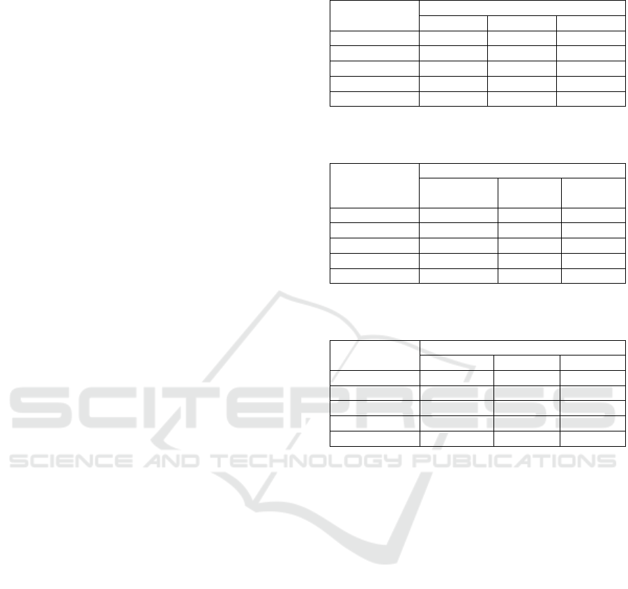
Figure 1 and 2 show the SNR value after the noise
exposure (day 2) in the 100 dB and 110 dB group was
decreased at frequencies of 1500-4000 Hz compared
to the SNR value before the exposure (day 0), this
occurs particularly on 3000 Hz and 4000 Hz
frequencies where the SNR value is 3.0 dB and 2.0
dB, respectively. At the 3000 Hz and 4000 Hz
frequencies, the SNR value was 2, 3 dB and 1.2 dB in
the 100 dB and 110 dB group, respectively. At the
5000 Hz frequency, there is a decrease of SNR but not
significant either in 100 dB or 110 dB group.
Meanwhile, after 3 days of noise-free period (day
5), the SNR value was slightly improved compared to
after exposure (day 2); the SNR value was decreased
and did not return to baseline value before the noise
exposure (day 0) on all frequencies in both group of
100 dB and 110 dB. This study showed that the noise-
break was able to improve the SNR value after the
noise exposure.
The Effect of Noise Exposure on Signal to Noise Ratio Changes of Distortion Product Otoacoustic Emissions (DPOAE) Examination in
Rattus Norvegicus
543
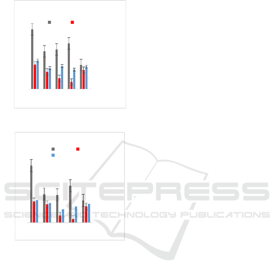
Figure 1. Average SNR changes in 100 dB group on
day 0, day 2, day 5.
Figure 2. Average SNR changes in 110 dB group on
day 0, day 2, day 5.
There is a difference of SNR value at the
frequency of 1500 Hz after noise exposure (day 2) (p
< 0.005) both in the 100 dB and 110 dB group. A
similar finding was found at the frequency of 2000,
3000 and 4000 Hz. At the frequency of 5000 Hz, there
were not any significant differences in SNR value (p>
0.005) both in the 100 dB and 110 dB group.
At all 1500-5000 Hz frequencies, no significant
differences in SNR value were found in days 0, day 2
and day 5 compared between groups of 100 dB with
110 dB (p> 0.005). This shows the difference in
intensity of 100 dB and 110 dB does not demonstrate
the difference of SNR value.
In this study, DPOAE examination showed a
decrease of the SNR after noise exposure at every
frequency, especially at 2000-4000 Hz. NIHL occurs
at high frequencies, especially at the frequency of
3000 Hz-6000 Hz; on audiometric examination, there
was a notch at 4000 Hz frequency (Dobie, 2014).
Several theories have been proposed to understand
why the 4000 Hz frequency is the more susceptible
region to noise exposure. The most popular theory is
that the anatomical structure in the area is weaker
(Dhingra, 2015). Damage to the cochlear structure
due to the noise was observed, especially in the initial
8-10 mm diameter at the cochlear base which is a
sensitive area caused by metabolic, vascular, and
anatomical factors, with corresponding topography to
the frequency of 4000 Hz (Yilmaz et al., 2015).
Salehi et al. (2012) assessed the performance of
outer hair cells in the noise-exposed rabbits, the result
showed that the noise exposure reduces the DPOAE
threshold at a frequency of 4-10 KHz (Salehi et al.,
2011). Nassiri et al. (2016) assessed the effect of
noise exposure on mice by assessing SNR changes at
the intensity of 65 dB, 95 dB and 105 dB concluded
that higher noise intensity will decrease DPOAE level
and sensitive DPOAE examination at various
frequencies (Nassiri et al., 2016).
Distortion product otoacoustic emission
(DPOAE) is an initial and rapid test to detect the
cochlear damage due to noise exposure. DPOAE
sensitively measures the activity of cochlear outer
hair cells recorded in the ear canal (Doosti et al.,
2014). Simultaneous stimulus response to the cochlea
with 2 pure tone frequencies f1 and f2, where
frequency f2 is slightly higher than f1 (2f1-f2)
(Moussavi-Najarkola et al., 2012; Janssen et al.,
2006;. The DPOAE examination measures the Signal
to noise ratio (SNR) which defines the emission
levels generated by noise levels as the background or
ratio of signal strength to noise expressed in decibels
(Nassiri et al., 2016).
The noise impact depends on some sound
characteristics i.e. intensity, spectrum/frequency,
duration and pattern of exposure (Demirel et al.,
2009;Agrawal et al., 2008). The damage to the outer
hair cells depends on the intensity of noise. High-
frequency noise exposure also causes damage to
stereocilia and hair cells that eventually cause
permanent damage. Primary exposure to noise will
damage cochlear hair cells. Initially, the damage
occurs to the outer hair cells, but if the exposure is
continuing, it could evolve to destruct the hair cell
(Nandi&Dhatrak, 2008). Noice-induced cochlear
injury not only affects outer and inner hair cells, but
also damage the fibroblasts of lateral cochlear wall
(Haryuna et al., 2015; Haryuna et al., 2016; Haryuna
et al., 2016; Haryuna et al., 2016). In this study, the
SNR value after the exposure and the noise-free
period was decreased compared to before exposure.
0
2
4
6
8
10
12
14
16
18
20
1500 2000 3000 4000 5000
SNR (dB)
Frequency (Hz)
Mean of 100 dB-intensity SNR
Day0 Day2
0
5
10
15
20
25
1500 2000 3000 4000 5000
SNR (dB)
Frequency ( Hz)
Mean of 110 dB-intensity SNR
Day0 Day2
Day5
ICOSTEERR 2018 - International Conference of Science, Technology, Engineering, Environmental and Ramification Researches
544

This confirms that there was a cochlear outer hair cell
injury measured by DPAOE at both groups.
This experimental study used mice as an animal
model. The mice also have similar inner ear structures
to humans and have been used as animal models for
genetic deafness studies (Vazquez et al., 2001; Salvi
& Boettcher, 2008). The most common gender in the
animal model is males. A study by Najarkola et. al.
using only male animal model showed that gender
affects the measurement of level distortion product
(Ldp). The Ldp is greater in females than in male.
Several studies have reported these differences due to
hormonal influences, and some reported due to the
difference in electromotility of outer hair cells, and
the mechanisms responsible for stereocilia motility,
due to gender difference of lipid membrane that alter
lipid-protein interactions (Moussavi-Najarkola et al.,
2012).
4 CONCLUSIONS
In this study, the SNR of DPOAE examination
decreased at each frequency after the noise exposure,
the decrease was especially seen at the frequency of
3000 and 4000 Hz. There was an improvement in
SNR values after the break period, and between the
100 dB and 110 dB group, there were not any SNR
differences in each frequency.
ACKNOWLEDGMENTS
The authors express their greatest gratitude to the
Research Institute of the Universitas Sumatera Utara
for their full support in this research. The support is
under the TALENTA of the Universitas Sumatera
Utara Year 2017 Number: 5338/UN5.1.R/PPM/2017.
REFERENCES
Agrawal, S.K, Schindler, D. N., Javkler, RK, Robinson, S.,
2008. Occupational Hearing Loss in Current Diagnosis
& Treatment Otolaryngology Head & Neck Surgery, 2
nd Edition, Newyork : Mc Graww Hill Lange, 732-743.
Balatsouras, D. G., 2004. The evaluation of noise-induced
hearing loss with distortion product otoacoustic
emissions, Medical science monitor : international
medical journal of experimental and clinical research,
10(5), 218-222.
Demirel, R. et al. 2009. Noise Induces Oxidative Stress in
Rat, Eur J Gen Med, 6(1), 20–24.
Derekoy, F. S. et al., 2004. Effects of Ascorbic Acid on
Oxidative System and Transient Evoked Otoacoustic
Emissions in Rabbits Exposed to Noise, Exposure,
(October), 1775–1779.
Dhingra, P.L. 2015. Anatomy of Ear. In: Diseases of Ear,
Nose & Throat 5
th
Edition, New Delhi: Elsevier, 3-15.
Doosti A, Lotfi, Y., Moosavi A, Bakhshi, E., Talasaz, A.H.
2014. Distortion Product Otoacoustic Emission
(DPOAE) as an Appropriate Tool in Assessment of
Otoprotective Effects of Antioxidants in Noise-Induced
Hearing Loss (NIHL), Indian Journal of
Otolaryngology and Head and Neck Surgery, 66(3),
325–329.
Dobie, R. A. 2014. Noise Induced Hearing Loss. Bailey BJ,
Johnson JT et al editors. Otolaryngology Head and
Neck Surgery, 4th Ed Vol 1. Philadelphia: Lippincott
SWilliams & Wilkins, 2190-2201.
Haryuna, T.S.H., Riawan, W., Reza, M., Saragih A.R.,
2015. Modulation of antioxidant status by curcumin
prevents cochlea damage after noise exposure. Jornal
of Chemical and Pharmaceutical Research,7 (11), 593-
597.
Haryuna, T.S.H., Riawan, W., Nasution, A., Ma’at, S.,
Harahap, J., Adriztina, I., 2016. Curcumin reduces the
noise-exposed Cochlear fibroblasts apoptosis.
International Archives of Otorhinolaryngology, 20(4),
370-376.
Haryuna, T.S.H., Lutan, R., Taufika, F.A.A., Anggraeni R.,
Zubaidah T.S., 2016. Effect of curcuma longa l. extract
on the AP1 expression in rat cochlear fibroblasts under
noise conditions. Journal of Chinese Pharmaceutical
Sciences, 25(9), 690-694.
Haryuna, T.S.H., Riawan, W., Reza, M., Purnami, N.,
Adnan, A., 2016. Curcumin prevents cochlear oxidative
damage after noise exposure. International Journal of
Pharmacy and Pharmaceutical Sciences, 8(1), 175-
178.
Jahani, L., Mehrparvar, A.H., Esmailidehaj, M.,
Moghbeolohossein, B., Razmjooei, Z., 2016. The effect
of atorvastatin on preventing noise- induced hearing
loss : an experimental study. Int. J Occup Environ Med,
7, 15-21.
Janssen T, Niedermeyer HP, Arnold W. 2006.Diagnostics
of the cochlear amplifier by means of distortion product
otoacoustic emissions. ORL, 68, 334-339.
Lee Preel, C.G., & Miller, M. 2016. The role of oxidative
stress in hearing loss. The Science of Free radical
Biology & Disease. USA: Jhon Willey & Son inc. 8,
115-128.
Moussavi-Najarkola SA, Khavanin A, Mirzaei R, Salehnia
M, Muhammanejad A, Akbari M. 2012. Noise Induced
Outer Hair Cells Dysfunction and Cochlear Damage in
Rabbits. Iran Red Cressent Med J, 14 (1), 647-656.
Nandi, S.S., Dhatrak, V. 2008. Occupational noise-induced
hearing loss in India. Indian Journal of Occupational
and environmental, 12, 53-56.
Nassiri, P., Zare S., Esmail, M.R.M., Pourbakhti, A., Azam,
K., Golmohammadi, T. 2016. Assessment of the Effects
of Different Sound Pressure Levels on Distortion
Product Otoacoustic Emissions ( DPOAEs ) in Rats. Int
Arch Otorhinolayngol, 8, 93-99.
The Effect of Noise Exposure on Signal to Noise Ratio Changes of Distortion Product Otoacoustic Emissions (DPOAE) Examination in
Rattus Norvegicus
545

Nassiri, P., Zare, S., Monazzam, M.R., Pourbakht, A.,
Azam, K., Golmohammadi, T. 2016. Modelling signal
to noise ratio of otoacoustic emissions in workers
exposed to different industrial noise levels. Noise &
Health, 18, 391-398.
Prieve, B. & Fitzgerald T., 2015. Otoaccoustic Emissions.
In : Handbook of Clinical Audiology 7
th
ed. Eds: Katz
J., Chasin M., English K., Hood L. J., Tillery K. L..
Philadelphia : Wolters Kluwer Health, 357-379.
Salehi N, Akbari M, Kashani M, Haghani H. 2011.
Protective effects of N-acetylcystein on the hearing of
rabbits exposed to noise and carbon monoxide.
Audiology, 20, 36-46.
Salvi, R. and Boettcher, F. A., 2008. Animal Models of
Noise-Induced Hearing Loss in Sourcebook of Models
for Biomedical Research, 289–301.
Souza Alcarás, P. A. et al., 2012. Evoked otoacoustic
emissions in workers exposed to noise: A review,
International Archives of Otorhinolaryngology, 16(4),
515-522.
Vazquez, A.E., Luebke, A.E., Martin, G.K., 2001.
Temporary and Permanent Noise Induced Changes in
Distortion Product Otoacoustic Emissions in CBA/CaJ
mice, Elsevier Hearing Research, 156(3), 31-43.
Yilmaz H, Aydin S, Sanli A, Erdogan BA, Kibar S, Sirvanci
S, et al. 2015. Evaluation of the effect of betahistin in
noise induce hearing loss using distortion product
otoacoustic emissions and scanning electron
microscopy. The Journal of International Advanced
Otology 11 (1), 6-11.
ICOSTEERR 2018 - International Conference of Science, Technology, Engineering, Environmental and Ramification Researches
546
