Light Amplification and Nonlinear Microscopy by Stimulated Raman
Scattering
M. A. Ferrara
1
, A. D’Arco
1,2
, M. Indolfi
1
, N. Brancati
3
, L. Zeni
2
and L. Sirleto
1
1
National Research Council (CNR) - Institute for Microelectronics and Microsystems, I-80131 Napoli, Italy
2
Second University of Naples (SUN), Department of Information Engineering, I-81031 Aversa, Italy
3
National Research Council (CNR) – Istituto di Calcolo e Reti ad Alte Prestazioni, I-80131 Napoli, Italy
Keywords: Nanophotonics, Biophotonics, Nonlinear Optics, Stimulated Raman Scattering.
Abstract: The Stimulated Raman Scattering has important connections with nanophotonics and biophotonics.
Concerning nanophotonics, one of the most recent challenges is the investigation of ‘nonlinear optical
phenomena at nanoscale’. Among them, stimulated Raman scattering is one of the most interesting, due to
its significant implications from both fundamental and applicative point of view. In this paper, comparison
among experimental investigations of stimulated Raman scattering in amorphous silicon nanoparticles and
in silicon micro- and nano-crystals, at the wavelengths of interest for telecommunications, are reported. In
addition, concerning biophotonics, first, the implementation of femtosecond Stimulated Raman
Spectroscopy (f-SRS), as a single point of scanning microscopy, is described. Then, the integration of f-SRS
in a laser scanning microscope and label free imaging of polystyrene-beads are demonstrated.
1 INTRODUCTION
In silicon photonics and silicon nanophotonics,
amplification and light emission are still the most
challenging goals to get. In Stimulated Raman
Scattering (SRS), a pump laser beam enters a
nonlinear medium and spontaneous generation and
amplification lead to a beam at a frequency different
from the pump. Raman amplification, demonstrated
in the early 1970s, is an interesting approach for
optical amplification, because it is only restricted by
the pump wavelength and Raman active modes of
the gain medium (Shen and Bloembergen, 1965).
Raman lasing can be achieved by using SRS
phenomenon, which permits, in principle, the
amplification in a wide interval of wavelengths,
from the ultraviolet to the infrared (Stolen, 2004).
SRS is used in tunable laser development, high
energy pulse compression, etc. Considering bulk
semiconductors, lasing by SRS was first discovered
in GaP (Nishizawa and Suto, 1980), whereas SRS
from spherical droplets and microspheres, with
diameters 5-20 µm, has been observed using both
pulsed and continuous wave probe beams (Spillane
et al., 2002). Raman lasers have been also
demonstrated in silicon micro-waveguides (Rong et
al., 2005).
The "ideal" material for Raman amplification
should have wide, flat and high Raman gain into all
the range of interest in telecommunications (from
1270 to 1650 nm). Unfortunately, as a general rule,
due to the physics behind the Raman effect, there is
a tradeoff for Raman amplification. In nature, we
have material, for example silicon, with high Raman
efficiency and small bandwidth, and material, for
example silica, with a large bandwidth but with
small amplification (see figure 1). The above-
mentioned tradeoff is a fundamental limitation
towards the realization of micro-/nano-sources with
large emission spectra. Therefore, the investigation
of new materials possessing both large Raman gain
coefficients and spectral bandwidth is becoming
mandatory in order to satisfy the increasing
telecommunications demands (Refi, 1999). In order
to overcome these limitations, a possible option is to
consider nanocomposite and nanostructured
materials.
Raman scattering in electrons-confined and
photons confined materials is a fascinating research
field of great importance from both fundamental and
applicative point of view. Concerning the
fundamental one, there have been a number of
investigations both experimental and theoretical, but
the question is still "open" (Gaponenko et al., 2002),
while from an applicative point of view, there are
some important prospective, for example to realize
micro/nano source, with improved performances,
based on SRS.
The phenomenon of strong resonant and local
enhancement of visible electro-magnetic (EM)
radiation when incident on the surface of metallic
particles and films resulting from surface plasmon
resonances, continues to attract significant attention
for fundamental and applied interests (Kawata,
2009). However, the possibility of EM radiation
enhancement from semiconducting and insulating
materials, particularly in silicon, is noteworthy for
silicon-based optoelectronic applications owing to
the potential for monolithically integrating photonic
technology and semiconductor electronics (Cao et
al., 2006). Except for a report of SRS from
individual single walled carbon nanotubes (Zhang et
al., 2006), and the observation of SRS from
semiconductor nanowires (Wu et al., 2009), we find
no other evidence for this important nonlinear optic
effect in nanostructured materials.
In biophotonics, confocal and multiphotons
fluorescence microscopy are important and powerful
techniques for imaging of biological samples.
However, these microscopic techniques show some
limitations, indeed, they require chemical labels that
could interfere with biological functionalities;
additionally the photo-bleaching introduces artefacts
and limits the measurement repeatability. Therefore,
it is necessary to introduce and implement a new
multiphotons microscopy technique suited for real
time imaging with high three dimensional spatial
resolution and chemical specificity of unlabeled
living cells. Raman microscopy can be used as a
contrast mechanism based on vibrational properties.
A typical Raman spectrum makes available
information on the molecular and chemical structure
of the sample, offering an intrinsic chemical
selectivity. Nevertheless, linear Raman microscopy
is limited to weak signals, so, to obtain an image
acquisition times are very long.
It is worth noting that, due to the recent
femtoseconds laser technological development,
nonlinear techniques have found application in soft
matter and in particular in biological materials.
Femtoseconds laser allows to obtain an average
power, incident onto the sample, lower than the
photodamage limit and a high enough pump peak
power to ensure the triggering of the nonlinear
effects. In addition, the range of pulses wavelengths,
generated into the range between 680 nm and 1300
nm, permits to work in the window of water
transparency, significantly reducing the absorption.
Coherent Raman Scattering (CRS) techniques
are sensitive to the same molecular vibrations
probed in spontaneous Raman spectroscopy, but
unlike linear Raman spectroscopy, CRS techniques
exhibit a nonlinear dependence on the incoming
light fields and produce coherent radiation. In CRS,
two collinear laser beams (pump and probe) at
different frequencies excite the sample. When the
difference in frequencies is equal to a molecular
vibration, a stimulated and coherent excitation of
molecular bond vibration modes (third order non-
linear process) occur and a significant increase of
Raman signal is observed. This latter property has
popularized CRS as a microscopy modality, as it is
intimately related to the technique’s strong optical
signals that enable fast imaging applications. CRS
microscopy makes it possible to achieve images
based on vibrational Raman contrast at imaging
speeds much faster than attained with conventional
Raman microscopes. Clearly, this attribute is very
attractive for biological imaging, where imaging
speed is an important experimental parameter
(Ploetz et al., 2007; Freudiger et al., 2008; Ozeki et
al., 2009; Nandakumar et al., 2009; Fu et al., 2013).
CRS includes two techniques: coherent anti-
Stokes Raman scattering (CARS) and SRS. We note
that a CARS spectrum is different from its
corresponding spontaneous Raman spectrum due to
a non-resonant background, which complicates
spectral assignment, causes difficulties in image
interpretation, and limits detection sensitivity (Ploetz
et al., 2007; Freudiger et al., 2008; Ozeki et al.,
2009; Nandakumar et al., 2009).
The recent development of SRS microscopy
overcame these limitations and provided better
imaging contrast mechanism (vibrational) contrast.
SRS eliminates the non-resonant background
problem because the generated third order SRS
nonlinear polarization is directly heterodyne mixed
and amplified by the input beam with the exact same
phase, therefore always resulting in a zero non-
resonant contribution. Definitely, SRS is free from
the non-resonant background, exhibiting an identical
spectrum as the spontaneous Raman it is linearly
proportional to the concentration of the analyte, and
therefore it allows straightforward quantification. In
such situations, it is natural to consider the
application of SRS to biological microscopy. When
SRS microscopy was proposed (Ploetz et al., 2007;
Freudiger et al., 2008; Ozeki et al., 2009), two
transform-limited picosecond (ps) lasers with narrow
spectral bandwidth were used to excite a single
Raman-active vibrational mode for fast imaging
with high spectral resolution. With this ps–ps
excitation sources it is not possible to distinguish
mixed chemical species with overlapped Raman
bands in the sample because other vibrational modes
of the sample are not excited. However, in many live
biological and biomedical applications, simultaneous
mapping of different chemical species in the same
sample is extremely important for the investigation
of the co-distribution or dynamic correlation
between pairs of biomolecules. Therefore,
multicolor imaging with multiple chemical contrasts
is considered necessary. We note that multicolor
imaging can be realized only taking advantage of
femtosecond laser source
(Nandakumar et al., 2009).
In this paper, in paragraph 2 a comparison
between our experimental results of SRS obtained
on different samples of nanostructured amorphous
silicon clusters, and of silicon micro-crystals (Si-µc)
and nano-crystals (Si-nc) are reported. The two main
figure of merit (Raman gain and bandwidth) are
compared to an ideal material and we highlight a
possible trend to get the best performance.
In paragraph 3, the details of experimental set up
and the main experimental issues of femtosecond
Stimulated Raman Spectroscopy (f-SRS)
implementation are reported. In addition, steps
towards nonlinear microscopy, i.e. the integration of
f-SRS in a laser scanning microscope, is described.
Finally, label free imaging of polystyrene-beads are
demonstrated.
2 LIGHT AMPLIFICATION BY
STIMULATED RAMAN
SCATTERING
In our previous papers (Sirleto et al., 2004 and 2006;
Ferrara et al., 2008), some advantages of silicon
nanostructure with respect to silicon were
demonstrated. Experimental results, proving
spontaneous Raman scattering in silicon
nanostructures at the wavelength of interest for
telecommunications (1.54 µm), were reported in
Refs. (Sirleto et al., 2004 and 2006; Ferrara et al.,
2008). According to phonon confinement model in
Refs. (Sirleto et al., 2004 and 2006; Ferrara et al.,
2008), two significant improvement of Raman
approach in silicon quantum dots with respect to
silicon were demonstrated: the broadening of
spontaneous Raman emission and the tuning of the
Stokes shift. Considering silicon quantum dots
having crystal size of 2 nm, a significant broadening
of about 65 cm
-l
and a peak shift of about 19 cm
-l
were obtained. Because the width of C-band
telecommunication is 146 cm
-1
, taking into account
the broadening and the shift of spontaneous Raman
emission, more than the half of C-band could be
cover using silicon quantum dots, without
implementing the multi-pump scheme.
Nanocomposities are random media containing
domains or inclusions that are on the nanometric
size scale. The optical properties of composite
materials can be adjusted by controlling the
constituents and morphology of the composite
structure. The optical nanocomposite approach
offers opportunities to produce high-performance
and relatively low-cost optoelectronic media suitable
for many applications.
In our previous papers (Sirleto et al., 2004 and
2006; Ferrara et al., 2008), SRS has been measured
using as pump a CW Raman laser operating at 1427
nm. SRS net gain (G) is given by:
(1)
where I
p
= P/A, with P is the power incident onto the
sample and A is the effective area of pump beam.
Since the sample is transparent to the incident light,
L is taken to be equal to the thickness of the sample
along the path of the incident light. G as a function
of signal laser wavelength was measured in three
different samples:
Silicon nanocomposites dispersed in SiO
2
matrix, with a probe signal at 1542.2nm
(Sirleto et al., 2009; Ferrara et al., 2011). The
mean radius of the silicon dots and the dot
density were respectively of 49nm (Si-µc) and
1.62x10
8
dots/cm
2
.
Amorphous silicon nanoclusters embedded in
Si-rich Nitride/Silicon superlattice structures
(SRN/Si-SLs), with a probe signal at 1540.6nm
(Sirleto et al., 2008). The structure of the
sample consists of 10 SRN layers and 9
amorphous Si (a-Si) layers for a total thickness
of 450 nm. Amorphous silicon nanoclusters
size was about 2nm.
Silicon nanocrystals embedded in silica matrix,
with a probe signal at 1541.3nm (Sirleto et al.,
2012). Si-nc size was about 4nm.
In particular, we focalized our study on two
different nanocomposities materials based on
amorphous or crystalline silicon. The difference
between them is related to their different
spontaneous Raman signal, indeed in amorphous
silicon Raman spectra is broadband but shows a low
intensity, while in crystalline silicon Raman spectra
is very narrowband but shows a high intensity.
Additionally, considering that Raman effect is a
volume effect, in the sense that the greater is the
volume of interaction, the higher is the Raman
signal, we aspect that different concentration of
nanoparticles could lead to different Raman gain.
Results obtained can be summarized as follows:
In silicon nanocomposites, an amplification of
Stokes signal up to 1.4 dB/cm is reported. This
result showed a preliminary valuation of
approximately a five-fold enhancement of the
Raman gain with respect to bulk silicon.
Moreover, a threshold power reduction of
about 60% is also reported (Sirleto et al., 2009,
Ferrara et al., 2011).
In SRN/Si-SLs, amplification of Stokes signal
up to 0.87 dB/cm was experimentally
demonstrated, consistent with a preliminary
valuation of approximately a four-fold
enhancement of the Raman gain with respect to
bulk silicon. Moreover, a threshold power
reduction of about 40% is also reported (Sirleto
et al., 2008).
A giant Raman gain from the silicon
nanocrystals is obtained that is up to four
orders of magnitude greater than in bulk
crystalline silicon (Sirleto et al., 2012).
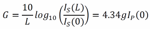
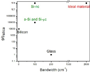
In figure 1 the Raman Gain coefficients and their
bandwidth for all the samples are reported and
compared with an ideal material. We note that si-nc
has a Raman gain value comparable to the ideal
materials, but the bandwidth is still small. Probably
if the nanostructures was embedded in a different
matrix, a greater bandwidth could be obtained, too.
Figure 1: Raman Gain coefficients and their bandwidth are
reported for different materials: silicon and silica (as 'bulk
material'), and silicon micro- and nano-particles
(amorphous and crystalline). Features for 'ideal materials'
for Raman amplification are reported, too.
3 STIMULATED RAMAN
MICROSCOPY
In SRS microscopy, pump and probe pulses at
angular frequencies of ω
1
and ω
2
(ω
1
> ω
2
) are
focused into a sample, and the intensity change of
the probe pulse due to SRS is detected. Intuitively,
SRS is caused by the optical phase modulation
induced by the time-dependent refractive index
reflecting the molecular vibration, which is
coherently driven by the intensity beat between
pump and probe pulses (Shen and Bloembergen,
1965).
Concerning SRS, an important issue is its
sensitivity, because the SRS signal is detected as a
small change of the intensity of excitation beam, and
hence is deteriorated by shot-noise and laser
intensity noise. The laser intensity noise is quite
important aspect to take into account in SRS
microscopy because it can easily surpass the shot
noise, an intrinsic property of the light source (Min
et al., 2011).
In the field of laser spectroscopy, SRS was
extensively studied as a highly sensitive tool of
vibrational spectroscopy (Owyoung, 1978) and the
detection of SRS with a shot-noise limited
sensitivity was achieved (Owyoung, 1978, Levine et
al., 1979; Heritage et al., 1980). The basic idea is to
take advantage of lock-in detection at high-
frequency. In this approach, a high-frequency
modulation transfer method to detect the signal is
used. The intensity of the pump beam is modulated
with an electro-optic modulator and the modulation
transfered to the probe beam is measured with a
lock-in amplifier (LIA) after blocking the pump
beam with an optical filter. It is preferable to
increase the modulation frequencies because the
relative intensity noise of laser pulses typically
decreases with frequency. Increasing modulation
frequency of the beam, at frequencies above 1 MHz,
it allows to reach the intrinsic limit of
photodetectors. The thermal noise can be negligible
compared to the shot noise when the optical power is
of the order of several milliWatts.
Fig. 2 shows the schematic layout of the
microscope; it requires two sources. The first one,
used as a pump beam, is a femtosecond Ti-sapphire
(Chameleon Ultra II) with a pulse duration of
approximately 100 fs with a repetition rate of
80MHz and emission wavelengths into the range
680-1080nm. The second one, used as probe beam,
is a femtosecond synchronized optical parametric
oscillator (SOPO-Chameleon Compact OPO) with a
pulse duration of approximately 200 fs with a
repetition rate of 80MHz that emits into the range of
wavelengths 1000-1600nm. An electro -optic
modulator (EOM 350-160 KD*P CONOPTICS) was
placed for the intensity modulation of the pump
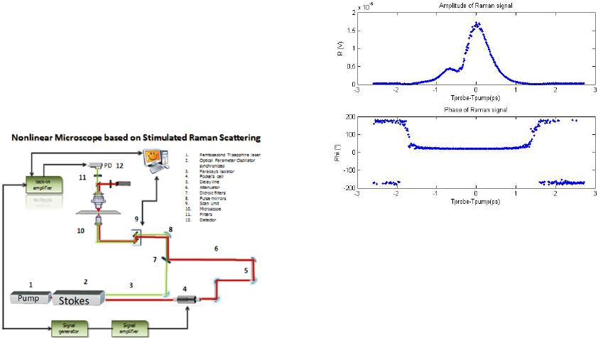
pulses at a modulation frequency of 9.1 MHz. These
two beams were collinearly combined with a
dichroic mirror (Semrock FF875-Di01-25x36),
temporally overlapped by a delay line (Newport
MOD M-ILS200CC) and focused inside a sample
through a scanning microscope (Ti-eclipse Nikon).
The integration with the microscope ensures greater
stability of the system and increase the spatial
resolution of the measurement, while the presence of
scanning unit allows the analysis on a large area, i.e.
the realization of an image.
Figure 2: Experimental setup for SRS microscopy.
A 20X objective focuses beams inside a sample,
and output pulses are collected by a 60X high
numerical aperture objective. After, pump pulses are
removed by a stack of optical filter, while probe
pulses are detected by a photodetector (Thorlabs
DET 10N/M) and measured by a lock-in amplifier
(SR844- 200 MHz dual phase). The electrical signal
coming out from the lock-in amplifier is digitalized
by a PCI card, which manages and synchronizes the
lock-in amplifier and the scanning unit of
microscope in order to collect information and to
obtain a 2D image.
A first measurement of stimulated Raman
spectroscopy was carried out on a single point of a
drop of a water solution with a very high density of
polystyrene beads, which is placed between a
microscope slide and a coverslip; the polystyrene
beads had a diameter of 15µm. In order to
investigate a typical C-H bond of polystyrene
(Raman shift of 3054 cm
-1
), the pump signal was set
at 799 nm with a focused power of 20mW, while the
probe signal was set at 1057nm with a focused
power of 10mW. The temporal overlap of these two
beams was obtained by scanning the delay line with
steps of 0.001mm corresponding to 13,3fs time-shift.
The time constant of the LIA was set to 3 ms with a
slope of 18 dB/oct and 30μV sensitivity. The
measured values from lock-in amplifier, in terms of
phase and amplitude of SRS signal as a function of
the probe-pump delay in ps, are reported in Fig.3.
Figure 3: Amplitude and Phase of SRS signal measured by
lock-in amplifier.
Because SRS uses near-infrared excitation light,
the standard optics of a laser-scanning multiphoton
microscope are compatible with SRS modalities. In
particular, SRS uses the same high numerical
aperture (NA) lenses that are employed in
multiphoton microscopes. In fact, a SRS imaging
modality shares many of the imaging properties of a
multiphoton microscope, including fast image
acquisition and sub-micrometer resolution.
Commercial laser-scanning microscopes can be
upgraded with a SRS module with some important
modification. In its simplest form, a SRS microscope
can be constructed from a fluorescence laser-
scanning microscope by equipping a forward
detector and with proper bandpass filters and
interfacing the scanning unit of microscope with the
detector. Moreover, although standard condensers,
which have a NA of ∼0.55, suffice to capture a
significant portion of the forward- propagating
signal, better collection efficiencies are obtained
with higher-NA condensers. This is especially
important for the SRS techniques, where
photothermal and position-dependent interference
effects may introduce artifacts in the image (Popov
et al., 2012;
Chung et al., 2013). Such effects can be
mitigated by choosing a high-NA condenser.
Fast image acquisition rates are the prime
advantage of SRS imaging over spontaneous Raman
microscopy. Using high repetition rate femtosecond
pulse trains with average powers in the 10 mW
range on the sample produces SRS signals with
acceptable SNR in a few microsecond per pixel. For
an image with 512 × 512 pixels, this translates in
acquisition rates of about a frame per second. Such
imaging speeds are at par with those of linear and
multiphoton fluorescence microscopy techniques.
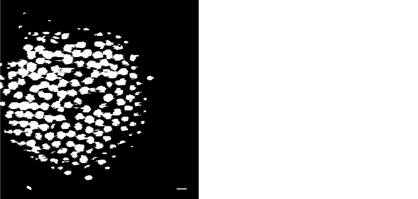
To demonstrate the feasibility of the proposed
device, the same sample, used for spectroscopic
investigation, is studied. The binary image shown in
fig. 4, is single recordings of 512x512 pixels with
acquisition time of 16 seconds. The time constant of
lock-in amplifier was set to 100μs with a slope of
18dB/oct and 3μV sensitivity. For the process of
binarization, an adequate threshold of gray level has
been selected with Otsu method (Otsu, 1979).
Figure 4: Binary SRS image of 15μm polystyrene beads at
3054cm
-1
at I
pump
at 7mW. Scale bar is 15μm.
4 CONCLUSIONS
In this paper, we describe some important
connections of SRS with nano- and bio-photonics.
As far as nanophotonics is concerned,
experimental investigations of stimulated Raman
scattering in nanostructured silicon based materials
are compared. Because a theoretical understanding
addressing the physical origin of enhanced Raman
gain in nanostructured materials remains to be
established, in this work we try to give some tiles for
the important open question about stimulated Raman
scattering at nanoscale. In addition, our results could
be an important step towards to silicon based Raman
laser.
As far as biophotonics is concerned,
femtosecond stimulated Raman spectroscopic and
microscopy have been implemented. As a
preliminary step for nonlinear microscopy, label free
imaging of polystyrene-beads is demonstrated. Next
step is to apply this nonlinear optical imaging
approach to biological research.
ACKNOWLEDGEMENTS
We really appreciate the useful discussions and
continuous support from Giacomo Cozzi, product
specialist - Nikon Instruments. We thank Vitaliano
Tufano from IMM-CNR, for his valuable technical
assistance.
This work was partially supported by Italian
National Operative Programs PONa3_00025
(BIOforIU) and by Euro-bioimaging large scale
pan
‐
European research infrastructure project.
REFERENCES
Cao L., Nabet B., Spanier J. E., 2006, Enhanced Raman
scattering from individual semiconductornanocones
and nanowires, Physical Review Letters, Vol. 96, no.
15, Article ID 157402, pp. 1-4.
Chung C. Y. et al., 2013, Controlling stimulated coherent
spectroscopy and microscopy by a position-dependent
phase, Phys. Rev. A 87(3), 033833.
Ferrara M. A., Donato M. G., Sirleto L., Messina G.,
Santangelo S., Rendina I., 2008, Study of Strain and
Wetting Phenomena in Porous Silicon by Raman
scattering, Journal of Raman Spectroscopy, Vol. 39,
Issue 2, pp. 199-204.
Ferrara M. A., Sirleto L., Nicotra G., Spinella C., Rendina
I., 2011, Enhanced gain coefficient in Raman
amplifier based on silicon nanocomposites, Photonics
and Nanostructures – Fundamentals and
Applications;pp. 1-7.
Freudiger C. W., et al., 2008, Label-free biomedical
imaging with high sensitivity by stimulated Raman
scattering microscopy, Science 322, 1857–1861.
Fu D., Holtom G., Freudiger C., Zhang X. & Xie X. S.,
2013, Hyperspectral imaging with stimulated Raman
scattering by chirped femtosecond lasers.J. Phys.
Chem. B 117, 4634–4640.
Gaponenko S. V., 2002, Effects of photon density of states
on Raman scattering in mesoscopic structures,
Phys.Rev. B, Vol. 65, 140303.
Heritage J. P. and Allara D. L., 1980, Surface Picosecond
Raman Gain Spectra of a Molecular Monolayer,
Chem. Phys. Lett. 74, 507–510.
Kawata S., Inouye Y., Verma P.,2009, Plasmonics for
near-field nanoimaging and superlensing, Nature
Photonics, Vol. 3, no. 7, pp. 388-394.
Levine B. F., Shank C. V., and Heritage J. P., 1979,
Surface Vibrational Spectroscopy Using Stimulated
Raman Scattering, IEEE J. Quantum Electron. QE_15,
1418–1432.
Min W. Freudiger C.W., Lu S., and Xie X. S., 2011,
Coherent Nonlinear Optical Imaging: Beyond
15μm
Fluorescence Microscopy, Annu. Rev. Phys. Chem.
62, pp. 507-530.
Nandakumar P., Kovalev A., and Volkmer A., 2009,
Vibrational Imaging Based on Stimulated Raman
Scattering Microscopy, New J. Phys. 11, 033026–
033035.
Nishizawa J., & Suto K., 1980, Semiconductor Raman
Laser, J. Appl. Phys., Vol. 51,2429-2431.
Otsu N. 1979, A Threshold Selection Method from Gray-
Level Histograms, IEEE Transactions on Systems,
Man, and Cybernetics, Vol. 9, No. 1, pp. 62-66.
Owyoung A., 1978, Coherent Raman Gain Spectroscopy
Using CW Laser Sources, IEEE J. Quantum
Electron.QE_14, 192–203.
Ozeki Y., Dake F., Kajiyama S., Fukui K., & Itoh K.,
2009, Analysis and experimental assessment of the
sensitivity of stimulated Raman scattering microscopy,
Opt.Express 17, 3651–3658.
Ploetz E., Laimgruber S., Berner S., Zinth W., & Gilch P.,
2007, Femtosecond stimulated Raman microscopy
.Appl. Phys. B 87,pp.389–393.
Popov K. I. et al., 2012, Image formation in CARS and
SRS: effect of an inhomogeneous nonresonant
background medium, Opt. Lett. 37(4), 473–475.
Refi J. J., 1999, Bell Labs Tech. J., Vol. 4, 246-261.
Rong H. S., Liu A. S., Jones R., Cohen O., Hak D.,
Nicolaescu R., Fang A., Paniccia M.,2005, Nature,
Vol. 433 (7023), 292-294.
Shen Y. R. and Bloembergen N., 1965, Theory of
Stimulated Brillouin and Raman Scattering, Phys.
Rev., Vol. 137, AI787.
Sirleto L., Raghunathan V., Rossi A. and Jalali B., 2004,
Electronics Letters 40 (19), 121-122.
Sirleto L., Ferrara M. A., Rendina I., Jalali B., 2006,
Applied Physics Letters 88, 211105.
Sirleto L., Ferrara M: A., Rendina I., Basu S. N., Warga J.,
Li R., Dal Negro L., 2008, Enhanced stimulated
Raman scattering in silicon nanocrystals embedded in
silicon-rich nitride/silicon superlattice structures,
Applied Physics Letters, 93, 251104.
Sirleto L., Ferrara M. A., Rendina I., Nicotra G., Spinella
C.,2009, Observation of stimulated Raman scattering
in silicon nanocompisties, Applied Physics Letters, 94,
221106.
Sirleto L, Ferrara M A, Nikitin T, Novikov S,
Khriachtchev L., 2012, Giant Raman gain in silicon
nanocrystals. NATURE COMMUNICATIONS,
ISSN: 2041-1723.
Spillane S. M., Kippenberg T. J., Vahala K. J., 2002,
Nature, Vol. 415 (6872), 621-623.
Stolen R. H., 2004, Raman Amplifiers for
Telecommunications 1, (Ed. M. N. Islam) Springer-
Verlag, New York, p35.
Wu J., Awnish K. Gupta, Humberto R. Gutierres, Eklund
P. C., 2009, Nano Letters, Vol. 9, N. 9, 3252-3257.
Zhang B. P., Shimazaki K., Shiokawa T., Suzuki M.,
Ishibashi K., Saito R., 2006 Appl. Phys. Lett., Vol. 88
(24),241101-241103.
