
EEG and Eye-Tracking Integration for Ocular Artefact Correction
P. Rente Lourenço
1
, W. W. Abbott
1
and A. A. Faisal
1,2
1
Department of Bioengineering, Brain and Behaviour Lab, Imperial College London, Exhibition Road, London, U.K.
2
Department of Computing, Brain and Behaviour Lab, Imperial College London, Exhibition Road, London, U.K.
Keywords: EEG, Eye-Tracking, Ocular Artefacts, ICA, Wiener Filter, Wavelet Decomposition.
Abstract: Electroencephalograms (EEG) are a widely used brain signal recording technique. The information
conveyed in these recordings can be an extremely useful tool in the diagnosis of some diseases and
disturbances, as well as in the development of non-invasive Brain-Machine Interfaces (BMI). However, the
non-invasive electrical recording setup comes with two major downsides, a. poor signal-to-noise ratio and b.
the vulnerability to any external and internal noise sources. One of the main sources of artefacts are eye
movements due to the electric dipole between the cornea and the retina. We have previously proposed that
monitoring eye-movements provide a complementary signal for BMIs. He we propose a novel technique to
remove eye-related artefacts from the EEG recordings. We couple Eye Tracking with EEG allowing us to
independently measure when ocular artefact events occur and thus clean them up in a targeted manner
instead of using a “blind” artefact clean up correction technique. Three standard methods of artefact
correction were applied in an event-driven, supervised manner: 1. Independent Components Analysis (ICA),
2. Wiener Filter and 3. Wavelet Decomposition and compared to “blind” unsupervised ICA clean up. These
are standard artefact correction approaches implemented in many toolboxes and experimental EEG systems
and could easily be applied by their users in an event-driven manner. Already the qualitative inspection of
the clean up traces show that the simple targeted artefact event-driven clean up outperforms the traditional
“blind” clean up approaches. We conclude that this justifies the small extra effort of performing
simultaneous eye tracking with any EEG recording to enable simple, but targeted, automatic artefact
removal that preserves more of the original signal.
1 INTRODUCTION
Electroencephalogram (EEG) recordings are widely
used nowadays for different neurological
applications, such as diagnosis of epilepsy or sleep
disorders, or brain machine interfaces. (Iber, Ancoli-
Israel, Chesson., et al., 2007; Giannitrapani and
Kayton, 1974; Saatchi, Oke, Allen, et al., 1995). The
EEG trace is known to be highly variable, in part
due to transient physiological conditions and state of
the brain as well as noise inside the nervous system
(e.g. Faisal, 2010, Sengupta et al, 2013, Neishaborui
and Faisal , 2014; for general overview see Faisal et
al., 2008) but mainly due to noise and artefacts from
any kind of non-neuronal genereated electro-
magnetic fields. Noise artefacts are caused by
external (e.g. AC line noise, mobile phones, electric
motors) or biological electromagnetic activity from
muscle contractions of the face and the eyes, as well
as movement of the eye-ball itself. Ocular artefacts
are most relevant since the influence of the eye
dipole (potential difference between the Retinal
Pigment Epithelium and the cornea) in the recording
is very high, due to the proximity to the electrodes.
The influence of eye blinks specifically is very high
as it causes a large change in the signal, both due to
the influence of the eye lid and the reflex rotation of
the eye ball downwards and inwards (Iwasaki,
Kellinghaus, Alexopoulos, et al., 2005).
Eye Tracking technology, and mostly the video-
based recording of eye gaze, have recently become
by a factor of up to 1,000 less costly (Abbott and
Faisal, 2012) and rapid “walk-up” calibration
(Abbott et al, 2013) is enabling this technology to be
more widely used in several applications (e.g.
medical diagnostics or robotic control). Moreover,
video-based eye tracking is not affected by external
electrical fields and as such is independent from
EEG noise sources.
Most of the current approaches to Ocular
79
Lourenço P., Abbott W. and Faisal A..
EEG and Eye-Tracking Integration for Ocular Artefact Correction.
DOI: 10.5220/0005094600790086
In Proceedings of the 2nd International Congress on Neurotechnology, Electronics and Informatics (NEUROTECHNIX-2014), pages 79-86
ISBN: 978-989-758-056-7
Copyright
c
2014 SCITEPRESS (Science and Technology Publications, Lda.)
Artefact removal are “blind” and include removal of
blink regions (Yoo, Basa and Lee, 2007), wavelet
decomposition (Kumar, Arumuganathan,
Sivakumar, et al., 2008), Independent Components
Analysis (Vigário, 1997) or use Electrooculogram
recordings (EOG) to then subtract this from the EEG
(Jervis, Coelho and Morgan, 1989). “Blind”
approaches have the downfall of the artefact removal
being performed generically to the whole signal, so
there is a step in identifying what is and what is not
an artefact, which is prone to error. By having an
eye tracking recording we eliminate this error and
are sure of when an artefact is ocurring. Moreover, it
enables the specific ocular artefacts to be
characterised for use in other removal approaches,
such as the Wiener filter.
In this study we use the Eye Tracking
information to detect regions of Ocular Artefacts and
use that to perform local correction, thus minimizing
the influence of the corrective measures in the rest of
the signal. This will provide a non corrupted but
clean signal, that can then be used in EEG
applications such as Brain Machine Interfaces or
Medical Diagnosis.
2 METHODS
A simple gaze fixation protocol was used to record
EEG and Eye-tracking signals simultaneously.
Subjects were instructed to stare at a white dot
presented on a screen without moving their head. No
instructions were given regarding blinking, allowing
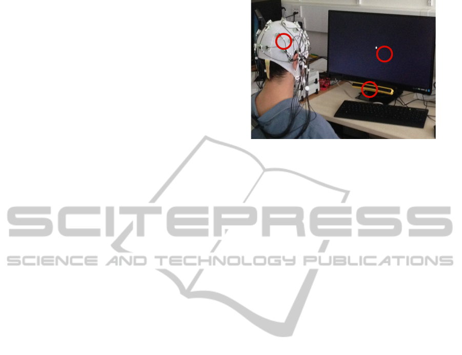
the subjects to blink freely. Figure 1 represents the
experimental setup.
Eye Tracking was performed with an SMI Red-
m Eye Tracker (SensoMotoric Instruments GmbH,
Teltow, Germany), a binocular, remotely mounted
Eye Tracker. EEG data was collected with a
BrainProducts ActiCHamp amplifier and a 32
active-electrode set with an ActiCap (Brain Products
GmbH, Gilching, Germany). Eye Tracking was
performed at 120 Hz and EEG recordings were
sampled at 500 Hz. Impedance of Electrodes against
the skin was reduced to levels always below 15 kΩ,
to ensure EEG signal quality. Eye Tracking was
performed at a distance of 50-70 cm from the
cameras.
The EEG data was then pre-processed by a
bandpass filter between 0.1-50 Hz, resampled to 120
Hz and Common Average Re-referenced. Eye Gaze
data (retrieved from the Eye Tracker) was used to
find blink regions and extract blink markers.
Figure 1: Experimental Setup. 1 is the Eye Tracker, 2 is
the stimuli screen and 3 is the electrode cap.
2.1 Experimental Setup
The task was set up in Matlab with the help of the
PsychoPhysics Toolbox (Brainard, 1997).
Participants were asked to sit at a distance of 50-70
cm from the Eye Tracker and Monitor, to ensure
tracking (as per the Eye Tracker’s technical
information sheet). Time to relax was given to
patients while performing the setup of the EEG
apparatus and participants were instructed to sit
comfortably and focus only on the screen. External
interference was minimized to avoid distractions that
could result in inadvertent saccadic movements.
Data was collected from 12 subjects with an
average age of 25 years.
2.2 Analysis Methods
Several methods were studied in order to find the
most suitable for ocular artefact correction,
including Independent Components Analysis (ICA),
Wavelet Decomposition and Wiener Filtering. The
traces resulting from these methods were then
analysed and compared.
2.2.1 Independent Components Analysis
ICA is an algorithm that maximizes the
independence of different components of a signal by
finding a linear coordinate system that creates
signals that are statistically independent (Lee, 1998).
ICA is used for Blind Source Separation. As ocular
artefacts do not correspond to neural activity (i.e.
they have a different source), ICA seemed a suitable
approach to ocular artefact correction in EEG
signals.
The ICA algorithm used is present in the
EEGLAB toolbox for Matlab and uses the infomax
learning rule (Bell and Sejnowski, 1995). This rule
1
2
3
NEUROTECHNIX2014-InternationalCongressonNeurotechnology,ElectronicsandInformatics
80

minimizes the mutual information in the components
in the output, thus maximizing their statistical
independence.
The original infomax condition fails to separate
sub-Gaussian sources due to the sigmoid function
used; a solution to this problem was proposed by
Bell and Sejnowski and consisted of a flexible
sigmoid function (Bell and Sejnowski, 1995), but
empirical results have shown that sometimes it is not
possible to find independent components with this
approach, alongside it being highly demanding in
terms of computational load.
To evaluate the Gaussianity of a distribution, a
measure of its kurtosis can be used. Kurtosis is
defined as the 4th order cumulant and gives a
measure of the shape of a distribution. A cumulant is
used to describe and in some cases approximate a
normal distribution; these are similar to moments in
the sense that two distributions with identical
moments will also have identical cumulants.
To overcome the problems of the original rule
proposed by Bell and Sejnowski, an extended
version of their algorithm was created: in this
version the algorithm switches according to the
kurtosis of the distribution of the data points. This
means that according to the sign of the kurtosis, the
learning rule is updated and this way it is possible to
overcome the original problem. Simulations run on
datasets with multiple sources and a variety of sub-
and super-Gaussian distributions show that this
extended version of the infomax algorithm is able to
separate the sources (Lee, Girolami and Sejnowski,
1999).
The original learning rule with a natural gradient
is defined as (Bell and Sejnowski, 1995):
∆ ∝
tanh
(1)
where represents the estimated sources, denotes
the identity matrix and , being the
mixed components signals. The extended learning
rule, proposed in (Lee, Girolami and Sejnowski,
1999) is defined as:
∆ ∝ tanh
′
(2)
where
are elements of the N-dimensional
diagonal matrix . This matrix is related to the
kurtosis of the data, so if
1 the data is sub-
Gaussian and if
1 the data is super-Gaussian.
2.2.2 Wiener Filter
The Wiener Filter approach creates an optimal linear
filter based on the signal and noise power spectra, as
stated in the equation:
(3)
where is the EEG neural signal and
is the
ocular artefact (both in time domain). Since we can
retrieve the artefact positions in the signal through
the Eye Tracker, an “average artefact” can be
obtained by averaging the signal pieces that contain
an artefact, and thus the Wiener Filter kernel can be
calculated and applied to the signal.
Let’s assume that and are stationary
and uncorrelated – a valid assumption, considering
these signals have different origins and therefore
should not have any strong correlation. This can be
translated into the fact that the expectation is zero:
.
,
0
(4)
The goal is to find an optimal filter that
minimizes the error between the signal and the
estimated signal :
min
(5)
and
∗
(6)
where
denotes the filter and ∗ represents
convolution. By using the orthogonality principle
(Papoulis and Pillai, 2002) it is possible to obtain the
filter that minimizes the mean square error:
,
,
,
∗
,
0
(7)
When converted to Fourier Space, the above
equation will turn into an algebraic equation:
(8)
where
represents the power spectral density
of the signal (with no artefacts),
is the power
spectral density of the artefact extracted and is
the filter function.
and
were computed
by extracting a mean artefact and mean clean signal
and then calculating the power spectral density of
each.
After the computation of this filter function and
in order to apply it to the whole signal, either the
filter function has to be inversely transformed to be
in a time basis or the signal has to be transformed to
be in Fourier space. The signal is then convolved
(time) or multiplied (Fourier) with the filter and the
noise should be removed.
2.2.3 Wavelet Decomposition
Wavelets and wavelet decomposition are tools used
in signal processing to analyse, correct and
EEGandEye-TrackingIntegrationforOcularArtefactCorrection
81
characterize signals. Wavelet functions define the
basis over which the signal is going to be
decomposed.
From the several different types of wavelets in
existence in signal processing it is possible to choose
some whose properties adjust better to a specific
purpose or case. In the case of artefact correction,
wavelets that mimic the artefact will be more
suitable, since the coefficients of the transform will
be higher in the artefact zones.
The Discrete Wavelet Transform (DWT) consists
of the decomposition of a signal into a wavelet basis,
thus attributing coefficients that relate the signal to
the wavelet form. The main equation that describes
this process is (Kumar, Arumuganathan, Sivakumar,
et al., 2008):
Ψ
,
2
Ψ2
(9)
where Ψ represents the wavelet function. The
process of obtaining the wavelet coefficients of a
signal can be performed at different levels, each one
of them defined by the binary decimation factor
(Nason and Silverman, 1995):
(10)
where represents the signal. This implies that
chooses every even number of a sequence.
The main issue of the Discrete Wavelet
Transform (DWT) is that it is not time-invariant, and
thus the translation invariance property is lost, i.e.
the translated DWT of a signal is not the same as the
DWT of a translated signal.
Stationary Wavelet transform is a variation of the
usual Discrete Wavelet transform. The advantage
relies on the independence of the choice of origin for
the wavelets, which is achieved by applying
appropriate high and low pass filters to the data at
each level, thus producing two sequences at the next
level. This way there is no decimation, instead the
filters are changing at each level by zero-padding in
a well-defined way. The details of the filter
adaptation are described in (Nason and Silverman,
1995). The Stationary Wavelet Transform (SWT)
contains the coefficients of the Discrete Wavelet
Transform but shifted according to the choice of the
origin of DWT. There is no restriction on the
localisation as the stationary wavelet transform fills
the gaps between coefficients in decimated DWT
(Nason and Silverman, 1995).
In the case of artefact correction of the EEG,
(Kumar, Arumuganathan, Sivakumar, et al.,
2008)show a simple way to correct the eye blink
artefacts from the EEG using Stationary Wavelet
Transforms and Symlet Wavelets (part of the.
Figure 2: EEG recording. Different colours represent
different channels; the spikes in the signal are blink
artefacts.
Figure 3: Average Blink for one subject. The artefact
extracted is quite large and thus can influence the use of
the data.
Daubechies (Daubechies, 1990) family) of level 3.
In this paper they show a method to correct the
artefacts with a simple threshold of the wavelet
coefficients.
3 RESULTS
In order to visualize the influence of the artefacts in
the signal, all 31 channels of the recording are
shown in Figure 2. The same recording is shown in
this paper for the sake of comparison, and it is only
illustrative of the data collected.
The Eye Tracker data was aligned with the EEG
recording and thresholded to yield a set of artefact
markers. The extraction and average of blink
artefacts through the use of these markers is
represented in Figure 3.
Event-driven Independent Components Analysis:
ICA was applied to 1500 points in the data around
the artefact; 30 channels were used to guarantee full-
NEUROTECHNIX2014-InternationalCongressonNeurotechnology,ElectronicsandInformatics
82
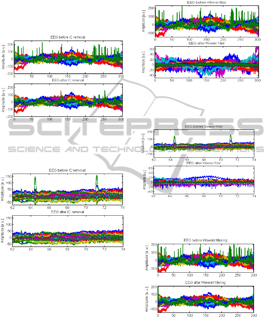
rank data. After projection of Independent
Components to the original data space, Artefact
components were identified and subtracted from the
data. The result is shown in Figure 4 and Figure 5.
Figure 4: Top: EEG signal before artefact correction;
Bottom: Same signal after correction of artefacts with
ICA. The artefacts that correspond to the spikes in the
upper plot are reduced in the bottom plot. The black
window represents the region that was zoomed for the
detail plot in Figure 5.
Figure 5: Detail plot of two blink artefacts. Top: before
correction; Bottom: after correction with ICA.
Event-driven Wiener Filtering:
To calculate the filter kernel, the EEG signal with
the artefacts and without artefacts was separated and
averaged; both signals were zero-padded to the
length of the signal and the power spectral density
was calculated and then used in the filter function
calculation (Izzetoglu, Devaraj, Bunce, et al., 2005;
Kailath, Sayed and Hassibi, 2000; Jingdong Chen,
Benesty, Yiteng Huang, et al., 2006). The result of
the filtering is shown in Figure 6 and Figure 7.
Figure 6: EEG signal before and after correction of
artefacts with Wiener filter. Top: signal before artefact
correction; Bottom: signal after artefact correction. The
black window represents the region of the signal that is
zoomed in the detail plot (Figure 7).
Figure 7: Detail plot of the EEG signal. Top: signal before
artefact correction; Bottom: signal after artefact correction
with Wiener filter.
Event driven Wavelet Decomposition:
Figure 8: EEG signal before and after correction of
artefacts with Wavelet Decomposition. Top: signal before
artefact correction; Bottom: signal after artefact
correction. The black window indicates the region of the
signal that is zoomed in the detail plot (Figure 9).
EEGandEye-TrackingIntegrationforOcularArtefactCorrection
83
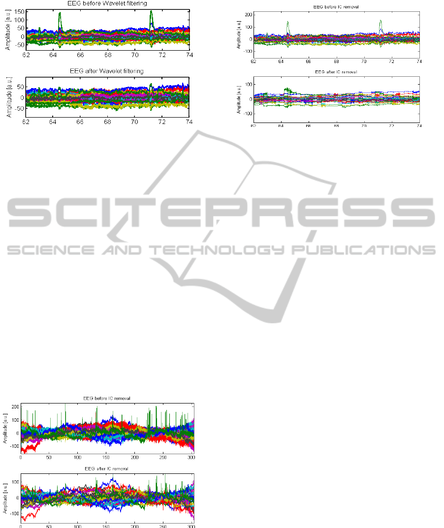
Figure 9: Detail plot of the EEG signal. Top: signal before
artefact correction; Bottom: signal after artefact correction
with Wavelet Decomposition.
Stationary wavelet decomposition was used to
correct the artefact; Symlet wavelets were chosen
due to their resemblance to the ocular artefact
(Kumar, Arumuganathan, Sivakumar, et al., 2008)
and 8 levels of decomposition were applied to 1500
data points around the ocular artefact. Figure 8 and
Figure 9 show the results of this method.
“Blind” Independent Components Analysis:
Another method applied to the data in order to prove
the pertinence of our methods, was a standard ICA
clean up, where we have a sliding window over the
data, calculating Independent Components and
eliminating those that resemble an artefact. This
approach is blind and as such has no knowledge of
how a blink artefact looks like or even their
locations. Results are shown in Figure 10 and Figure
11.
Figure 10: EEG signal before and after correction of
artefacts with Blind ICA. Top: signal before artefact
correction; Bottom: signal after artefact correction. The
black window indicates the region of the signal that is
zoomed in the detail plot (Figure 9).
Figure 11: Detail plot of the EEG signal. Top: signal
before artefact correction; Bottom: signal after artefact
correction with Blind ICA.
4 DISCUSSION
In this work we studied the effect of using an Eye
Tracker in Ocular Artefact correction of EEG data.
We implemented standardised signal processing
methods such as ICA or Wavelet Decomposition, as
well as a Wiener Filter, a method not generally used
in EEG artefact correction.
Our results show that all 4 methods are
successful in correcting the artefacts, although
Event-Driven ICA seems to yield the best signal
after correction. This is an expected finding
considering that the origins of the artefact and the
signal are different, and thus Blind Source
Separation techniques such as ICA have great
potential in achieving the best signal output. When
compared to the other methods, Blind ICA clearly is
stricter with the data and sometimes leads to an
over-correction. In Figure 11 we can clearly see that
the data, although it might preserve most of its
frequency spectrum, has been severely affected by
the corrective measure.
There is high inter- and intra-subject variability
on the EEG recordings; shape of head, changes in
electrode impedance or subject behaviour can
influence the data recordings, by introducing
artefacts and non-linear trends in the signal.
Moreover, attention or drowsiness can influence the
Eye Tracking (Di Stasi, McCamy, Catena, et al.,
2013).
The Wiener Filter is the method that is more
prone to failure, as it relies on an effective extraction
of the average artefact. Moreover it will filter out all
the frequencies represented in the artefact, which are
low (duration of about 200 milliseconds) (Caffier,
Erdmann and Ullsperger, 2003) and thus can
eliminate relevant information from the signal
NEUROTECHNIX2014-InternationalCongressonNeurotechnology,ElectronicsandInformatics
84

(Harmony, Fernández, Silva, et al., 1999; Whitham,
Pope, Fitzgibbon, et al., 2007; Iber, Ancoli-Israel,
Chesson., et al., 2007).
One improvement that could be performed to the
Wavelet Decomposition method is the use of a more
complex adaptive thresholding technique, since the
one used for this analysis combines only the mean
and variance of the signal to obtain a threshold;
other methods have been tested in “blind”
approaches (Stein, 1981; Krishnaveni, Jayaraman,
Anitha, et al., 2006) and thus could be implemented
in this study.
The ICA technique could be implemented as an
online correction technique, though it would lead to
some delay in the output of results. Wavelet and
Wiener filter methods can only be used for post-
processing and not for online correction with the
approaches described in this work.
As further work we would like to appoint the
validation of these techniques and their pertinence in
artefact correction. A validation approach was
attempted, with a Movement Imagery task and a
simple K- Nearest Neighbours classifier. The goal
was to examine the classifier’s accuracy for different
methods of ocular artefact correction, but in the
experiments the number of ocular artefacts was
correlated with the Movement Imagery epochs
(number of blinks increased in Movement Imagery
and lowered in Rest epochs), thus proving this
validation method as unable to accurately find the
best corrective algorithm.
The potential benefits of a clean EEG signal that
can be expected are among a better understanding of
neural signals and better use for these, such as in
Brain Machine Interfaces that can be used to help
patients suffering from Locked in Syndrome, as an
example. Online implementation is although
required for this purpose, but the usage of an eye
tracker that is not affected by external
electromagnetic fields (unlike, for example,
electrooculograms or magnetic search coils (Schlag,
Merker and Schlag-Rey, 1983)). Our work suggests
simple steps towards a cleaner EEG signal,
hopefully with more usable neural information being
conveyed in it and useable in real-time.
REFERENCES
Abbott, W.W. and Faisal, A.A. (2012) Ultra-low-cost 3D
gaze estimation: an intuitive high information
throughput compliment to direct brain–machine
interfaces. Journal of Neural Engineering. 9 (4),
046016.
Abbott, W.W., Zucconi, A. and Faisal, A.A. (2013) Large-
field study of ultra low-cost, non-invasive task level
BMI, 6
th
Intl IEEE/EMBS Conference on Neural
Engineering (NER), 2013, 97-100
Bell, A.J. and Sejnowski, T.J. (1995) An Information-
Maximization Approach to Blind Separation and Blind
Deconvolution. Neural Computation 7 (6), 1129-1159.
Brainard, D.H. (1997) The psychophysics toolbox. Spatial
Vision. 10 (4), 433–436.
Caffier, P.P., Erdmann, U. and Ullsperger, P. (2003)
Experimental evaluation of eye-blink parameters as a
drowsiness measure. Eur. J. App. Physiol. 89 (3), 319–
325.
Daubechies, I. (1990) The wavelet transform, time-
frequency localization and signal analysis. IEEE Trans
on Information Theory, 36 (5), 961–1005.
Faisal, A. A. (2010). Stochastic simulation of neurons,
axons and action potentials. Stochastic Methods in
Neuroscience, 297-343.
Faisal, A.A., Selen, L.P.J. and Wolpert, D.M. (2008)
Noise in the nervous system, Nature Rev Neurosci., 9
(4),292-303
Faisal, A.A, Fislage, M., Pomplun, M., Rae, R. and Ritter,
H. (1998) Observation of human eye movements to
simulate visual exploration of complex scenes, SFB
Report 360, 1-34
Neishabouri, Ali and Faisal, A.A. (2013) ; Axonal Noise
as a Source of Synaptic Variability, PLoS
computational biology,10 (5), e1003615,
Giannitrapani, D. and Kayton, L. (1974) Schizophrenia
and EEG spectral analysis. Electroencephal and Clin.
Neurophysiol. 36377–386.
Harmony, T., Fernández, T., Silva, J., Bosch, J., et al.
(1999) Do specific EEG frequencies indicate different
processes during mental calculation? Neuroscience
letters. 266 (1), 25–28.
Iber, C., Ancoli-Israel, S., Chesson., A.L. and Quan, S.F.
(2007) The AASM Manual for the Scoring of Sleep and
Associated Events: Rules, Terminology and Technical
Specifications. 1st edition. Westchester, Illinois,
American Academy of Sleep Medicine.
Iwasaki, M., Kellinghaus, C., Alexopoulos, A.V., Burgess,
R.C., et al. (2005) Effects of eyelid closure, blinks,
and eye movements on the electroencephalogram.
Clinical Neurophysiology. 116 (4), 878–885.
Izzetoglu, M., Devaraj, A., Bunce, S. and Onaral, B.
(2005) Motion Artifact Cancellation in NIR
Spectroscopy Using Wiener Filtering. IEEE
Transactions on Biomedical Engineering. 52 (5), 934–
938.
Jervis, B.W., Coelho, M. and Morgan, G.W. (1989) Effect
on EEG responses of removing ocular artifacts by
proportional EOG subtraction. Medical and Biol Eng.
and Computing. 27 (5), 484–490.
Jingdong Chen, Benesty, J., Yiteng Huang and Doclo, S.
(2006) New insights into the noise reduction Wiener
filter. IEEE Transactions on Audio, Speech and
Language Processing. 14 (4), 1218 - 1234
Kailath, T., Sayed, A.H. and Hassibi, B. (2000) Linear
Estimation. Prentice Hall (Upper Saddle River, NJ)
EEGandEye-TrackingIntegrationforOcularArtefactCorrection
85

Krishnaveni, V., Jayaraman, S., Anitha, L. and Ramadoss,
K. (2006) Removal of ocular artifacts from EEG using
adaptive thresholding of wavelet coefficients. Journal
of Neural Engineering. [Online] 3 (4), 338–346.
Kumar, P.S., Arumuganathan, R., Sivakumar, K. and
Vimal, C. (2008) Removal of Ocular Artifacts in the
EEG through Wavelet Transform without using an
EOG Reference Channel. Int. J. Open Problems
Compt. Math. 1 (3), 188–200.
Lee, T.-W. (1998) Independent Component Analysis -
Theory and Applications. 1st edition. Springer
Science+Business Media (Dordrecht)
Lee, T.-W., Girolami, M. and Sejnowski, T.J. (1999)
Independent Component Analysis using an Extended
Infomax Algorithm for Mixed Sub-Gaussian and
Super-Gaussian Sources. Neural Computation. 11 (2):
417-441
Nason, G.P. and Silverman, B.W. (1995) The Stationary
Wavelet Transform and some Statistical Applications.
Lecture Notes in Statistics. 103281–299.
Papoulis, A. and Pillai, U. (2002) Probability, Random
Variables and Stochastic Processes. 4th edition.
McGraw-Hill (New York, NY)
Saatchi, M.R., Oke, S., Allen, E.M., Jervis, B.W., et al.
(1995) Signal processing of the contingent negative
variation in schizophrenia using multilayer
perceptrons and predictive statistical diagnosis. IEE
Proceedings-Science, Measurement and Technology.
142 (4), 269–276.
Schlag, J., Merker, B. and Schlag-Rey, M. (1983)
Comparison of EOG and search coil techniques in
long-term measurements of eye position in alert
monkey and cat. Vision Research. 23 (10), 1025–1030.
Sengupta, B., Faisal, A.A., Laughlin, S.B., Niven, J. E.
(2013) The effect of cell size and channel density on
neuronal information encoding and energy efficiency,
J. of Cerebral Blood Flow and Metabolism,33 (9),
1465-1473
Di Stasi, L.L., McCamy, M.B., Catena, A., Macknik, S.L.,
et al. (2013) Microsaccade and drift dynamics reflect
mental fatigue. Eur. J. Neurosci. 38 (3), 2389–2398.
Stein, C.M. (1981) Estimation of the Mean of a
Multivariate Normal Distribution. Annals of Statistics.
9 (6), 1135–1151.
Vigário, R.N. (1997) Extraction of ocular artefacts from
EEG using independent component analysis.
Electroencephalography and Clin. Neurophysiol. 103
(3), 395–404.
Whitham, E.M., Pope, K.J., Fitzgibbon, S.P., Lewis, T., et
al. (2007) Scalp electrical recording during paralysis:
Quantitative evidence that EEG frequencies above
20Hz are contaminated by EMG. Clinical
Neurophysiology. 18(8), 1877-88.
Yoo, K.-S., Basa, T. and Lee, W.-H. (2007) Removal of
Eye Blink Artifacts From EEG Signals Based on
Cross-Correlation. Intl. Conf. on Convergence
Information Technology. pp. 2005–2014.
NEUROTECHNIX2014-InternationalCongressonNeurotechnology,ElectronicsandInformatics
86
