
ANALYTICAL IMAGING OF CULTURAL HERITAGE BY
SYNCHROTRON RADIATION AND VISIBLE LIGHT- NEAR
INFRARED SPECTROSCOPY
Jay Arre Toque, Yuji Sakatoku, Julia Anders, Yusuke Murayama and Ari Ide-Ektessabi
Advanced Imaging Technology Laboratory, Graudate School of Engineering
Kyoto Univeristy, Yoshida-honmachi, Sakyo-ku, 606-8501, Kyoto, Japan
Keywords: Analytical imaging, Spectral reflectance, Synchrotron radiation, Visible light, Near infrared, Multispectral
imaging, Cultural heritage.
Abstract: Imaging is an important tool for analyzing cultural heritage. Due to its delicate nature, the analysis presents
numerous technical challenges, probably the most important of which is its requirement for non-destructive
and non-invasive investigation. In this study, two techniques used in the analysis of cultural heritage are
presented. The first one, synchrotron radiation x-ray fluorescence, is an advanced analytical technique with
high accuracy and good spatial resolution. On the other hand, spectroscopic technique based on visible
light-near infrared spectrum is becoming popular due to some information that it can provide, which are not
available even in advanced analytical techniques. These two techniques were used to analyze real cultural
heritage such as an ancient Mongolian textile, traditional Korean painting and commonly used pigments in
Japanese paintings. The results revealed that using synchrotron radiation-based techniques is sometimes not
enough in providing critical information (e.g. spectral reflectance, color, etc.) necessary for understanding
of cultural heritage. This can be complemented using visible light-near infrared technique.
1 INTRODUCTION
Cultural heritage refers to artifacts and intangible
features inherited from previous generations, which
are preserved or maintained for the benefit of future
generations. The cultural heritage which we
inherited, show the techniques, the way of life, the
social values and the way people were thinking in
old days. In the past, interests in cultural heritage
are mainly based on its artistic and historic values.
However during the recent years, it has been
attracting the attention of scientists and engineers
because of the technical challenges it presents during
analysis, restoration and preservation. Its delicate
nature requires that the investigation should be non-
destructive and non-invasive (Faubel, et al., 2007;
Toque, et al., 2008).
Among the available analytical techniques,
synchrotron radiation-based analysis offers high
precision and high accuracy in addition to being
non-destructive. Synchrotron radiation (SR) is
emitted in the tangential direction when electrons
and positrons are accelerated at relativistic speed
subjected to a magnetic field (Margaritondo, 1988).
The energy of SR covers a broad spectrum. X-ray
fluorescence analysis using synchrotron radiation is
a powerful technique in terms of detecting ultra trace
elements and studying them in detail. Its unique
features include local area analysis by using micro
beam; capability of doing measurement in air or
water; non-contact and non-destructive assay; rapid
measurements; and precise assay of trace elements
(Ide-Ektessabi, 2007). However, even though SR-
based techniques are powerful, there are information
they cannot provide, such as spectral reflectance,
color information and others. These are important in
analyzing cultural heritage.
To compensate for some inadequacies of SR-
based techniques, imaging at the visible light-near
infrared (VL-NI) spectrum is employed. The basic
idea is to use polychromatic images (with RGB
tristimulus values) or multispectral images to extract
spectroscopic data. It has been reported that
materials emit specific spectral features within a
121
Toque J., Sakatoku Y., Anders J., Murayama Y. and Ide-Ektessabi A. (2009).
ANALYTICAL IMAGING OF CULTURAL HERITAGE BY SYNCHROTRON RADIATION AND VISIBLE LIGHT- NEAR INFRARED SPECTROSCOPY.
In Proceedings of the First International Conference on Computer Imaging Theory and Applications, pages 121-128
DOI: 10.5220/0001788201210128
Copyright
c
SciTePress
certain range (Balas, et al., 2003). In other
wavelength range, this is more pronounced.
However, at the VL-NI range the interaction is more
complex. Nonetheless, there are some unique
features that are only observable within this range.
This makes it valuable in cultural heritage analysis.
2 EXPERIMENT
2.1 Imaging and Spectroscopic
Synchrotron Radiation Analysis
Synchrotron radiation X-ray fluorescence (SRXRF)
was performed on a 13
th
century Mongolian textile
and dislodged fragments collected from an old
Korean painting. Measurements were done at beam
line 4A of Photon Factory. The electron beam
energy in the storage ring was 2.5 GeV, with a
maximum current of 400 mA. Incident X-ray
energy was 15 keV. The cross-section of the beam
was approximately 1(v) x 1(h) mm
2
on the sample.
The synchrotron radiation was monochromated by a
multilayered reflecting mirror. Precise beam size of
monochromated X-rays was adjusted using slits. The
incident and transmitted X-rays were monitored by
ionization chambers that were set in front of and
behind the sample. The fluorescent X-rays were
collected by a solid-state detector at 90 degrees to
the incident beam. Measurements were performed in
air. Point spectra were measured for obtaining
consistent elements of the samples. The spectra were
obtained by using a multi-channel analyzer. The
measurement time was 100 seconds for each
spectrum. XRF imaging technique was applied in
order to investigate the distributions of main
elements. X-Y step pulse motors moved the sample
stage. The measurement areas were divided into
matrices of 20 x 20 pixels. At each pixel, the XRF
yields for each element were integrated by single
channel analyzers. The measurement time was three
seconds for each pixel.
Synchrotron radiation was also used to
investigate the relationship between fine structural
change and color fading in natural mineral,
specifically azurite and malachite. Heating of natural
pigments is well practiced among traditional
Japanese painters to modify the shade and color of
the pigments. It is of great interest to understand the
factors involved in this process. Ten samples of each
pigment were heated at 260°C with holding time
from 10 minutes up to 90 minutes at 10-minute
increments. The spectral reflectance of the heated
and unheated pigments was measured to track the
changes in color. X-ray fluorescence and X-ray
absorption fine structure (XAFS) were used to
characterize the pigments. The incident X-ray was
15 keV for XRF analysis while the energy was
scanned at the Cu K absorption edge from 8.90 to
9.09 keV for XAFS.
2.2 Imaging at the Visible-Near
Infrared Range
In order to use VL-NIS for the analytical imaging of
cultural heritage, a technique for image analysis was
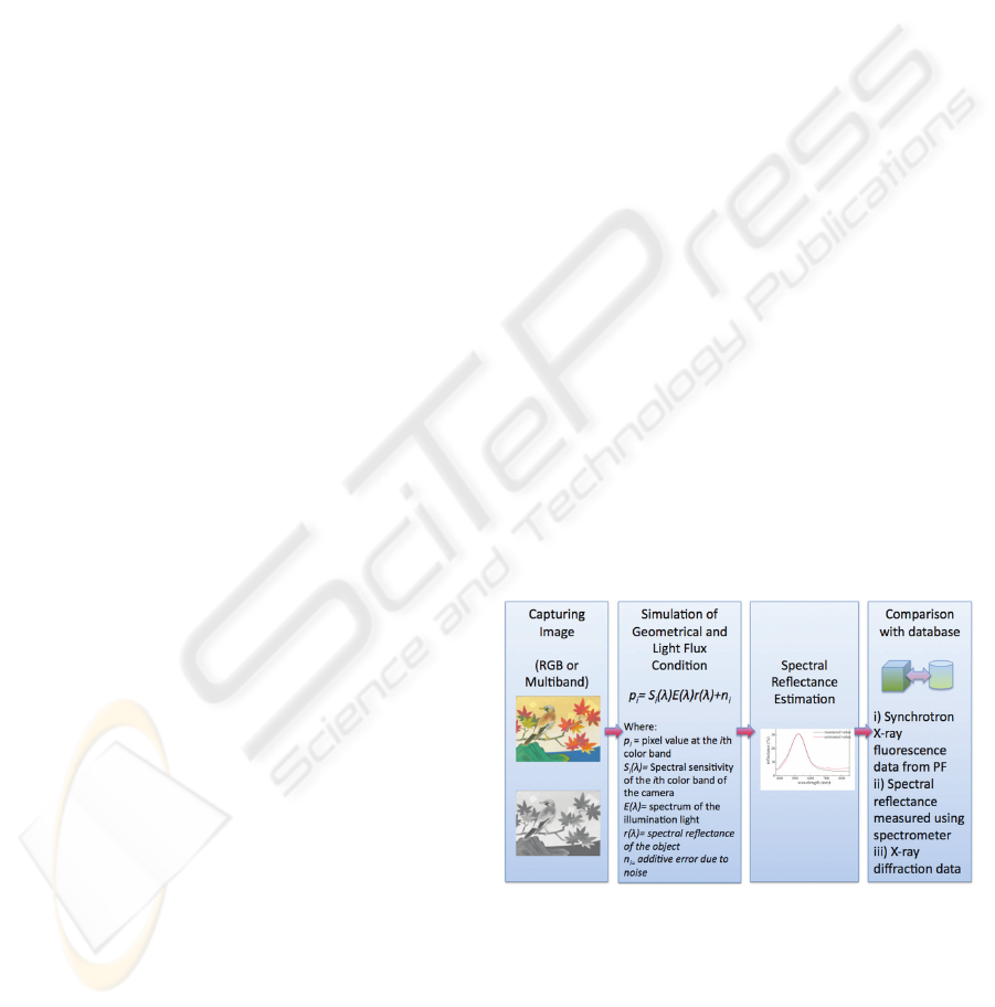
developed. Figure 1 illustrates the basic scheme of
the analysis. It begins by capturing an image of the
object to capture spectral and color information. The
picture can be an RGB or a multispectral image. An
RGB image refers to an image captured using
tristimulus values corresponding to red, green and
blue colors. A multispectral image, on the other
hand, is an image captured by using 6-7 bands of
color and infrared filters. The images are captured
within a certain wavelength band. The color
information is then used to simulate the geometrical
and light flux conditions. The simulation enables the
estimation of the spectral reflectance, which is used
for comparison with a database to provide useful
information about the image. The database includes
more than 1000 mineral pigments and is
continuously being updated. The database also
includes additional data such as SRXRF spectra,
XRD spectra as well as information about the
artwork’s history.
Figure 1: Image analysis scheme using VL-NIS.
IMAGAPP 2009 - International Conference on Imaging Theory and Applications
122
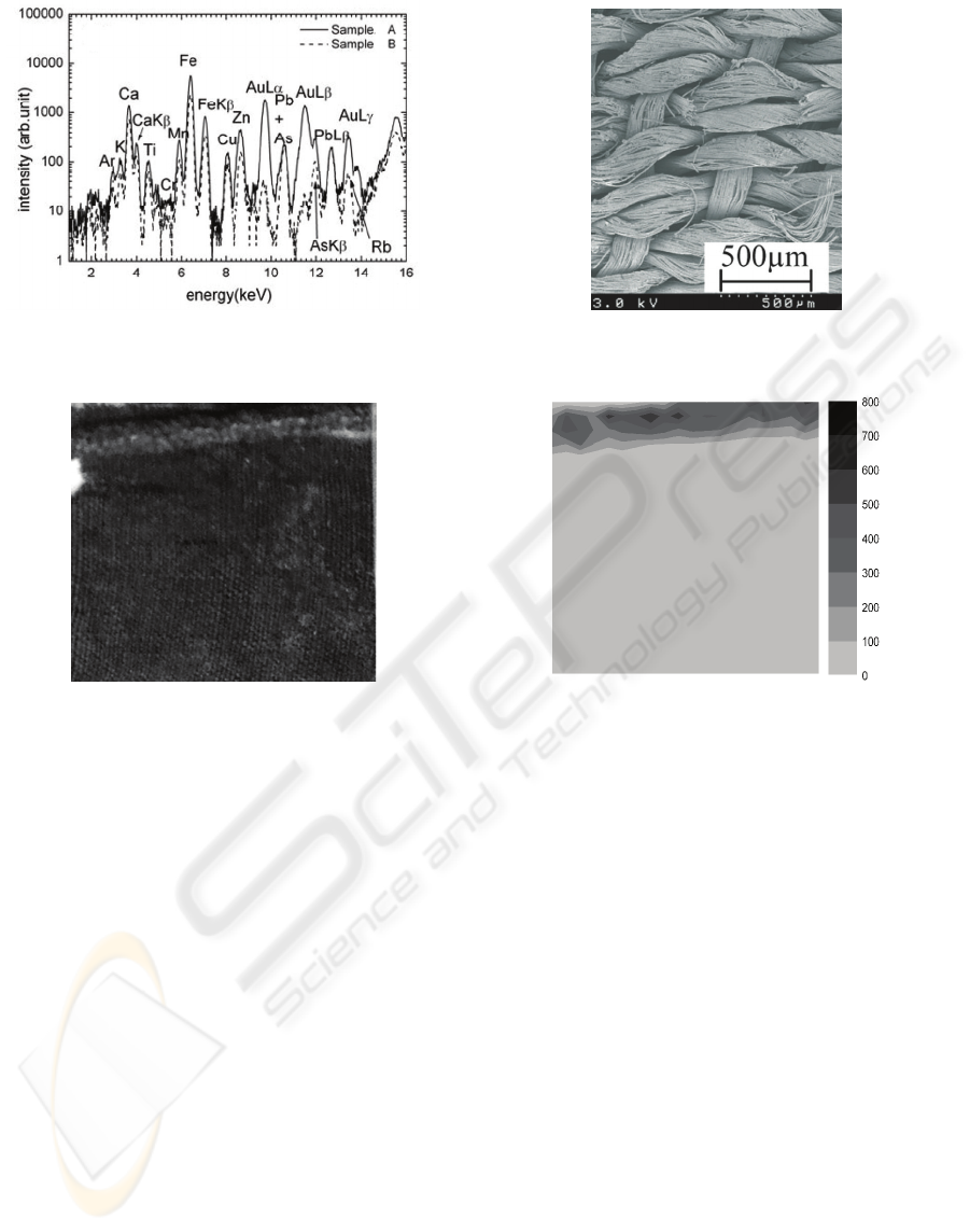
Figure 2: (a) SRXRF spectra of an ancient Mongolian textile. Sample A refers to the gold thread while Sample B refers to
the textile; (b) Field-emission SEM showing textile structure.
Figure 3: (a) Optical image of an ancient Mongolian textile laced with gold thread; (b) Elemental distribution image of gold
derived from the SRXRF spectra.
3 RESULTS AND DISCUSSION
3.1 Synchrotron Radiation of Actual
Cultural Heritage
3.1.1 Ancient Mongolian Textile
SRXRF was used to detect the constituent elements
used for the manufacturing of the ancient textile as
well as obtaining elemental distribution images.
Figure 2a shows the XRF spectra of the Mongolian
textile. The spectra revealed the presence of
significant amount of gold and iron. The textile was
dated to be about 700-800 years old. It is believed
that the textile was produced during the 13
th
century
at the height of power of the Mongolian empire
under Genghis Khan and Kublai Khan. This may
explain why gold was detected from the XRF
spectra. Lacing and decorating the textile with gold
was a symbol of wealth and prosperity in the old
days. The spectra also show traces of Cu and Ti.
These elements are well-known metallic mordant
and are believe to be widely used during that period.
Fig. 2b shows a field-emission SEM image of the
Mongolian textile sample. It depicts textile structure
and weaving condition. In addition, Figure 3a shows
an optical image of a textile laced with gold thread.
An elemental image distribution of gold (Figure 3b)
is derived from the XRF spectra.
3.1.2 Old Korean Painting
SRXRF was used to investigate a traditional Korean
painting. Figure 4 shows the spectra of several
natural pigments used (white, blue, green and red).
The main elements detected were Pb, Cu, Hg and
Fe. It is interesting to note that the spectra of the
blue and green pigments are very similar to popular
pigments used in traditional Japanese painting
known as gunjo (azurite) and ryokusho
(malachite). These pigments are copper-based
pigments [2CuCO
3
•Cu(OH)
2
and CuCO
3
•Cu(OH)
respectively]. The difference lies with the significant
trace of Pb found in the Korean painting. It is
ANALYTICAL IMAGING OF CULTURAL HERITAGE BY SYNCHROTRON RADIATION AND VISIBLE LIGHT-
NEAR INFRARED SPECTROSCOPY
123
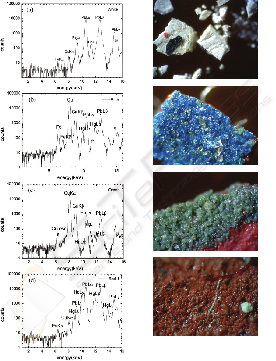
Figure 4: SRXRF of different pigments used in a
traditional Korean painting. Note: (a) white pigment; (b)
blue pigment; (c) green pigment; (d) red pigment.
Figure 5: Optical images of the pigments used on a
traditional Korean painting: (a) white pigment; (b) blue
pigment; (c) green pigment; (d) red pigment.
IMAGAPP 2009 - International Conference on Imaging Theory and Applications
124
attributed to the white pigment used in the painting.
It is believed that the white pigment was used as a
base medium. This may explain why all the
pigments studied contain significant traces of Pb.
Figure 5 shows the optical images of the pigments
used on the Korean painting investigated. It also
shows that the pigments used are granulated.
3.1.3 Effect of Heating on Pigment
Discoloration
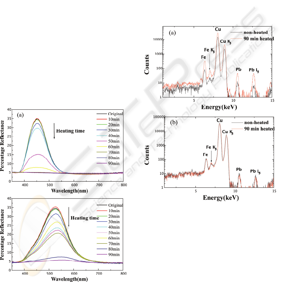
Figure 6 shows the spectral reflectance of azurite
and malachite pigments as a function of heating
time. These two pigments were selected as test
samples because they are the most commonly used
blue and green pigments in traditional Japanese
paintings. The pigments predictably changed its
color upon heating. It is interesting to note however
that the discoloration did not change the position of
the spectral peak. This implies that the color
wavelength did not change, only the luminosity
(Toque, et al., 2008). The samples got darker with
the increase in heating time. In addition, abrupt
change in color was observed between 40-60
minutes of exposure to elevated temperature.
Figure 6: Spectral reflectance of (a) azurite and (b)
malachite as a function of heating time. Prolonged
exposure to elevated temperature lowers the reflectance of
the pigments.
In order to understand the mechanism of
discoloration of the pigments when subjected to
elevated temperature, they were subjected to
synchrotron XRF and XAFS analysis. Figure 7
shows the XRF spectra of azurite and malachite. It
shows minimal change in the spectra. This implies
that the color change was not due to the trace
elements of the pigments. The trace elements of both
the pigments are similar but their colors are quite
different. The spectral reflectance of azurite leans
toward the short-wavelength range giving it a shade
of blue while malachite is around the mid-
wavelength range giving at a shade of green.
Figure 7: XRF spectra of the heated and non-heated: (a)
azurite and (b) malachite. Both the pigments have similar
spectra. In addition, minimal change is observable
between the burnt and unburned pigments.
The changes in color of both the pigments may
be more attributed to the change in chemical
bonding state of the main element Cu. Figure 8
shows the XAFS spectra of the pigments. It was
found that the absorption edge of the samples heated
for 80 and 90 minutes shifted to lower energy level.
This may also explain the abrupt change in color as
the pigments were heated for a long time.
ANALYTICAL IMAGING OF CULTURAL HERITAGE BY SYNCHROTRON RADIATION AND VISIBLE LIGHT-
NEAR INFRARED SPECTROSCOPY
125
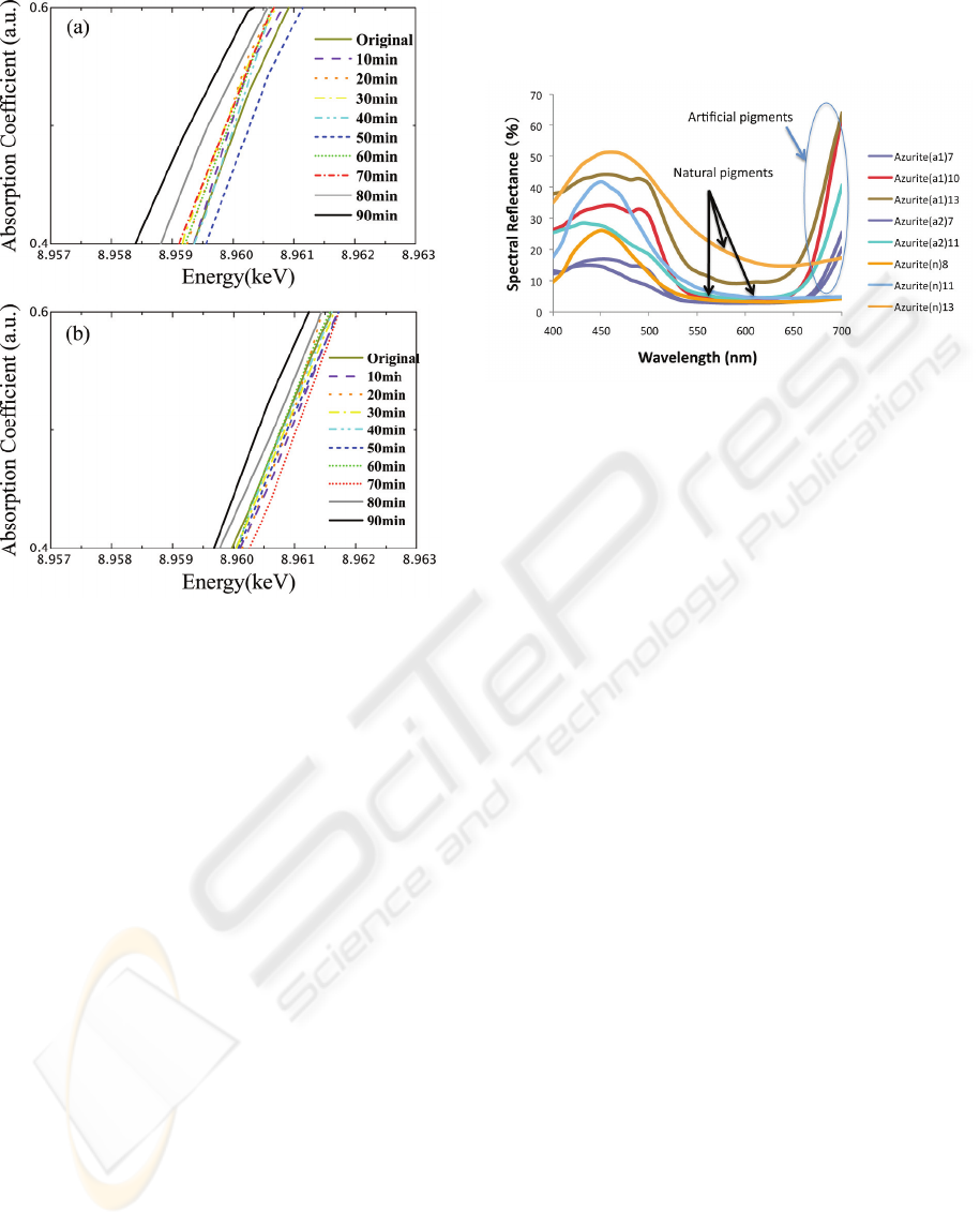
Figure 8: Position of Cu K absorption edge of heat-
discolored (a) azurite and (b) malachite. Unlike the XRF
spectra, XAFS provide more insights about the
discoloration of the pigments.
3.2 Visible Light-Near Infrared
Spectroscopy of Cultural Heritage
Visible light- near infrared spectroscopy (VL-NIS)
gives information about an image, which can
provide better understanding of the mechanism of
degradation and useful insights for the restoration
and preservation of cultural heritage. Some
information about a material can be extracted by
analyzing the interaction between matter and light at
the visible to near infrared radiation spectrum, which
are not noticeable in other range.
The analysis covers the electromagnetic radiation
wavelength from 380-850 nm. This range includes
the visible to near infrared range. IR is included
since recent investigations have shown that unique
spectral characteristics are observable, especially
from 650-800nm wavelengths. Figure 9 shows the
spectral characteristics of different azurite pigments.
They are grouped into natural and artificial mineral
pigments. It is noticeable that all the pigments have
similar peak positions around 430-480 nm, which
gives them bluish hue. However, starting from about
650 nm, the percentage reflectance of the artificial
pigments starts to increase drastically while the
natural pigments did not show significant change.
These spectral signatures may be used to identify the
nature of the pigments used in an artwork.
Figure 9: Spectral reflectance of natural and artificial
azurite mineral pigments. The alphanumeric symbol
enclosed in parentheses indicates whether the pigment is
natural or artificial while the other indicates relative
particle size: the higher the number, the smaller the
particle size.
3.3 Analysis of Cultural Heritage
The analysis of cultural heritage presents several
technical challenges. First, its delicate nature
requires non-destructive and non-invasive analysis.
The techniques presented in this paper satisfy the
requirement. However, there are some issues that
cannot be fully covered by a single technique. For
example, SRXRF method offers high accuracy and
precision but it can only give information about the
chemical composition of the pigments used in the
artwork. When it is used in the study of
discoloration of heated pigments, it cannot give
sufficient information. VL-NIS can give more
information in this case; the changes in color due to
heating can be tracked by the changes in spectral
reflectance. XAFS can also provide insight on the
discoloration by analyzing the chemical state of
copper absorption edge. However, neither SRXRF
nor XAFS can distinguish between the differences in
pigment size. It was observed that pigment particle
size affects the hue. Small pigments have lighter hue
than large pigments even though they have the same
chemical composition. This is illustrated by Figure
10 and confirmed by the SRXRF spectra given by
Figure 11.
IMAGAPP 2009 - International Conference on Imaging Theory and Applications
126
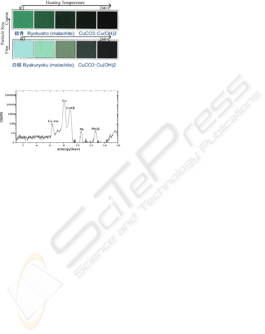
Figure 10: Pigment hues as a function of relative particle
size and heating temperature.
Figure 11: Corresponding SRXRF spectra.
Another concern that poses technical challenge
on the analysis of cultural heritage is the preparation
of sample. In advance analytical techniques such as
synchrotron radiation, the size of the sample is quite
small. In order to satisfy this, preparing a small
fraction of the sample is necessary. In this regard,
this can be considered destructive. In most cases,
this is unacceptable since a cultural heritage is
irreplaceable. On the other hand, VL-NIS can
address this concern. The size of the sample is
basically irrelevant. In VL-NIS, the major concern is
the accuracy. It is worth mentioning that the
limitation in sample size in X-ray techniques can be
addressed by using portable devices, such as
portable XRF. This is widely practice in analyzing
artworks. However, its accuracy is inferior
compared with large-scale X-ray devices. Therefore,
in the analysis of cultural heritage, the techniques
presented should be used in conjunction with one
another and not independently. This ensures that the
information extracted from the images can be
maximized.
4 CONCLUSIONS
Analysis of cultural heritage using analytical
imaging is a burgeoning field. This is attributed to
the numerous technical challenges it presents (i.e.
non-destructive and non-invasive). In this study,
synchrotron radiation and visible light-near infrared
techniques were used to analyze real cultural
heritage. It is considered that detailed analysis of
such historic objects (in this case, an ancient
Mongolian textile and old Korean painting) is of
prime importance. In addition, the mechanism of
color discoloration as a result of heating was
investigated. Synchrotron radiation analysis revealed
that it was due to the change in chemical bonding
state. VI-NI analysis also revealed some results. It
was shown that the change in chemical bonding state
results in increase or decrease in magnitude of the
spectral reflectance but does not affect the position
of the peaks. Having a clearer understanding of
pigment degradation can help in cultural heritage
preservation and restoration efforts. The results have
also shown that no single technique is capable of
providing all the necessary analytical information.
The available techniques should be used to
complement one another. It was also shown in this
study that analysis using visible light- near infrared
radiation could be an indispensable tool for
investigating cultural heritage. This is due to some
unique spectral features of materials that are only
observable in this range. Since VL-NIS does not
require complex instrumentation, robust and flexible
systems are achievable.
ACKNOWLEDGEMENTS
This work has been done as part of the project “An
Integrated System for Secure and Dynamic Display
of Cultural Heritage” sponsored by Japan Science
and Technology Agency, Regional Resources
Development Program. This collaborative project
was organized by Kyoto University Graduate School
of Engineering, S-tennine Kyoto (Ltd) and Kyushu
National Museum. The Authors would like to
express their thanks to Imazu Setsuo of Kyushu
National Museum and other staff of the museum
and, Oshima of S-tennine Kyoto and his group for
supporting this work. The authors are also grateful to
Prof. Atsuo Iida of Photon Factory (Tsukuba,
Japan). Thanks is also due to Mr. G. Enkhbat of
Center of National Heritage, Mongolia who supplied
the Mongolian textiles used as samples.
ANALYTICAL IMAGING OF CULTURAL HERITAGE BY SYNCHROTRON RADIATION AND VISIBLE LIGHT-
NEAR INFRARED SPECTROSCOPY
127

REFERENCES
Balas, C., Papadakis, V., Papadakis, N., Papadakis, A.,
Vazgiouraki, E., Themelis, G., 2003. A novel hyper-
spectral imaging apparatus for the non-destructive
analsyis of objects of artistic and histori values.
Journal of Cultural Heritage, 4, pp 330s-337s.
Fauble, W., Staub, S., Simon, R., Heissler, S., Pataki, A.,
Banik, G., 2007. Non-destructive analysis for the
investivation of decompodition phenomena of
historical manuscripts and prints. Spectrochimica Acta
Part B, 62, pp 669-676.
Ide-Ektessabi, A., 2007. Applications of Synchrotron
Radiation. Springer- Verlag Berlin Heidelgberg.
Margaritondo, G, 1988. Introduction to Synchrotron
Radiation. New York Oxford, Oxford Univeristy
Press.
Toque, J.A., Nishimura, R., Ide-Ektessabi, A., 2007,
Analysis of cultural heritage by synchrotron radiation
and visible light-near infrared spectroscopy. PF
Activity Report 2007, 4A, 25B/2006G110.
Toque, J.A., Sakatoku, Y., Komori, M., Murayama, Y.,
Ide-Ektessabi, A., 2008, Analytical imaging of cultural
heritage by UHRS and MBI, 1
st
AUN/SEED-Net
Regional Conference in Manufacturing Engineering,
Manila Philippines, November 24-25, 2008.
IMAGAPP 2009 - International Conference on Imaging Theory and Applications
128
