
SEGMENTATION OF MULTISPECTRAL IMAGES USING
MATHEMATICAL MORPHOLOGY AND AUTOMATIC
CLASSIFICATION
Application to Microscopic Medical Images
Sarah Ghandour, Eric Gonneau and Guy Flouzat
LERISM, Toulouse University,118 Route de Narbonne, Toulouse, France
Keywords: Segmentation, Watershed Algorithm, Region Adjacency Graph, Mathematical Morphology, Generalized
Likelihood Ratio, Clustering, Hypercube Classification.
Abstract: In this paper, a new color segmentation scheme of microscopic color images is proposed. The approach
combines a region growing method and a clustering method. Each channel plane of the color images is
represented by a set of regions using a watershed algorithm. Those regions are represented and modeled by
a Region Adjacency Graph (RAG). A novel method is introduced to simplify the RAG by merging
candidate regions until the violation of a stopping aggregation criterion determined using a statistical
method which combines the generalized likelihood ratio (GLR) and the Bayesian information criterion
(BIC). From the resulting segmented and simplified images, the RGB image is computed. Structural
features as cells area, shape indicator and cells color are extracted using the simplified graph and then stored
in a database in order to elaborate meaningful queries. A regularization step based on the use of an
automatic classification will take place. Results show that our method that does not involve any a priori
knowledge is suitable for several types of cytology images.
1 INTRODUCTION
The image segmentation is an essential step of low-
level processing of imagery. It aims to split the
image into disjoint regions that are generally
homogenous in terms of color and texture. Various
algorithms and segmentation methods can be found
in the literature and can be divided into several
categories: clustering methods, edge-based
techniques, region growing process and
mathematical morphology. Please refer to (Lucchese
and Mitra, 2001) for more details about those state-
of-the-art techniques. Approaches combining some
of these methods were also proposed in (Lucchese
and Mitra, 2001) and (Lezoray and Lecluse, 2007).
In the context of automated analysis of medical
microscopic images, we are interested on studying
the color image segmentation in order to decompose
those images into meaningful entities.
Many of the existing medical images
segmentation methods involve a priori knowledge
as the desired number of classes and use many
parameters which are generally difficult to tune
(Lezoray, 2003). In this work, a novel unsupervised
method of microscopic color images segmentation is
presented in order to cure those weaknesses by
reducing the number of parameters. We propose to
combine different approaches of segmentation
towards this goal.
In this paper, we focused on studying the region
growing process based on morphological operations
applied to Region Adjacency Graph (RAG). The aim
of the region growing process is to simplify the
over-segmented images by merging the candidate
adjacent regions using morphological operations
until the violation of a stopping aggregation
criterion. This criterion is determined using the
combination of the Generalized Likelihood Ratio
(GLR) and the Bayesian Information Criterion (BIC)
used in the segmentation method proposed in (El-
Khoury et al., 2007).
This paper is organized as follows: in section 2,
we describe our morphological segmentation
process. In section 3 an original clustering method is
proposed. In section 4 we present the hypercube
classification. Then, results on cytological images
237
Ghandour S., Gonneau E. and Flouzat G. (2009).
SEGMENTATION OF MULTISPECTRAL IMAGES USING MATHEMATICAL MORPHOLOGY AND AUTOMATIC CLASSIFICATION - Application to
Microscopic Medical Images.
In Proceedings of the Fourth International Conference on Computer Vision Theory and Applications, pages 237-240
DOI: 10.5220/0001753702370240
Copyright
c
SciTePress

are shown in section 5. Conclusions and future
works are described in section 6.
2 MORPHOLOGICAL
SEGMENTATION
As the segmentation of color images may be time-
consuming and due to the numerous gray-scale
methods developed in the literature, we choose to
segment independently each RGB channel. First, the
images are represented and modeled by a set of
regions using a watershed algorithm (Lucchese and
Mitra, 2001). An important step in morphological
segmentation is to detect the edges of the objects in
the image to be segmented. Thus, we proposed to
use the color gradient image as input image for
watershed algorithm in order to provide the first set
of homogenous regions. Instead of using grayscale
gradient techniques to individual channels which
seems to be inadequate, we decide to apply a RGB
color gradient which provides more accurate
description of the image.
2.1 Color Gradient Watershed
Watershed algorithm constitutes an image
segmentation tool based on the mathematical
morphology (Lucchese and Mitra, 2001). This
process considers the image as a topographic surface
on which a flooding action starting from its minima
is applied. The basic idea of the watershed
construction is to create an influence zone for each
regional minima of the image. Generally, the
watershed transformation is applied on the gradient
of the image representing pixels altitudes.
Different gradients of color images were defined
in the literature (Hirata et al., 2000) to detect edges.
In our work, we used the supremum of the
morphological gradient computed on the red, green
and blue images. It is defined as follow:
sup R G B
BBBB
,,
⎡⎤
∇=∨∇∇∇
⎣⎦
(1)
where
B
∇
is the classical Beucher gradient and B the
structuring element (Beucher and Lantuéjoul, 1979)
applied on every spectral channel and defined as the
arithmetic difference between dilation and erosion:
BBB
∇=δ−ε
(2)
This operator yields a grayscale image where each
point is the difference between the maximum and
the minimum gray levels of the image inside the
structuring element.
The resulting gradient image
sup
B
∇
is used as an
input image for the watershed algorithm. The
application of watershed algorithm provides an over-
segmented image represented by a set of disjoint
homogeneous regions
{}
12 n
RR,R,...,R= of any
sizes and shapes.
The proposed process will constitute a good
starting point to carry out the morphological process
on RAG.
2.2 Region Adjacency Graph
The Region Adjacency Graph (RAG) is an efficient
way to manipulate image information because it
provides a spatial adjacency view of the regions.
One way to represent a RAG consists of associating
a node
i
P
to each region
i
R
and an edge
ji
A
,
to
each pair of adjacent regions
),(
ji
RR
. Two regions
are defined to be adjacent if they share the same
boundary. For more details about the RAG
construction, please refer to (Mestar et al., 2007).
To each node
i
P
are associated the relevant
attributes of the region it represents such as area,
perimeter, the mean gray level values of the region,
the length of the boundaries shared by adjacent
regions and a compactness factor of the regions.
2.2.1 Morphological Region Growing
Process on the RAG
In order to simplify the over-segmented regions and
obtain only the meaningful ones, we develop an
algorithm based on region growing process applied
on the RAG. In our case, the region growing process
starts with a region
i
R
already provided by the
watershed algorithm and then iteratively adds to
i
R
neighboring regions
(
)
Ai
VR which share some
spectral and spatial properties.
(
)
Ai
VR is defined as follow:
()
(
)
{
}
Ai j ij
VR R R,R,R A=∈ ∈
(3)
where A is the set of edges separating pairs of
adjacent regions.
VISAPP 2009 - International Conference on Computer Vision Theory and Applications
238
This region growing process on RAG is based on
applying morphological operations such as opening
and closing operations ((Mestar et al.,
2007),(Pesaresi and Benediktsson, 2001)) defined
respectively as follow:
iij
(RAG)(R ) Min(RAG(R ),Max(RAG(R )))γ=
(4)
iij
(RAG)(R ) Max(RAG(R ),Min(RAG(R )))ϕ=
(5)
where
{}
jAj i
RV(R)R=−
(6)
The geometrical action of the opening and
closing operations
i
(RAG)(R )γ
and
i
(RAG)(R )ϕ
respectively, consists of merging two candidate
adjacent regions
i
R
and
j
R
on the RAG in only one
area
()
ji
RR ∪
if the aggregation criterion is
verified. This criterion is determined using a
clustering method.
3 CLUSTERING
To determine the aggregation criterion, we propose
to perform a thresholding using a statistical method
proposed in (El-Khoury et al., 2007)
.This approach
splits the histogram of the grayscale images into
several sections by computing automatically the
thresholds that separate the different representative
classes of pixels in the image without introducing
any a priori knowledge.
This method supposes that the probability
density function of each cluster is Gaussian and then
finds the most probable spectral points of change
that separated two consecutive clusters by using the
generalized likelihood ratio (GLR) and the Bayesian
information criterion (BIC). In our case, the
hypothesis test is defined as:
0
H
:
i
R
and
j
R
, two adjacent regions belong to
the same cluster.
H
1
:
i
R
and
j
R
, two adjacent regions belong to
different clusters separated by a point of change C.
The GLR is computed as followed:
)P(H
)P(H
GLR
0
1
=
(7)
Once the points of change are detected a re-
adjustment step takes place in which GLR is applied
several times until stabilization. Finally the
definitive change detection step is processed using
BIC.
The clustering result is used to control the
morphological operations on the RAG. Therefore
two adjacent regions
i
R
and
j
R
that belong to the
same cluster will be merged on the RAG using the
opening and closing operations as described above.
The three segmented maps are fused together
giving the final segmented RGB image. A final stage
yields a segmentation refinement using the
hypercube classification.
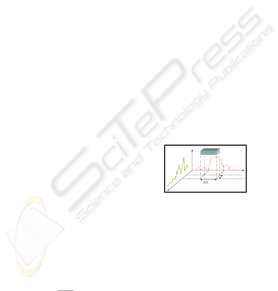
4 HYPERCUBE
CLASSIFICATION
This method suppresses the isolated pixels and
filters the classes that contain less than 3 pixels that
are incorporated into the larger adjacent class with
which the minimum difference in color is verified. It
consists in detecting the valley on the gradient of the
histogram 1D of R, G and B on which an interval
[]
SSS Δ+
λλ
;
is defined for each of the three
components. The intersection of all intervals defines
classes. This process is shown in Figure 1.
Figure 1: Hypercube classification.
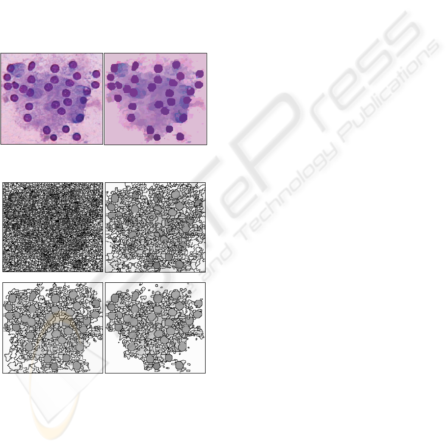
The final segmented RGB image is shown in
Figure 2.
5 EXPERIMENTAL RESULTS
In this section we present some results obtained by
applying our segmentation scheme on microscopic
medical test images. Here we present the process of
the iterative RAG processing on a cytological image
that decreases the number of regions by 80%
without introducing clustering errors. Segmentation
results are presented in Figure 3 by showing the
SEGMENTATION OF MULTISPECTRAL IMAGES USING MATHEMATICAL MORPHOLOGY AND AUTOMATIC
CLASSIFICATION - Application to Microscopic Medical Images
239
region borders showing the ability of the proposed
segmentation scheme to simplify the original image.
The use of morphological process as opening and
closing operations applied on the RAG yields an
interesting feature extraction as the photometric
value, the area, the perimeter, the compactness
factor, the number of neighbors of a region and their
relationship for each region. This description
represents the simplified information and contains
potentially an elaborate knowledge. After the
interpretation of the image, a list of retained objects
and their associated features are stored in an XML
(eXtensible Markup Langage) file and ready to be
integrated into a medical information system.
Figure 2: The original RGB image and its corresponding
segmented image.
(a)
(b)
(d)
(c)
Figure 3: Hierarchical RAG levels shown the step of
region merging.
6 CONCLUSIONS
We have proposed a new method of microscopic
medical images segmentation using mathematical
morphology applied on RAG and an automatic
clustering method followed by a regularization step
using an automatic hypercube classification. Due to
the unsupervised nature of the procedure especially
the use of automatic thresholds detection, it can be
reliable to the huge variability of intensities and
shapes of the image regions and will be tested as a
part of future work in other color space without
introducing
a priori knowledge and pre-processing
stages.
Results show the effectiveness of our method for
medical image applications as cytology images and
the impact that it introduces on the semantic high
level search for any disease or abnormal cells.
In this paper, the morphological operations
consider only the extrema of region neighborhood.
For future works, we will pursue the aggregation
operations beyond the limits presented by the
morphological processing avoiding the refinement
segmentation step that uses the hypercube
classification.
REFERENCES
Lucchese, L., Mitra, S.K., 2001. Color image
segmentation: A state-of-the art survey. In Image
Process. Vis. Pattern Recog. Proc. Indian Nat. Sci.
Acad. (INSA-A). Vol67 i2.pp. 207-221.
Lezoray, O., Lecluse, M., November 2007. Automatic
segmentation and classification of cells from broncho
alveolar lavage. In Image Analysis and Stereology,
Vol.26, pp. 111-119.
Lezoray, O., Juin 2003. Supervised automatic histogram
clustering and watershed segmentation. Application to
microscopic medical images. In Image Analysis and
Stereology, Vol. 22, Numéro 2, pp.113-120.
El-Khoury, E., Senac, C., André-Obrecht, R., 2007.
Speaker Diarization: Towards a more Robust and
Portable System. Int. Conf. On Acoustics, Speech, and
Signal Processing (ICASSP’2007), Honolulu, Hawaii,
USA.
Hirata Jr., R., César Flores, F., Barrera, J., Lotufo, R.,
Meyer, F., 2000. Color Image Gradients for
Morphological Segmentation. In XIII Brizilian
Symposium on Computer Graphics and Image
Processing (SIBGRAPI'00), pp.316-326.
Beucher, S., Lantuéjoul, C., Sept. 1979. Use of watersheds
in contour detection. In Int.workshop on image
processing, real-time edge and motion detection.
Mestar, A., Vannoorenberghe, P., Flouzat, G., April 2007.
Mathematical Morphology applied to Very High
Resolution Spatial images interpretation. In Urban
Remote Sensing Joint Event, pp. 1-7.
Pesaresi, M., Benediktsson, J.A., Feb. 2001. A new
approach for the morphological segmentation of high-
resolution satellite imagery. In Geoscience and
Remote Sensing,IEEE Transactions, Vol 39, Issue 2.
VISAPP 2009 - International Conference on Computer Vision Theory and Applications
240
