
Faster R-CNN Approach for Diabetic Foot Ulcer Detection
Artur Leandro da Costa Oliveira
a
, Andr
´
e Britto de Carvalho
b
and Daniel Oliveira Dantas
c
Departamento de Computac¸
˜
ao, Universidade Federal de Sergipe, S
˜
ao Crist
´
ov
˜
ao, SE, Brazil
Keywords:
Deep Learning, Convolutional Neural Networks.
Abstract:
Diabetic Foot Ulcer (DFU) is one of the major health concerns about Diabetes. These injuries impair the
patient’s quality of life, bring high costs to public health, and can even lead to limb amputations. The use
of automatic tools for detection can assists specialists in the prevention and treatment of the disease. Some
methods to address this problem based on machine learning have recently been presented. This article proposes
the use of deep learning techniques to assist the treatment of DFUs, more specifically, the detection of ulcers
through photos taken from the patient’s feet. We propose an improvement of the original Faster R-CNN
using data augmentation techniques and changes in parameter settings. We used a training dataset with 2000
images of DFUs annotated by specialists. The training was validated using the Monte Carlo cross-validation
technique. Our proposal achieved a mean average precision of 91.4%, a F1-score of 94.8%, and an average
detection speed of 332ms which outperformed traditional detector implementations.
1 INTRODUCTION
Diabetes is a serious complication with a high long-
term impact on the population. The incidence of di-
abetes has grown globally in the last decades causing
high health costs. It is among the top 10 causes of
death in adults (Saeedi et al., 2019). Diabetic Foot
Ulcer (DFU) is one of the major complications of Di-
abetes. The patients have a probability of 12-25%
of developing DFU during their lifetime. This rate
can reach 19-34% depending on the data used (Arm-
strong et al., 2017). Such ulcers have become a major
problem in public health because of the increase in
morbidities, decreased quality of life, and because the
treatment is expensive. Due to inadequate conduct in
the treatment of foot ulcers, there is a delay in the im-
provement of the injury and the possibility of lower
limb amputation (Leung, 2007).
In the early stages of the DFU, it is important to
quickly detect and to keep track of the disease. To
make a diagnostic, specialists take into account differ-
ent evaluation criteria, such as the medical history of
the patient, examination of the diabetic foot, and ad-
ditional tests like CT scans, MRI, and X-Ray (Goyal
et al., 2018a). The use of computer vision techniques
can lead to an improvement in the diagnosis of the
a
https://orcid.org/0000-0002-0165-6699
b
https://orcid.org/0000-0002-6498-9706
c
https://orcid.org/0000-0002-0142-891X
disease and in the agility of the entire clinical pro-
cess. Image processing is used in the medical field
in several types of systems and has been successful
in different medical applications. These systems are
used in treatment planning, surgery, and biological
images. Databases can have two, three, or more di-
mensions. These dimensions carry a vast amount of
information that can be used in the clinical area or
application research (Bankman, 2008). Initially, low-
level pixel processing methods (edge detection, line
detection filters, and region growth) and mathemat-
ical models were used to solve specific problems in
the medical field. In the late 90s, supervised learn-
ing techniques, where training data is used to develop
a system, started to become popular. A crucial step
in the design of such systems is the extraction of dis-
criminant features from the images. Later, the use of
deep learning techniques arises, allowing computers
to learn the features that optimally represent the data
of the problem at hand (Litjens et al., 2017).
Generally, from a computer vision and medical
image perspective, three different tasks are performed
to detect anomalies in medical images: classifica-
tion, localization, and segmentation (Goyal et al.,
2018b). Classification is to recognize the type of
the anomaly. Localization is to point out the region
of the anomaly. Segmentation is to define precise
limits of the anomaly. To solve these tasks, convo-
lutional neural network (CNN) based object detec-
tors have been used, such as the faster region-based
Oliveira, A., Britto de Carvalho, A. and Dantas, D.
Faster R-CNN Approach for Diabetic Foot Ulcer Detection.
DOI: 10.5220/0010255506770684
In Proceedings of the 16th International Joint Conference on Computer Vision, Imaging and Computer Graphics Theory and Applications (VISIGRAPP 2021) - Volume 4: VISAPP, pages
677-684
ISBN: 978-989-758-488-6
Copyright
c
2021 by SCITEPRESS – Science and Technology Publications, Lda. All rights reserved
677

Figure 1: Samples of the DFU Dataset.
convolutional network (Faster R-CNN) (Ren et al.,
2015), region-based fully convolutional networks (R-
FCN) (Dai et al., 2016), single shot multibox de-
tector (SSD) (Liu et al., 2016), and you only look
once (YOLO) (Redmon et al., 2016). These methods
are accurate enough to be used in consumer applica-
tions (Huang et al., 2017) and are usually evaluated by
their mean average precision (mAP), but other metrics
can also be used, such as specificity, sensitivity, exe-
cution time, and memory usage.
Studies that assist the treatment of DFU using
computational methods are rarely found in the liter-
ature. Liu et al. (Liu et al., 2015) and Saminathan
et al. (Saminathan et al., 2020) proposed automatic
methods that use the temperature characteristic in in-
frared images to perform DFU detection. Their pa-
pers show good results in detecting DFU in images of
feet. However, they present difficulties in terms of dif-
ferent symmetries and positioning of feet in images.
Deformed feet and/or amputated limbs can also im-
pair detection by these methods. Goyal et al. (Goyal
et al., 2017) proposes to segment DFU lesions us-
ing Fully Convolutional Networks (FCNs). Its re-
sults demonstrate a high accuracy which can help in
the detection and treatment of the disease. Goyal
proposes a convolutional neural network architecture
called DFUNet to improve the classification of DFU
images (Goyal et al., 2018a). Its good performance
in classifying parts of skin with DFU allows it to be
used also for classifying other skin diseases. The
DFUNet obtains a better performance compared to
GoogLeNet. GoogleNet is a convolutional neural net-
work also known as Inception and was responsible for
achieving the state-of-the-art in detection and classi-
fication in the ImageNet Large-Scale Visual Recog-
nition Challenge 2014 (ILSVRC14) (Szegedy et al.,
2015).
Goyal et al. (Goyal et al., 2018b) proposes a real-
time detection tool of DFUs for mobile devices. The
usage of such a tool on a mobile device assists spe-
cialists in quickly detecting and diagnosing the dis-
ease. The major challenge of automatic methods for
DFU is to optimize metrics such as specificity, sen-
sitivity, execution time, and memory usage. An im-
provement in these metrics allows greater reliability
in the use of this type of application for the treatment
of DFUs. In contrast to traditional machine learning,
deep learning methods have demonstrated superior-
ity in object localization and segmentation of DFUs,
which suggests that the robust fully automated detec-
tion of DFUs may be viable (Goyal et al., 2018b).
This work proposes a tool for detecting foot ulcers
in individuals with diabetes based on the Faster R-
CNN object detection (Ren et al., 2015). The purpose
of this work is to help the prevention and treatment of
the disease. The main task of the tool is to locate ar-
eas of interest in the image and classify them as ulcers
or not. This work was motivated by the Diabetic Foot
Ulcers Grand Challenge 2020 (DFUC 2020) (Cassidy
et al., 2020), challenge that aims to improve the ac-
curacy of DFU detection in real environments. Our
main contribution is the improvement of the Faster
R-CNN for DFU detection. In our experiments, we
achieved better mAP, F1-score, and detection speed
in comparison to the state-of-the-art detectors. Our
strategy reduced the number of false positives, which
lead to an improvement in precision.
The rest of the work is organized as follows: Sec-
tion 2 summarizes basics concepts needed to under-
stand the work. Section 3 describes the methodology
used to create the tool. Section 4 presents the experi-
ments and the results, and Section 5 presents the final
considerations.
2 BACKGROUND
Created to approach the problem of object detection
by region proposal, the Faster R-CNN (Ren et al.,
2015) is an evolution of the Fast R-CNN (Girshick,
2015). Unlike its predecessor, the Faster R-CNN con-
sists of two modules. The first module is the region
proposal network (RPN), a deep convolutional neural
network. The second module is the Fast R-CNN de-
tector. Both the RPN and the object classifier share
convolutional layers. The region proposal network is
intended to guide the detection, determining the best
regions among different scales and proportions. Basi-
cally, the RPN tells the classification module where to
look. The classification module, composed of a deep
VISAPP 2021 - 16th International Conference on Computer Vision Theory and Applications
678
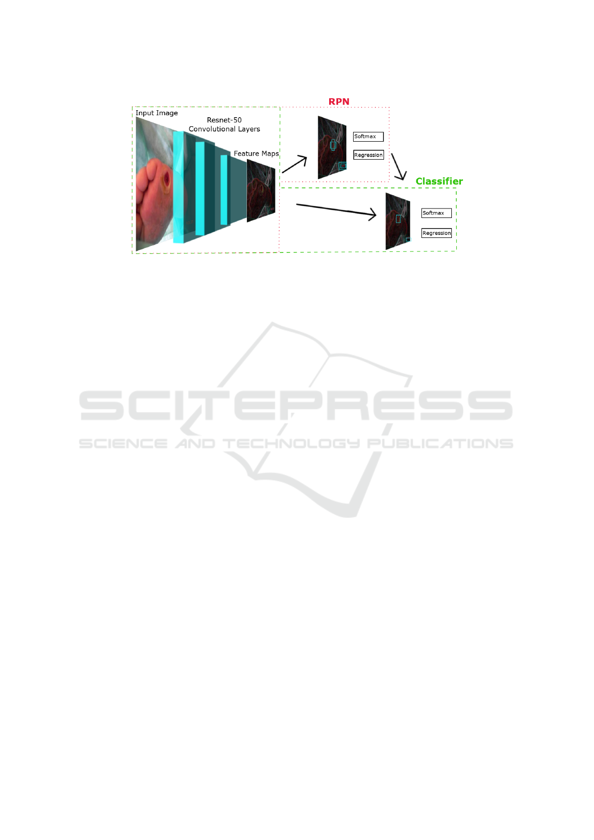
Figure 2: Faster R-CNN DFU Architecture.
convolutional network, receives different regions of
the image and classifies them.
The motivation for using the Faster R-CNN is due
to its high accuracy in object detection. This network
achieved the best accuracy in PASCAL VOC (Ever-
ingham et al., 2007; Everingham et al., 2015) both in
2007 and in 2012 and was the basis for the winners
of Imagenet detection and localization in ILSVRC
2015 and COCO detection in the COCO 2015 com-
petition (Ren et al., 2015). Regardless of the CNN
used for detection, Faster R-CNN is still superior to
other detection methods in terms of accuracy. Re-
garding the detection speed, the SSD method comes
out ahead (Huang et al., 2017). Despite being slower,
the Faster R-CNN guarantees an adequate speed to be
used in this work.
Deep convolutional neural networks require a
large set of training data to avoid overfitting, but large
sets are often difficult to obtain. One approach to
avoid overfitting is to use regularization techniques
such as Dropout (Srivastava et al., 2014) and Batch
Normalization (BN) (Ioffe and Szegedy, 2015). An-
other regularization technique is data augmentation,
which consists of creating new examples from the
training base (Lemley et al., 2017). It increases the
training base by using various transformations in the
image: translation, rotation, flipping, cropping, addi-
tion of noise etc.
Knowledge transfer is often used, and is shown to
be successful by several machine learning works (Pan
and Yang, 2009). Traditional machine learning tech-
niques learn from scratch, whereas transfer learning
train a previously trained model with new data. Using
models and weights trained in generic bases such as
ImageNet and MS-COCO for detection in the medical
field helps improving the performance of the convo-
lutional network (Goyal et al., 2017).
3 METHODOLOGY
This section details the image dataset and the detector
used to solve the problem. Also exposes the adap-
tations in the detector, parameter settings, and other
functions to improve the performance in the detection
of foot ulcers in patients with diabetes.
3.1 The DFU Dataset
The image dataset used in this work is part of the Di-
abetic Foot Ulcers Grand Challenge 2020 challenge
(DFUC 2020) (Cassidy et al., 2020). There are 2000
images for training, 200 for validation, and in the
end, 2000 images were released as test dataset. The
images were collected over the years at Lancashire
Teaching Hospital (LTH). These images are close-
ups of feet with ulcers from patients with diabetes.
Figure 1 shows image examples of the dataset. All
images have 640×480 pixels. The images were ac-
quired without flash as primary light source, and in-
stead, room lights were used to ensure consistent col-
ors. The ulcers were marked on the images as a rect-
angular region of interest (ROI) by specialists who
used a specific software for this task (Cassidy et al.,
2020).
3.2 Implementation Details
In this work, we propose an adapted version of the
Faster R-CNN architecture for DFU detection, called
Faster R-CNN DFU. Figure 2 describes the entire ar-
chitecture of our approach. The RPN and the Classi-
fier are the two main modules that share a common set
of convolutional layers. The feature maps extracted
by the convolution layers serve as input for the RPN
Faster R-CNN Approach for Diabetic Foot Ulcer Detection
679

and the Classifier. The RPN outputs a set of rectan-
gular object proposals, each one with an objectness
score, which also serves as input for the Classifier.
Each rectangular object is classified into a set of pre-
defined labels, each one with a score. Our adaption
of this architecture aims to improve the precision of
the ROIs, enhance the detection of different sizes of
ulcers, minimize the detection of false positives, and
speed up the detection time. We also propose a vari-
ant of the detector which can be used for general kind
of problems, called Faster R-CNN FP. Its focus is on
reducing false positives and improving detection per-
formance.
The original Faster R-CNN implementation used
ZF (Zeiler and Fergus, 2014) and VGG (Simonyan
and Zisserman, 2014) as part of the RPN and of the
classifier. However, Ren et al. (Ren et al., 2018) have
experimentally proved that the pre-trained ResNet-
50 model achieves a better performance when com-
pared to other popular CNNs such as VGG and In-
ception (Zeiler and Fergus, 2014; Szegedy et al.,
2015; Szegedy et al., 2016). Therefore, in this work,
ResNet-50 was chosen as the deep convolutional neu-
ral network for the Faster R-CNN DFU and Faster R-
CNN FP.
In the Fast R-CNN detector (Girshick, 2015) the
negative ROI (Region of Interest) samples that are
sent for classification are those that have an IoU (In-
tersection over Union) in the range of [0.1, 0.5). The
IoU is an evaluation metric, also known as the Jaccard
Index, given by Equation 1:
IoU =
|A ∩ B|
|A ∪ B|
(1)
where A and B are respectively the detected and
ground truth bounding boxes. An IoU greater than
or equal to 0.1 causes the classifier not to be trained
with regions of the image without ulcers, which can
favor the appearance of false positives. We changed
the interval to [0.0, 0.5), so that true negatives, regions
without ulcers, are also used in training. This range is
used by Ren et al. (Ren et al., 2015) and is shown to
improve the accuracy of the detector. We used this
strategy in both approaches in order to minimize false
positives.
Detailed analysis of the dataset reveals a wide va-
riety in the size of the ulcers. The original Faster
R-CNN implementation uses 9 standard anchors, de-
fined by all the possible combinations of the sizes
128×128, 256×256, and 512×512 with the aspect ra-
tios 1:1, 1:2, and 2:1. Due to this fact, the network
fails to detect very small lesions. In our implementa-
tion of the Faster R-CNN DFU, we added the 64×64
anchor size to the set of standard anchor scales, main-
taining the original aspect ratios. Therefore, a total of
12 anchors were used, which improved the accuracy
in the detection of small lesions (Ren et al., 2015; Sun
et al., 2018).
One of the great advantages of the Faster R-CNN
is due to the use of shared convolutional layers with
the RPN, which significantly reduces the region pro-
posal cost. RPN suggests regions of the image for
the classifier. The number of regions suggested in the
standard implementation is 300. But Fan et al. (Fan
et al., 2016) verified that a decrease of this number,
besides improving the response time, can also im-
prove precision. In the training of our approaches,
the value of 100 ROI suggestions was used.
The Faster R-CNN FP approach is an improve-
ment on the Faster R-CNN. It uses the ResNet-50 as
CNN, an IoU sample range of [0.0, 0.5) for the neg-
ative ROIs, and 100 ROI suggestions. The Faster R-
CNN DFU approach uses the same Faster R-CNN FP
configurations and improves it with specific strate-
gies for DFU detection. The main strategy was to
use 12 different anchors for the detection of a greater
variety of ulcer formats. Our algorithms were imple-
mented using the Tensorflow API (Abadi et al., 2016),
which provides an open-source framework that assists
in the implementation of several detection models.
The code is written in Python and is publicly avail-
able
1
.
3.3 Training
To augment the training and validation datasets, hori-
zontal and vertical flips, rotations by 180
◦
, and Gaus-
sian blur, to emulate the blur caused by cell phone
cameras, were used. The Dropout and BN regular-
ization techniques were used in the neural network.
We used the weights of ResNet-50 (He et al., 2016),
pre-trained with the image database ImageNet (Rus-
sakovsky et al., 2015). This dataset contains millions
of images with annotations of different classes of ob-
jects. The regularization techniques showed to im-
prove detection in our experiments.
We randomly divided the whole dataset of 2000
images provided by the challenge into 1600 images
(80%) for the training set and 400 images (20%) for
the test set. During training, we used the Monte Carlo
cross-validation methodology (Xu and Liang, 2001),
which randomly partitions the training set into 85%
for training and 15% for validation. At each new
training iteration, new images are selected for train-
ing and validation. Faster R-CNN requires the scal-
ing of the training images based on the smallest side
1
https://github.com/ArturLeandro/dfu faster rcnn
VISAPP 2021 - 16th International Conference on Computer Vision Theory and Applications
680

of the image. The 640×480 size was maintained for
the training and validation images.
We used 100 epochs to perform the training of our
algorithm. This number of epochs is enough for the
loss function to converge to its lowest value. At each
epoch, 1000 images were selected to train the RPN
and the classifier. The learning rate used was 0.00001
in the first 60 epochs and 0.000001 for the others. The
loss function implementation follows the same defini-
tions of multi-task loss minimization proposed by Ren
et al. (Ren et al., 2015).
4 EXPERIMENTS AND RESULTS
We tested four different approaches to detect DFUs
in our experiments. First of all, the Faster R-CNN
DFU detector with all the implementation details de-
scribed in the methodology section. The second is
the Faster R-CNN FP, our implementation that re-
duces false positives. The third one is the standard
SSD300 (Liu et al., 2016) approach with the convo-
lutional network VGG. And the fourth is the standard
Faster R-CNN (Ren et al., 2015) approach with the
pre-trained convolutional network ResNet-50. The
objective is to identify positive and negative points in
our strategies compared to the standard implementa-
tions of the detectors. We used a total of 100 epochs
for training the SSD300 and the three versions of
Faster R-CNN detectors. The experiments were done
by detecting ulcers in the 400 images of the test set.
The machine used in the experiments has a CPU In-
tel i3-8100 @ 3.6GHz, GPU NVIDIA GeForce GTX
1050 Ti SC 4GB, and 16GB DDR4 of RAM.
Table 1 shows the mean average precision (mAP)
and F1-Score of each detector. The mAP is a met-
ric widely used in detection works and is given by
the area under the precision/recall (PR) curve of the
detector. This metric needs an overlap criterion that
specifies the minimum value of the intersection over
union (IoU) to be considered a correct detection. The
value of 0.5 was chosen for this criterion as it is a
value widely used in the literature. The F1-score is
a metric defined by the harmonic mean of precision
and recall. The precision, recall, and F1-score can be
Table 1: Performance of DFU detection techniques on the
DFU Dataset. Proposed techniques are denoted with *.
Technique mAP F1-score
Faster R-CNN DFU* 91.4 94.8
Faster R-CNN FP* 86.5 91.9
Faster R-CNN 80.7 76.3
SSD300 52.7 65.7
calculated using the followings expressions:
precision =
TP
TP+FP
(2)
recall =
TP
TP+FN
(3)
F
1
= 2 ∗
precision∗recall
precision+recall
(4)
where TP represents the number of true positive de-
tections, FP the false positives, and FN the false neg-
atives.
Table 1 shows the results obtained by the tested
techniques. The proposed Faster R-CNN DFU out-
performed the other techniques. It has the best mAP
and F1-score for the DFU dataset, which indicates
that the regions found by our approach are closer to
the regions of ulcers marked by specialists. Because
of the high value of the F1-score, many true positive
regions are detected and a very low number of false
positives are detected. Figure 3 shows the approx-
imation of all detection techniques regions with the
ground-truth boxes. The returned values of classifica-
tion accuracy are labeled on top of the region mark.
It is possible to notice in the images the improvement
in the detection of DFUs with the two proposed tech-
niques. Unlike the standard version of Faster R-CNN,
the Faster R-CNN FP decreases the false positive de-
tection, and the Faster R-CNN DFU, besides increas-
ing the precision of the detection, is also successful
in detecting small ulcers. SSD300 has good results,
but it fails to find DFUs and does not achieve a good
precision.
Data augmentation and the changes proposed to
decrease false positives increased the mAP in 10.7
percentage points, and the F1-Score in 18.5 percent-
age points. Figure 4 (a) evidences this improvement
by showing the ROC curve of all detectors. The num-
ber of false positives decreases considerably after us-
ing the proposed techniques, particularly when com-
pared to the results of Faster R-CNN. Likewise, as
shown in Figure 4 (b), precision and recall both re-
main at high values, increasing the area under the
curve. A high recall is related to a low number of
Table 2: Detection average speed (DAS) in milliseconds
and model size in megabytes of DFU detection neural net-
works. Proposed techniques are denoted with *.
Technique DAS (ms) Size (MB)
SSD300 48 92.9
Faster R-CNN DFU* 332 111.1
Faster R-CNN FP* 362 111.1
Faster R-CNN 807 111.1
Faster R-CNN Approach for Diabetic Foot Ulcer Detection
681
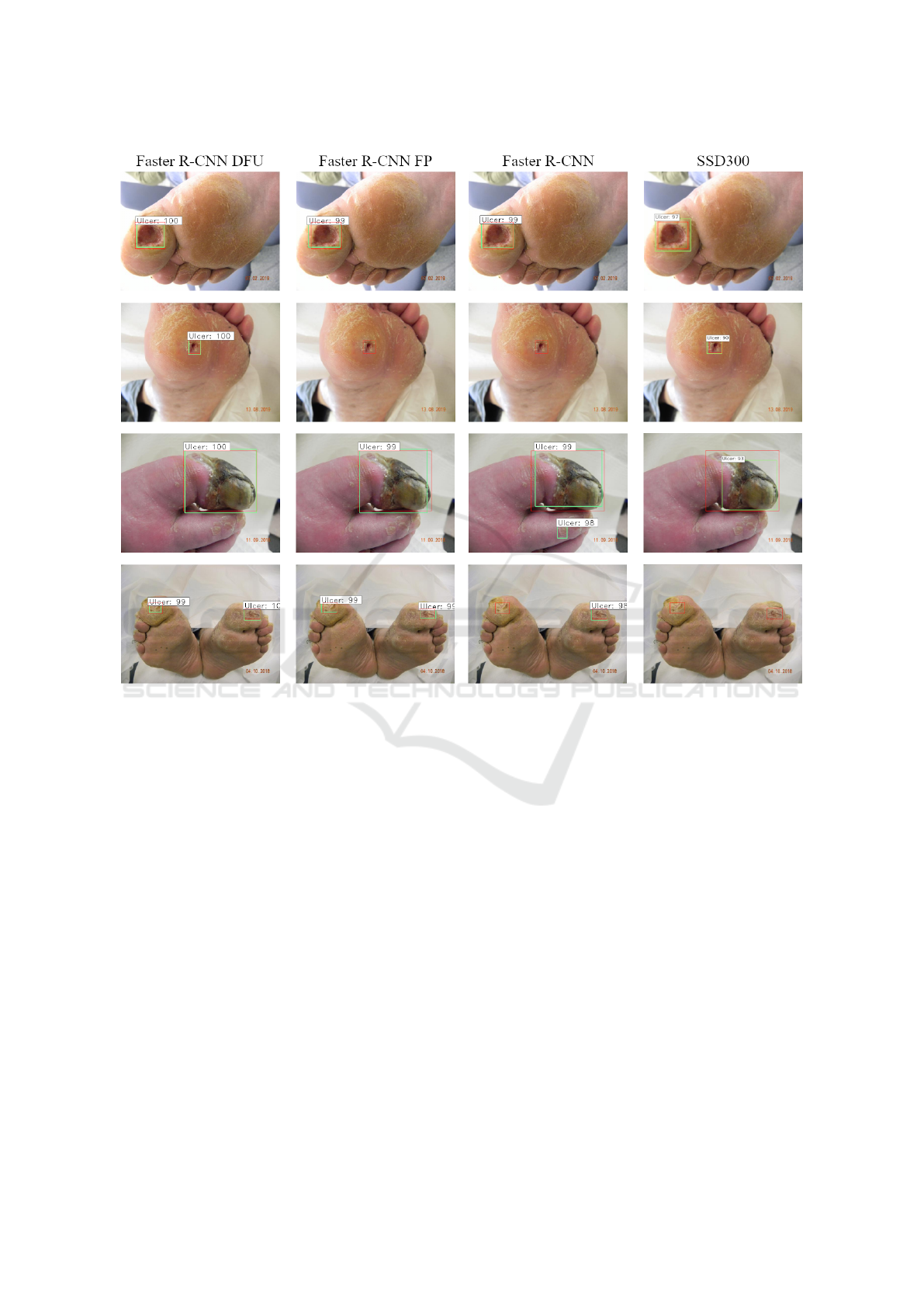
Figure 3: Detection results from the DFU detection techniques. In green are the detected regions and in red the ground-truth
boxes.
false negatives, which is usually desirable in medical
context.
Table 2 shows the results of the detection average
speed, and model size of each detector. The SSD300
obtained the best average speed and the smallest size
of the model in comparison to the other detectors.
This is mainly due to the simpler architecture to gen-
erate anchor boxes (Liu et al., 2016). However, its
precision is lower than the other techniques. The av-
erage detection time of our two proposals is smaller
than the standard Faster R-CNN implementation due
to the use of a smaller number of ROI suggestions.
The size of all Faster R-CNN variants is the same,
as all of them are based on the Resnet-50 CNN. Their
sizes are slightly larger than SSD300. Our approaches
can be used on devices that have limited resources due
to the small size of the model and a lower process time
consumption.
5 CONCLUSIONS
In this work, we propose an automatic approach to de-
tect DFUs using deep learning techniques. We have
implemented an extended version of the Faster R-
CNN approach. We have adopted several strategies to
achieve high precision in detecting ulcers, to decrease
the number of false positives, and to speed up the de-
tection time. We changed the numbers of regions, the
anchor scales, used data augmentation in the dataset,
and adopted a CNN that has better detection results
than previous approaches. Finally, we carried out ex-
periments with the chosen detectors, training each one
with 100 epochs. Results showed that our strategies
improve the mAP and F1-score when compared to
standard detector implementations known as the state-
of-the-art. Better mAP, F1-score and detection speeds
have been achieved which allows not only for better
detection of the DFUs, but also a better confidence to
use the Faster R-CNN DFU in real applications.
VISAPP 2021 - 16th International Conference on Computer Vision Theory and Applications
682
(a)
(b)
Figure 4: Comparisons of ROC curves for different experi-
mental settings for DFU detection.
REFERENCES
Abadi, M., Barham, P., Chen, J., Chen, Z., Davis, A.,
Dean, J., Devin, M., Ghemawat, S., Irving, G., Isard,
M., et al. (2016). Tensorflow: A system for large-
scale machine learning. In 12th {USENIX} sympo-
sium on operating systems design and implementation
({OSDI} 16), pages 265–283.
Armstrong, D. G., Boulton, A. J., and Bus, S. A. (2017). Di-
abetic foot ulcers and their recurrence. New England
Journal of Medicine, 376(24):2367–2375.
Bankman, I. (2008). Handbook of medical image process-
ing and analysis. Elsevier.
Cassidy, B., Reeves, N. D., Joseph, P., Gillespie, D.,
O’Shea, C., Rajbhandari, S., Maiya, A. G., Frank, E.,
Boulton, A., Armstrong, D., et al. (2020). Dfuc2020:
Analysis towards diabetic foot ulcer detection. arXiv
preprint arXiv:2004.11853.
Dai, J., Li, Y., He, K., and Sun, J. (2016). R-fcn: Object de-
tection via region-based fully convolutional networks.
In Advances in neural information processing sys-
tems, pages 379–387.
Everingham, M., Eslami, S. A., Van Gool, L., Williams,
C. K., Winn, J., and Zisserman, A. (2015). The pascal
visual object classes challenge: A retrospective. Inter-
national journal of computer vision, 111(1):98–136.
Everingham, M., Van Gool, L., Williams, C. K., Winn, J.,
and Zisserman, A. (2007). The pascal visual object
classes challenge 2007 (voc2007) results.
Fan, Q., Brown, L., and Smith, J. (2016). A closer look
at faster r-cnn for vehicle detection. In 2016 IEEE
intelligent vehicles symposium (IV), pages 124–129.
IEEE.
Girshick, R. (2015). Fast R-CNN. In Proceedings of the
IEEE international conference on computer vision,
pages 1440–1448.
Goyal, M., Reeves, N. D., Davison, A. K., Rajbhandari, S.,
Spragg, J., and Yap, M. H. (2018a). Dfunet: Convo-
lutional neural networks for diabetic foot ulcer clas-
sification. IEEE Transactions on Emerging Topics in
Computational Intelligence.
Goyal, M., Reeves, N. D., Rajbhandari, S., and Yap, M. H.
(2018b). Robust methods for real-time diabetic foot
ulcer detection and localization on mobile devices.
IEEE journal of biomedical and health informatics,
23(4):1730–1741.
Goyal, M., Yap, M. H., Reeves, N. D., Rajbhandari, S., and
Spragg, J. (2017). Fully convolutional networks for
diabetic foot ulcer segmentation. In 2017 IEEE inter-
national conference on systems, man, and cybernetics
(SMC), pages 618–623. IEEE.
He, K., Zhang, X., Ren, S., and Sun, J. (2016). Deep resid-
ual learning for image recognition. In Proceedings of
the IEEE conference on computer vision and pattern
recognition, pages 770–778.
Huang, J., Rathod, V., Sun, C., Zhu, M., Korattikara, A.,
Fathi, A., Fischer, I., Wojna, Z., Song, Y., Guadar-
rama, S., et al. (2017). Speed/accuracy trade-offs for
modern convolutional object detectors. In Proceed-
ings of the IEEE conference on computer vision and
pattern recognition, pages 7310–7311.
Ioffe, S. and Szegedy, C. (2015). Batch normalization: Ac-
celerating deep network training by reducing internal
covariate shift. arXiv preprint arXiv:1502.03167.
Lemley, J., Bazrafkan, S., and Corcoran, P. (2017). Smart
augmentation learning an optimal data augmentation
strategy. Ieee Access, 5:5858–5869.
Leung, P. (2007). Diabetic foot ulcers—a comprehensive
review. The Surgeon, 5(4):219–231.
Litjens, G., Kooi, T., Bejnordi, B. E., Setio, A. A. A.,
Ciompi, F., Ghafoorian, M., Van Der Laak, J. A.,
Van Ginneken, B., and S
´
anchez, C. I. (2017). A survey
on deep learning in medical image analysis. Medical
image analysis, 42:60–88.
Liu, C., van Netten, J. J., Van Baal, J. G., Bus, S. A., and
van Der Heijden, F. (2015). Automatic detection of di-
abetic foot complications with infrared thermography
by asymmetric analysis. Journal of biomedical optics,
20(2):026003.
Liu, W., Anguelov, D., Erhan, D., Szegedy, C., Reed, S.,
Fu, C.-Y., and Berg, A. C. (2016). Ssd: Single shot
multibox detector. In European conference on com-
puter vision, pages 21–37. Springer.
Pan, S. J. and Yang, Q. (2009). A survey on transfer learn-
ing. IEEE Transactions on knowledge and data engi-
neering, 22(10):1345–1359.
Faster R-CNN Approach for Diabetic Foot Ulcer Detection
683

Redmon, J., Divvala, S., Girshick, R., and Farhadi, A.
(2016). You only look once: Unified, real-time object
detection. In Proceedings of the IEEE conference on
computer vision and pattern recognition, pages 779–
788.
Ren, S., He, K., Girshick, R., and Sun, J. (2015). Faster R-
CNN: Towards real-time object detection with region
proposal networks. In Advances in neural information
processing systems, pages 91–99.
Ren, Y., Zhu, C., and Xiao, S. (2018). Object detection
based on fast/faster rcnn employing fully convolu-
tional architectures. Mathematical Problems in En-
gineering, 2018.
Russakovsky, O., Deng, J., Su, H., Krause, J., Satheesh, S.,
Ma, S., Huang, Z., Karpathy, A., Khosla, A., Bern-
stein, M., et al. (2015). Imagenet large scale visual
recognition challenge. International journal of com-
puter vision, 115(3):211–252.
Saeedi, P., Petersohn, I., Salpea, P., Malanda, B., Karu-
ranga, S., Unwin, N., Colagiuri, S., Guariguata, L.,
Motala, A. A., Ogurtsova, K., et al. (2019). Global
and regional diabetes prevalence estimates for 2019
and projections for 2030 and 2045: Results from the
international diabetes federation diabetes atlas. Dia-
betes research and clinical practice, 157:107843.
Saminathan, J., Sasikala, M., Narayanamurthy, V., Rajesh,
K., and Arvind, R. (2020). Computer aided detec-
tion of diabetic foot ulcer using asymmetry analysis
of texture and temperature features. Infrared Physics
& Technology, 105:103219.
Simonyan, K. and Zisserman, A. (2014). Very deep con-
volutional networks for large-scale image recognition.
arXiv preprint arXiv:1409.1556.
Srivastava, N., Hinton, G., Krizhevsky, A., Sutskever, I.,
and Salakhutdinov, R. (2014). Dropout: a simple way
to prevent neural networks from overfitting. The jour-
nal of machine learning research, 15(1):1929–1958.
Sun, X., Wu, P., and Hoi, S. C. (2018). Face detection us-
ing deep learning: An improved faster rcnn approach.
Neurocomputing, 299:42–50.
Szegedy, C., Liu, W., Jia, Y., Sermanet, P., Reed, S.,
Anguelov, D., Erhan, D., Vanhoucke, V., and Rabi-
novich, A. (2015). Going deeper with convolutions.
In Proceedings of the IEEE conference on computer
vision and pattern recognition, pages 1–9.
Szegedy, C., Vanhoucke, V., Ioffe, S., Shlens, J., and Wo-
jna, Z. (2016). Rethinking the inception architecture
for computer vision. In Proceedings of the IEEE con-
ference on computer vision and pattern recognition,
pages 2818–2826.
Xu, Q.-S. and Liang, Y.-Z. (2001). Monte carlo cross vali-
dation. Chemometrics and Intelligent Laboratory Sys-
tems, 56(1):1–11.
Zeiler, M. D. and Fergus, R. (2014). Visualizing and under-
standing convolutional networks. In European confer-
ence on computer vision, pages 818–833. Springer.
VISAPP 2021 - 16th International Conference on Computer Vision Theory and Applications
684
