
Curve based Fast Detail Enhancement for Biomedical Images
Ran Fei
1,2
, Ying Weng
1,2,*
, Yiming Zhang
1,2
and Jonathan Lund
1,2
1
School of Computer Science, University of Nottingham Ningbo China, China
2
School of Medicine, University of Nottingham, U.K.
Keywords: Image Enhancement, Contrast Enhancement, Histogram Equalization, Biomedical Imaging.
Abstract: Biomedical images are widely collected from various applications, which are used for patients' screening,
diagnosis and treatment. The dark regions of biomedical images may play as an important role as the bright
regions. The enhanced details in the dark regions of biomedical images simultaneously maintain the quality
of the rest of the images and reveal more information for doctors and surgeons in medical procedures. This
paper proposes a fast method to adaptively enhance the details in the dark regions of biomedical images,
including X-rays, video frames of laparoscopy in minimally invasive surgery (MIS).
1 INTRODUCTION
Biomedical image processing is a broad and complex
field. In order to diagnose and treat patients,
biomedical images are essential. Low quality and
contrast of biomedical images will reduce the doctor's
ability to analyze the images, causing subsequent
processing difficulties. Moreover, due to medical
devices and procedures, doctors may have limited
controls in acquiring medical images leaving
biomedical images with non-homogeneity of
luminance and contrast levels. Hence it is vital to
enhance the biomedical image quality as well as
improve the image contrast. For instance, X-rays may
be low in contrast and details. Frames obtained during
minimally invasive surgery may have a large shaded
region due to less adequate light introduced into the
cavity; dark-colored tissue may lack details in high
contrast frames. Then it is essential to recognize
images that need enhancement then adaptively select
the targeted dark regions for further processing and
image contrast enhancement.
Many image enhancement techniques have been
developed by researchers including fuzzy set theory
image enhancement method (Preethi & Rajeswari,
2013), histogram equalization image enhancement
method (Agaian et al., 2007), histogram matching
image enhancement method (Irmak & Ertas, 2016)
and equalized histogram equalization image
enhancement method (Kadhum, 2012). Besides, there
*
Corresponding author.
are other image enhancement techniques, such as
nonlinear image enhancement technique (Singh et al.,
2015; Yaping et al., 2012) and wavelet transform
technique (Singh et al., 2015; Premkumar et al., 2014;
Ehsani et al. 2011).
Also, in image enhancement, one of the most
generally used and essential technique is contrast
enhancement. The contrast enhancement's primary
purpose is to adjust the local contrast to bring out the
image's exact regions. Contrast stretching is a contrast
enhancement technique used to extend an image's
dynamic range (Zakaria et al., 2010). Other image
contrast enhancement methods, including
homomorphic filtering (Zakaria et al., 2010), retinex
(Chen & Beghdadi, 2010), and histogram
equalization (Kim, 1997). Our proposed method is
based on histogram equalization. The proposed
method consists of two parts: the first part divides
images into different intensity regions; the second
part will further process the dark regions of the
targeted images.
The rest of this paper is organized as follows.
Section 2 deals with the previous work of the
histogram equalization (HE) methods. Section 3
presents our proposed curve based fast detail
enhancement method. The experiment results are
shown in section 4, and section 5 gives the conclusion
of the paper.
Fei, R., Weng, Y., Zhang, Y. and Lund, J.
Curve based Fast Detail Enhancement for Biomedical Images.
DOI: 10.5220/0010250203370344
In Proceedings of the 16th International Joint Conference on Computer Vision, Imaging and Computer Graphics Theory and Applications (VISIGRAPP 2021) - Volume 4: VISAPP, pages
337-344
ISBN: 978-989-758-488-6
Copyright
c
2021 by SCITEPRESS – Science and Technology Publications, Lda. All rights reserved
337
2 PREVIOUS WORK
Histogram equalization (HE) based algorithms,
adjusting gray-level distributions are commonly
deployed in contrast enhancement as it is simple to
use on biomedical images. It increases the dynamic
range expansion by increasing each pixel's value,
thereby enhancing the input image's contrast and
brightness (Kim, 1997). This section summarizes the
work on histogram equalization (HE) based
algorithms previously proposed by various
researchers.
Global histogram equalization stretches the image
intensity level to the whole range of 8-bit values, 0 –
255, effectively increasing the dark-bright contrast of
images (Mokhtar et al., 2009). Global HE is
discriminative. It may introduce undesired intensity
changes in a large block and may increase the noise
level of images. Local HE was proposed to get rid of
specific Global HE issues, but the method a) requires
high computational cost, b) the output appearance
depends on the size of selected local areas (Abdullah-
Al-Wadud et al., 2007). Similarly, adaptive HE
algorithms, taking advantage of global and local HE,
were introduced to enhance broader applications
(Singh et al., 2016).
Another research brightness preserving bi-
histogram equalization (BBHE) is conducted by Kim
(1997), which can preserve brightness and avoid false
coloring. Moreover, dualistic sub-image histogram
equalization (DSIHE) is similar to BBHE that input
histogram is decomposed into two subsections (Patel
et al., 2013). DSIHE performs better than BBHE
regarding entropy and brightness preservation
(Kalhor et al., 2019). Recursive mean separated
histogram equalization (RMSHE) is an extended
version of BBHE (Patel & Muthu, 2020). Compared
to BBHE, RMSHE can preserve the original
brightness of the image. Besides, dynamic histogram
equalization (DHE) assists in the control of the effect
of an image without losing important information in
the image (Abdullah-Al-Wadud et al., 2007).
Besides, a method named contrast limited
adaptive histogram equalization (CLAHE) is
proposed by Reza (2004) used with Ostu to enhance
biomedical image. A novel contrast enhancement
algorithm Histogram equalization with adaptive
gama correction and homomorphic filtering
(QWAGC-FIL) was developed by Monika Agarwal
et al. (2017). This algorithm can enhance the image
with low contrast while maintaining maximum
entropy and enhancement control.
3 THE PROPOSED METHOD
The method proposed in this paper take advantages of
global histogram equalization (GHE). As shown in
Figure 2, an adaptive intensity mapping is applied
before GHE to compensate issues of noise and
unwanted colour boundaries, Figure 1.
(a) Input (b) After GHE
(c) Input (d) After GHE
Figure 1: Issues of global histogram equalization, (a) and
(b), unwanted colour boundaries in dark regions; (c) and
(d), noise around edges are amplified after global histogram
equalization.
The intensity mapping is done in the V channel of
HSV colour space, where V = max (R, G, B). To
calculate the mapping function, the input V channel
should be divided into 3 sub-images, a noise part to
determine the offset of the function, a relevant dark
part to be mapped to a brighter value region and the
bright part to determine how bright the dark part can
be mapped to. To obtain the three parts, Ostu
threshold is applied to the input channel, which output
2 sub-images where the inter-class variance of the
histogram of the two sub-images are maximised.
After the initial thresholding:
1) If the dark part average is smaller than 32
(thresh_low) and the bright part average is
greater than 64, the dark part will be used to
measure the noise level; if the bright part
average is smaller than 64, the image is an
almost black image and is not currently
considered in this paper.
2) If the dark part is recognised as the noise
part, the bright part will be further divided
into 2 sub-images using Ostu method. The
averages and derivatives of the relevant dark
and bright parts (ave
D
, det
D
and ave
B
, det
B
)
and the maximum of the dark region, max
D
,
are recorded as respectively.
VISAPP 2021 - 16th International Conference on Computer Vision Theory and Applications
338
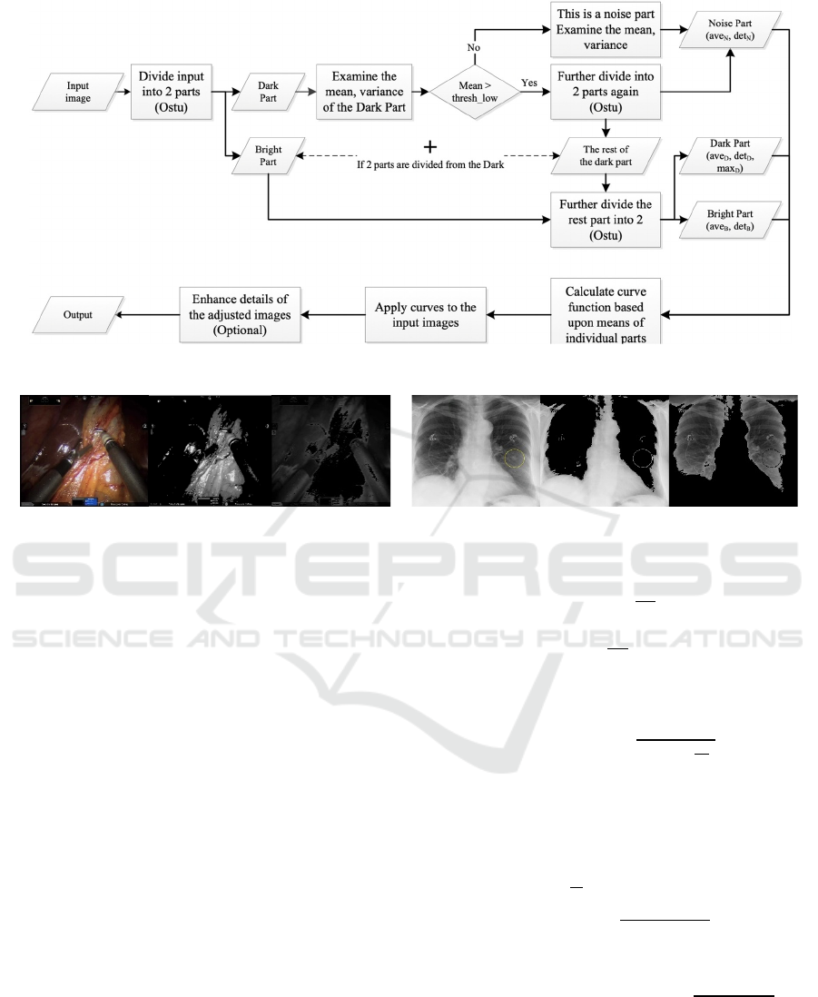
Figure 2: Workflow of the proposed algorithm.
Figure 3: Ostu thresholding.
3) If the initial divided dark part has averages
larger than 32, this dark part is further
divided until the noise part with mean value
smaller than 32 is separated, the average of
noise part is noted as ave
N
.
4) After that, the rest of the image will be
threshold (Ostu) into bright and dark parts.
The average, variance, maximum, ave
D
,
det
D
, max
D
and ave
B
, det
B
will be calculated
accordingly.
Averages and derivatives of separated dark and
bright parts are used to determine the shape of curve
applying to the input images. Curves are designed
following rules:
a) After applying the curves, pixels in the dark parts
should have smaller values than pixels in the
bright parts;
b) Logarithm curves g
x
N
i
N
i
ln
x
N
i
is
applied to pixels larger than N
i
to improve the
perceptive linearity of the relevant dark region
c) Near saturation region is supressed using sinuous
function to reduce the area of near saturated
region.
The result mapping function, f(x), is:
f
x
⎩
⎪
⎨
⎪
⎧
r
2
-1
xN
1
x in 0, N
1
r
2
N
1
N
1
ln
x
N
1
x in N
1
, N
2
r
3
xb
3
-Asin
2π
T
x-N
2
x in N
2
, 255
1) The curve starts from linear, x runs from 0 to
N
1
=max(ave
N
-det
N
, 0), with the curve output in
[N
1
, r
2
N
1
], where r
2
OutMaxV
2
N
1
N
1
ln
N
2
N
1
is the ratio
amplifying values in dark region from (N
1
,
N
2
=ave
D
+det
D
] to (r
2
N
1
, OutMaxV
2
].
2) To avoid oversaturation at the same time maintain
higher values in previous brighter part, OutMatV
2
is set to
min
N
1
N
1
ln
N
2
N
1
, max
ave
D
*rmax
D
*1-r,160
and r in [0, 1] is set to
mindet
D
, det
B
maxdet
D
, det
B
;
3) The rest of the part connects the maximum output
of the second (OutMatV
2
) to the maximum of 8-
bit output 255. Then, r
3
255-OutMaxV
2
255-N
and
b
2551-r
3
. To supress the near saturated
4) region, the half period of a sinuous function is
subtracted, where T= 2(255-N
2
) is the period of
5) the sinuous function, A is the magnitude of the
sinuous function. In experiment, A is set to 20.
Curve based Fast Detail Enhancement for Biomedical Images
339
0
64
128
192
256
0 64 128 192 256
When ave
N
5, ave
D
det
D
130 and the
maximum value of dark region, OutMatV
2
, is set to
130, the curve is demonstrated in Figure 4. Apply the
mapping process to 8-bit colour will result in Figure
5(b), where the very dark and very bright pixels are
mapped into the middle value range.
Figure 4: Curve with offset = 5, dark/bright division = 130.
(a) 8-bit color
(b) 8-bit color after curve processing
Figure 5: Curve and histogram equalization applied to 8-bit
colours of gray (x, x, x), red (x, 0, 0), green (0, x, 0) and
blue (0, 0, x) in order of (R, G, B), where x = [0, 255].
The design of the intensity mapping function and the
threshold selection is based on the non-linear
perception
of eyes in brightness and colour to the
Figure 6: Grey scale 8-bit colour. From block (a) to block
(h), intensity is gradually increased from 0 to 255. In each
block, the intensity in each adjacent slice is increased by 4.
input pixel values. As the 8-bit gray scale colour bar
demonstrated in Figure 6, the perception of intensity
change varies in each group. Eyes are more sensitive
to the colour difference in the mid-dark range (32 –
224) but not in the near black (0 – 32) and near
saturated (224 – 255) range. Mapping the range of
image intensities to the range of eye sensitive region
will help eyes to perceive more details from the input.
After intensity mapping, global histogram
equalization will have wider range of input images
with different intensity distributions. i.e. high contrast
laparoscopic surgical frames with have peaks in dark
and/or bright regions or X-ray images with pixels
distributed in mid-range. As demonstrated in Figure
6, apply the algorithm to a high contrast laparoscopic
image, the intensity of dark region will be increased
to reveal more details and the amplification of
brightness part is supressed without saturation. Apply
the same strategy to X-ray image, dark and bright
pixels from the input will be more evenly distributed
to obtain better eye perceived contrast.
(a) Laparoscopic
frame
(b) histogram
(c) X-ray
(f) histogram
(e)
p
rocessed (a) (
g
)
p
rocessed (c) (h) histo
g
ra
m
Figure 7: X-ray, laparoscopic surgical video frames (a, c) and their histogram (b, d) of intensity (V) calculated in HSV colour
space.
VISAPP 2021 - 16th International Conference on Computer Vision Theory and Applications
340
4 EXPERIMENTAL RESULTS
To examine the algorithm, images from websites are
tested. Following criteria are selected to evaluate the
performance of the algorithm:
‐ Amount of details revealed before / after the
application
‐ The colour consistency and truthfulness
before and after the application
‐ Amount of noise, alien boundaries
introduced through the application of the
algorithm
‐ Perceived change in brightness
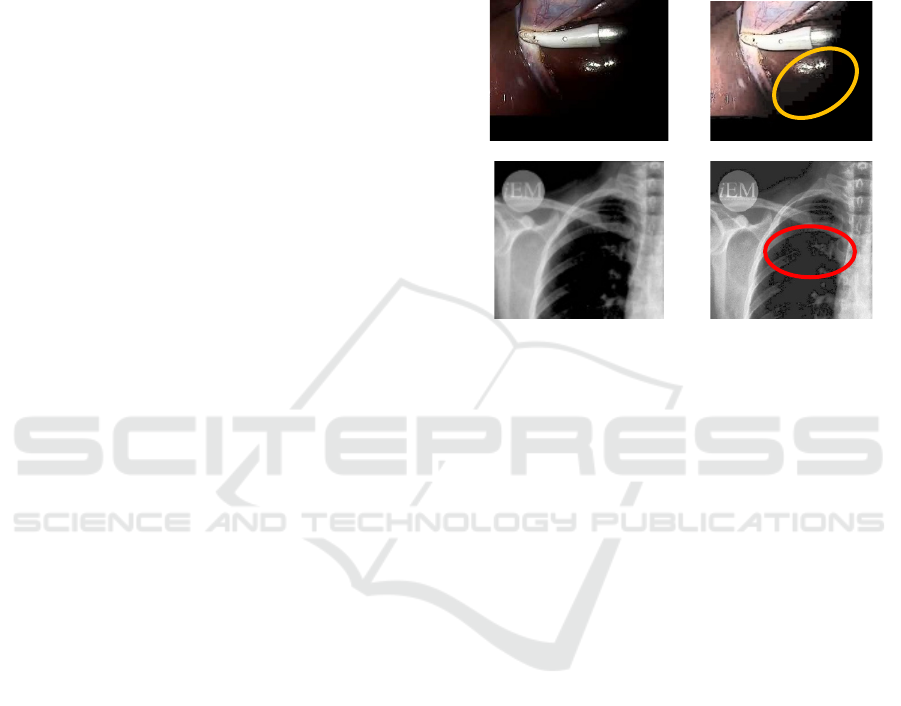
As demonstrated Figure 8, when applied to high
contrast laparoscopic surgical frames, details
including blood vessels and tissue patterns are better
presented at the same time maintained the colour
perception. Results of the algorithm applying to X-
ray images are similar to results of GHE with less
introduced noise level.
The algorithm is real-time capable for HD, 60fps,
1080*1920 video frames with the processing speed
approx. 8ms per frame, profiled through 4-core,
2.8GHz CPU. For similar size X-ray images, the
speed is around 3ms per image as only 1 channel is
processed, and no colour space conversion is
required.
5 CONCLUSIONS
This paper introduced a curve mapping and histogram
equalization-based method to enhance perceived
contrast and details of input biomedical images,
colour or gray-scale. The method takes advantages of
Ostu histogram analysis to adaptively separate
images into sub-regions with similar intensity
distributions, which intensively save the
computational loads. Curve based mapping adopted
from camera sensor processing map the image
intensity to eye sensitive range to help global
histogram equalization achieves better colour
truthfulness and reduce noise in near black region.
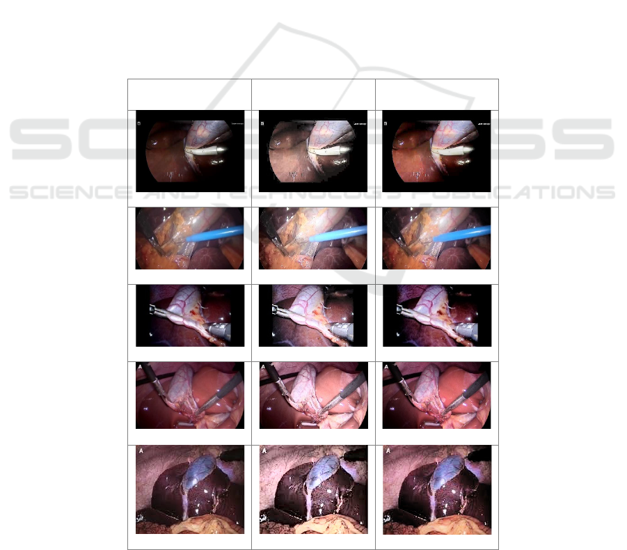
Original images Global Histogram
Equalization
Processed results
Figure 8: More results applied on website images.
Curve based Fast Detail Enhancement for Biomedical Images
341
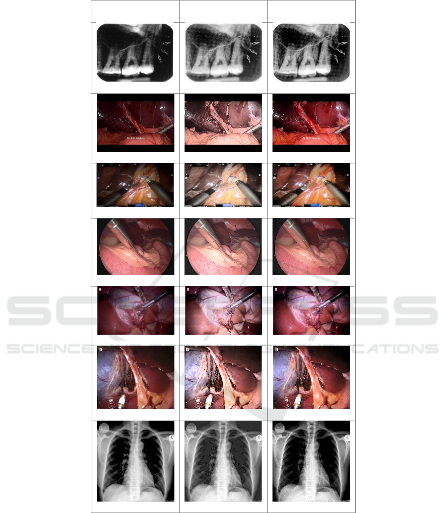
Original images Global Histogram
Equalization
Processed results
Figure 8: More results applied on website images (cont.).
VISAPP 2021 - 16th International Conference on Computer Vision Theory and Applications
342

ACKNOWLEDGEMENTS
We thank the R&D projects NBCP 2019C50052 and
NCHI I01200100023 for funding.
REFERENCES
Abdullah-Al-Wadud, M., Kabir, M. H., Dewan, M. A. A.,
& Chae, O. (2007). A dynamic histogram equalization
for image contrast enhancement. IEEE Transactions on
Consumer Electronics, 53(2), 593-600.
Agaian, S. S., Silver, B., & Panetta, K. A. (2007).
Transform coefficient histogram-based image
enhancement algorithms using contrast entropy. IEEE
transactions on image processing, 16(3), 741-758.
Agarwal, M., & Mahajan, R. (2017). Medical images
contrast enhancement using quad weighted histogram
equalization with adaptive gama correction and
homomorphic filtering. Procedia computer
science, 115, 509-517.
Chen, S., & Beghdadi, A. (2010). Natural enhancement of
color image. EURASIP Journal on Image and Video
Processing, 2010(1), 1-19.
Ehsani, S. P., Mousavi, H. S., & Khalaj, B. H. (2011,
November). Chromosome image contrast enhancement
using adaptive, iterative histogram matching. In 2011
7th Iranian Conference on Machine Vision and Image
Processing (pp. 1-5). IEEE.
Irmak, E., & Ertas, A. H. (2016, August). A review of
robust image enhancement algorithms and their
applications. In 2016 IEEE Smart Energy Grid
Engineering (SEGE) (pp. 371-375). IEEE.
Kadhum, Z. A. (2012). Equalize the histogram equalization
for Image enhancement. Journal of Kufa for
Mathematics and Computer, 1(5), 14-21.
Kalhor, M., Kajouei, A., Hamidi, F., & Asem, M. M. (2019,
January). Assessment of Histogram-Based Medical
Image Contrast Enhancement Techniques; An
Implementation. In 2019 IEEE 9th Annual Computing
and Communication Workshop and Conference
(CCWC) (pp. 0997-1003). IEEE.
Kim, Y. T. (1997). Contrast enhancement using brightness
preserving bi-histogram equalization. IEEE
transactions on Consumer Electronics, 43(1), 1-8.
Mokhtar, N. R., Nor Hazlyna, H., Yusoff, M., Mashor, P.,
Roseline, H., Nazahah, M., ... & Nasir, M. (2009).
Image enhancement techniques using local, global,
bright, dark and partial contrast stretching for acute
leukemia images.
Patel, O., Maravi, Y. P., & Sharma, S. (2013). A
comparative study of histogram equalization-based
image enhancement techniques for brightness
preservation and contrast enhancement. arXiv preprint
arXiv:1311.4033.
Patel, S., & Muthu, R. K. (2020). Medical Image
Enhancement Using Histogram Processing and Feature
Extraction for Cancer Classification. arXiv preprint
arXiv:2003.06615.
Preethi, S. J., & Rajeswari, K. (2013). Membership function
modification for image enhancement using fuzzy
logic. International Journal of Emerging Trends &
Technology in Computer Science, 2(2), 114.
Premkumar, S., & Parthasarathi, K. A. (2014, July). An
efficient approach for colour image enhancement using
Discrete Shearlet Transform. In Second International
Conference on Current Trends In Engineering and
Technology-ICCTET 2014 (pp. 363-366). IEEE.
Reza, A. M. (2004). Realization of the contrast limited
adaptive histogram equalization (CLAHE) for real-time
image enhancement. Journal of VLSI signal processing
systems for signal, image and video technology, 38(1),
35-44.
Singh, A., Yadav, S., & Singh, N. (2016, December).
Contrast enhancement and brightness preservation using
global-local image enhancement techniques. In 2016
fourth international conference on parallel, distributed
and grid computing (PDGC) (pp. 291-294). IEEE.
Singh, P. K., Agarwal, D., & Gupta, A. (2015, March). A
systematic review on software defect prediction.
In 2015 2nd International Conference on Computing
for Sustainable Global Development (INDIACom) (pp.
1793-1797). IEEE.
Singh, P. K., Panda, R., & Sangwan, O. P. (2015). A critical
analysis on software fault prediction techniques. World
applied sciences journal, 33(3), 371-379.
Yaping, L., Jinfang, Z., Fanjiang, X., & Xv, S. (2012,
November). The recognition and enhancement of traffic
sign for the computer-generated image. In 2012 Fourth
International Conference on Digital Home (pp. 405-
410). IEEE.
Zakaria, M. F., Ibrahim, H., & Suandi, S. A. (2010, April).
A review: Image compensation techniques. In 2010 2nd
International Conference on Computer Engineering and
Technology (Vol. 7, pp. V7-404). IEEE.
Curve based Fast Detail Enhancement for Biomedical Images
343

APPENDIX
Table 1: Source of images (in display order of Figure 8).
https://www.youtu
be.com/watch?v=f
s_hJO1RZMs
Lap chole basic,
around 3:12
https://medtube.net/general-
surgery/medical-videos/24250-
laparoscopic-cholecystectomy-
with-mishra-knot
https://www.youtu
be.com/watch?v=
SpSNewRpdW0
Full length HD
Laparoscopic
Cholecystectomy
with Critical View,
around 3:44
https://www.youtube.com/watch?
v=O4pO_RXELvE
Single incision robotic
cholecystectomy, around 1:10
http://drkashi.scie
nce/?p=3211,
Cefuroxime as a
prophylactic
antibiotic in
laparoscopic
cholecystectomy
https://smallanimal.vethospital.uf
l.edu/clinical-services/internal-
medicine/endoscopy/abdominal-
endoscopy/, Abdominal
Endoscopy
World J Gastrointest Surg. Feb 27, 2019; 11(2): 62-84,
Figure 13
Voermans, Rogier P., et al. "Hybrid NOTES transgastric
cholecystectomy with reliable gastric closure: an animal
survival study." Surgical endoscopy 25.3 (2011): 728-
736. Figure 1
https://www.flickr.
com/photos/iem-
student/29110322
657
https://www.waybuilder.net/swee
thaven/MedTech/Dental/DentalR
ad/default.asp?iNum=0303
VISAPP 2021 - 16th International Conference on Computer Vision Theory and Applications
344
