
Deep Learning Residual-like Convolutional Neural Networks for Optic
Disc Segmentation in Medical Retinal Images
Amir Hossein Panahi
1 a
, Reza Askari Moghadam
1 b
and Kurosh Madani
2
1
Faculty of New Sciences and Technologies, University of Tehran, Tehran, Iran
2
LISSI Lab, Senart-FB Institute of Technology, University Paris Est-Creteil (UPEC), Lieusaint, France
Keywords:
Deep Learning, Residual-like CNN, Computer Vision, Image Segmentation, Glaucoma Detection, Eye
Fundus, Optic Disc Segmentation, Medical Application.
Abstract:
Eye diseases such as glaucoma, if undiagnosed in time, can have irreversible detrimental effects, which can
lead to blindness. Early detection of this disease by screening programs and subsequent treatment can prevent
blindness. Deep learning architectures have many applications in medicine, especially in medical image pro-
cessing, that provides intelligent tools for the prevention and treatment of diseases. Optic disk segmentation is
one of the ways to diagnose eye disease. This paper presents a new approach based on deep learning, which is
accurate and fast in optic disc segmentation. By Comparison proposed method with the best-known methods
on publicly available databases DRIONS-DB, RIM-ONE v.3, the proposed algorithm is much faster, which
can segment the optic disc in 0.008 second with outstanding performance concerning IOU and DICE scores.
Therefore, this method can be used in ophthalmology clinics to segment the optic disc in retina images and
videos as online medical assistive tool.
1 INTRODUCTION
Digital retinal fundus images are used for the primary
exploration of ophthalmic. Glaucoma is amongst the
main retinal illness, which is the cause of vision loss
and blindness in the world (Federation, 2013). Early
detection of this disease by screening programs and
subsequent treatment can prevent blindness. Com-
puter systems are beneficial for diagnostic retinal im-
age analysis and can be the first phase in automated
screening (Fraz et al., 2015). Glaucoma is the sec-
ond most important reason of blinding in recent years.
Based on research about 80 million persons to be dis-
turbed with glaucoma by the year 2020 (Gao et al.,
2019; Quigley and Broman, 2006). The optic nerve
fibers damaged by glaucoma cannot be recovered. So
the most effective way is early detection to avoid in-
jury of retina vessels and nerve fibers. The reason
of glaucoma is commonly dependent on the increase
of Intraocular Pressure (IOP) in the eye, which re-
sults from obstruction of intraocular fluid (Xu et al.,
2012). The correct reason of this obstruction in most
of the time is unknown, but the other factors like old
age, steroid medication will affect the disease (Jack-
a
https://orcid.org/0000-0002-4624-1219
b
https://orcid.org/0000-0001-8394-7256
son and Radhakrishnan, 2014). The optic nerve car-
ries the data from the eye to the brain. By increasing
the IOP, the optic nerve damaged. Glaucoma does not
represent any signs until it has developed to advanced
steps (Bajwa et al., 2019). Nevertheless, if glaucoma
is recognized early, it is possible to reduce the disor-
der. World Health Organization (WHO) announces
glaucoma as the second biggest cause of blindness
in the world whose effects lead to irreversible vision
(Bourne et al., 2017). Glaucoma is usually deter-
mined by taking the medical history of a sick person
and assessment manually of Optic Disc (OD) using
ophthalmology to evaluate the configuration and col-
oration of the optic nerve (Chen et al., 2015). Op-
tic Disc is the region of the optic nerve connecting
to the retina of each eye. In the case of glaucoma,
the intraocular pressure damages the nerve fibers, and
the optic disc begins to deform, and color changes
to pale (Xu et al., 2012). In Figure 1 a healthy op-
tic disc with three various steps of glaucoma shown
sequentially. Cup-to-Disc Ratio (CDR), Optic disc
size, Ratio of Neuroretinal Rim, etc., are some of the
significant architectonic signs of glaucoma in retinal
fundus images (Abbas, 2017). These signs are usu-
ally around the optic disc, which is Region Of Interest
(ROI). Thus, segmentation of this region, which is de-
Panahi, A., Moghadam, R. and Madani, K.
Deep Learning Residual-like Convolutional Neural Networks for Optic Disc Segmentation in Medical Retinal Images.
DOI: 10.5220/0009799100230029
In Proceedings of the 1st International Conference on Deep Learning Theory and Applications (DeLTA 2020), pages 23-29
ISBN: 978-989-758-441-1
Copyright
c
2020 by SCITEPRESS – Science and Technology Publications, Lda. All rights reserved
23
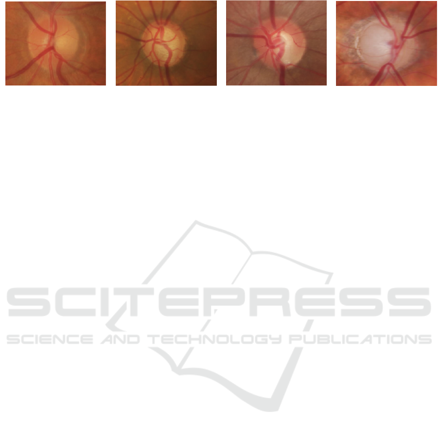
(a) (b) (c) (d)
Figure 1: Glaucoma in retinal fundus images. (a)normal disc, (b)glaucoma onset, (c)critical glaucoma, (d)advance glaucoma.
tecting the optic disc, is helpful for clinical evaluation
by the ophthalmologists. Nevertheless, automated op-
tic disc segmentation methods that used for glaucoma
detection should be sensitive. Cause a small error in
recognize Of optic disc may affect the diagnosis and
treatment seriously (Mookiah et al., 2012). Image
segmentation is a fundamental part of many optical
understanding systems that includes division images
into numerous segments or regions (Szeliski, 2010).
Image segmentation has been used in many applica-
tions, especially in medical image analysis such as
tumor boundary extraction and optic disc segmenta-
tion(Forsyth and Ponce, 2002). The other method for
optic disc segmentation is utilizing a novel vibrational
level set function on the red channel of the retinal fun-
dus images (Wong et al., 2008). In another algorithm,
localized the optic disc was applied by using template
matching; after that, morphological filtering removed
the blood vessels. At last, the boundary information
combines with the local edge vector to operate the de-
formable contour was used to detect the optic disc re-
gions (Yu et al., 2012; Zhang et al., 2008). For op-
tic disc segmentation task, a method based on math-
ematical morphology is proposed to detect and seg-
ment the optic disc in images (Welfer et al., 2010).
This method is expanded by combining a multiscale
morphological approach (Welfer et al., 2013). A
template-based approach for OD segmentation is uti-
lized edge detection and morphological methods con-
formed by circular Hough transformation to estimated
circular objects (Aquino et al., 2010). However, in the
past few years, deep learning networks have used as
a new efficiency method in image segmentation tasks
with an extraordinary performance that attaining the
highest accuracy and speed rates. The deep convo-
lutional neural network can extract indicated features
from the input images automatically. There are var-
ious models developed for medical image segmenta-
tion, which based on FCNs (Long et al., 2015) mod-
els. A U-shaped convolutional neural network was
proposed to segment optic disc, and advancement was
achieved by in comparison with the exert of old meth-
ods (Sevastopolsky, 2017). For optic disc segmenta-
tion task, the polar transformation and multi-label loss
function method were applied in a U-shaped (Fu et al.,
2018). The team extended this algorithm one year
later and suggested a Stack-U-Net network architec-
ture (Sevastopolsky et al., 2018), which is based on a
U-Net (Ronneberger et al., 2015) network. The other
optic disc segmentation method has a U-Shape with
Densely connected convolutional blocks (Al-Bander
et al., 2018), based on DenseNet (Huang et al., 2017).
In optic disc segmentation tasks, low time and high
accuracy are essential. In this paper, a network de-
signed based on deep learning and segmented the op-
tic disc with the low time, which can help the oph-
thalmology clinic to evaluate the retina disease like
glaucoma.
2 THE PROPOSED METHOD
This paper develops a deep learning algorithm for op-
tic disc segmentation and designs a new network ar-
chitecture based on a residual model. In this paper, a
new approach called residual-like convolutional neu-
ral network applied for optic disc segmentation in reti-
nal color fundus images. In the structure of this net-
work, there are some layers which based on residual
layers in ResNet (He et al., 2016). For optic disc seg-
mentation, in pixel-level where features can be take
out from various sized windows, but at the identical
time, passing some features from first layers to resul-
tant layers, as residual layers do, should be useful.
Residual block structure seems to be suitable for such
roles when needed to construct data that be similar
to the input image. Another benefit of using residual
blocks is the amplify and improved gradient circula-
tion, which has a positive effect on network conver-
gence. Figure 2 is the residual layer that proposed in
this paper. As shown in Figure 1 in this paper, in this
convolutional network, the RELU activation function
is used after the end of each convolution layer, and
after that batch normalization is used. The Relu func-
tion gives an output x if x is positive and 0 otherwise.
Also, the Sigmoid activation function is applied to the
DeLTA 2020 - 1st International Conference on Deep Learning Theory and Applications
24
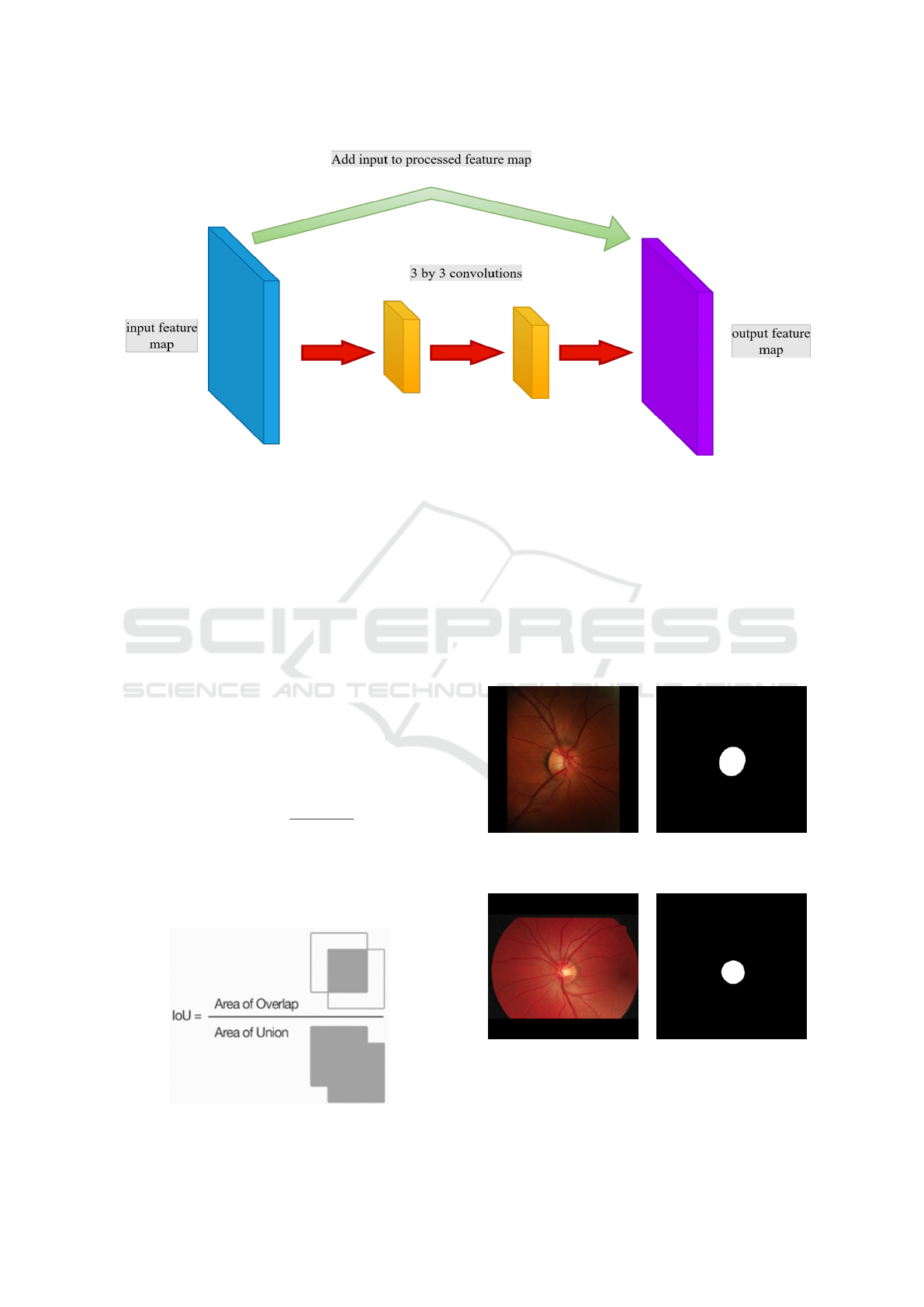
Figure 2: Residual layer used in proposed network.
last layer. As shown in Figure 2 the network at first,
extracts the features from an input image and then up-
samples the feature map. In this paper, the output of
the proposed network is a binary image shown in Fig-
ure 5. For evaluating the results, loss function defined
as:
l(A, B) = −logd (A, B) (1)
A is a predicted output, comprising probabilities
that each predicted pixel appertains to the foreground,
and also B is a correct binary output. For binary im-
ages d(A; B) is an expansion of Dice score. Dice
score calculates the expanse of overlapping regions
between any two images. Dice (Dice, 1945) to gauge
the similarity of two samples, such as image and
ground truth, is defined as:
Dice(A, B) =
2|A ∩ B|
(|A| + |B|)
(2)
The ranges value of the Dice coefficient is be-
tween 0 and 1. In this study RMSprop (Tieleman
and Hinton, 2012) optimizer is used with a learn-
ing rate of 0.0002. There are different datasets
Figure 3: Intersection-over-Union (IOU).
for optic disc segmentation. In this paper, we have
used two well-known datasets and then compare the
results of the proposed method with the other meth-
ods that have used these datasets. As shown in Fig-
ure 4 DRIONS-DB (Carmona et al., 2008) and RIM-
ONE v.3 (Fumero et al., 2011) (110 and 159 im-
ages,respectively) datasets used to evaluate the re-
sults, which comprise the manual segmentation of
the optic disc. Another parameter for the quality of
the trained algorithm evaluated by Intersection-over-
Original image Ground truth
(a) An example of DRIONS-DB dataset.
Original image Ground truth
(b) An example of RIM-ONE v.3 dataset
Figure 4: Some examples from RIM-ONE v.3 and
DRIONS-DB datasets.
Deep Learning Residual-like Convolutional Neural Networks for Optic Disc Segmentation in Medical Retinal Images
25
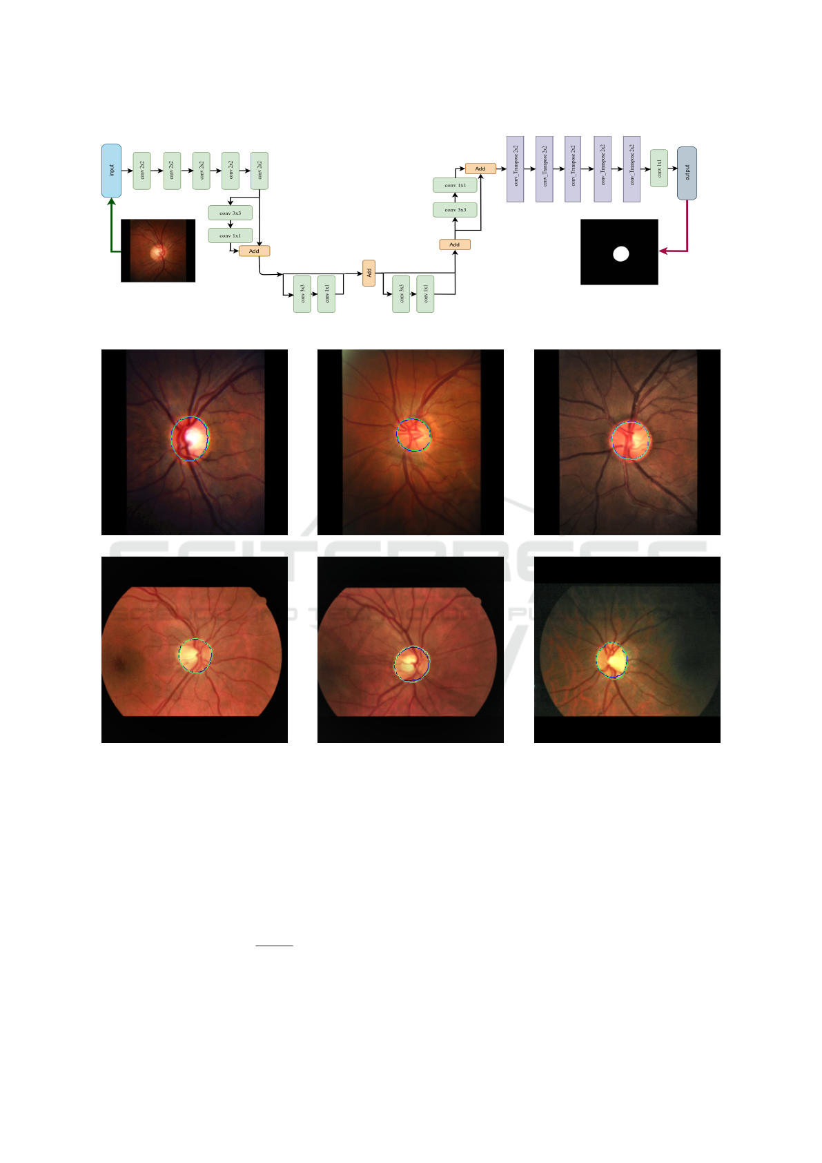
Figure 5: Proposed network structure.
(a)
(b)
Figure 6: (a) Examples from RIM-ONE v.3 for OD segmentation by the proposed method. The green contour refers to the
ground truth, and blue is prediction, (b) Examples from RIM-ONE v.3 for OD segmentation by the proposed method. The
green contour refers to the ground truth, and blue is prediction.
Union, as well as called the Jaccard Index, is one
of the well-known metrics in image segmentation.
Intersection-over-Union(IOU) is the region of overlap
between the ground truth and predicted segmentation,
and defined as:
IOU =
|A ∩ B|
|A ∪ B|
(3)
3 EXPERIMENTS AND RESULTS
The Dice coefficient and IOU score do not depend on
the object scale and image scale.
IOU is a score that used in image segmentation.
As shown in Figure 3 IOU score is necessary to eval-
uate the percentage of overlap between ground-truth
and predicted segmentation. IOU score is similar to
the Dice coefficient that often used for loss function
DeLTA 2020 - 1st International Conference on Deep Learning Theory and Applications
26

Table 1: Comparison of the proposed method with existing methods(on DRIONS-DB).
DRIONS-DB Dataset
Methods Dice IOU predict time (s)
Proposed method 0.9452 0.8853 0.008
(Walter et al., 2002) 0.6813 - -
(Morales et al., 2013) 0.9084 - -
(Abdullah et al., 2016) 0.9102 0.851 43.2
(Rehman et al., 2019) 0.8990 0.8210 31.10
(Zahoor and Fraz, 2017) - 0.8862 1.60
(Fan et al., 2017) 0.9137 0.8473 -
(Ramani and Shan-
thamalar, 2020)
0.8962 0.8217 1.41
DRIU (Maninis et al.,
2016)
0.94 0.89 0.1
(Sevastopolsky, 2017) 0.97 0.88 0.13
Table 2: Comparison of the proposed method with existing methods(on RIM-ONE v.3).
RIM-ONE v.3 Dataset
Methods Dice IOU predict time (s)
Proposed method 0.9371 0.87 0.008
(Zilly et al., 2017) 0.94 0.98 5.3
(Maninis et al., 2016) 0.96 0.89 0.13
(Joshua et al., 2019) 0.96 0.88 0.03
(Sevastopolsky, 2017) 0.95 0.89 0.1
(Civit Masot et al.,
2019)
0.97 - -
in the training network. IOU ranges from 0-1, which
1 (100) indicate fully overlapping segmentation. For
training this algorithm, we use free GPU service of
the Google Colab framework.
4 CONCLUSIONS
In this paper, we prove that our algorithm based on
Residual-like CNN can detect OD in shorter time,
better than other reported methods on retinal fun-
dus images. The best preponderance of the proposed
method, simple programming, accurate, and the low-
est prediction time, which is 0.008 per second. The
results by IOU and DICE scores were evaluated and,
great performances for optic disc segmentation were
achieved. The lowest prediction time and experiment
results express that optic disc segmentation can be
done automatically in ophthalmology clinics as on-
line predictions medical assistive tool.
ACKNOWLEDGEMENTS
The authors thanks Mr.Hooman Misaghi for his sup-
port and helps.
REFERENCES
Abbas, Q. (2017). Glaucoma-deep: detection of glaucoma
eye disease on retinal fundus images using deep learn-
ing. Int J Adv Comput Sci Appl, 8(6):41–5.
Abdullah, M., Fraz, M. M., and Barman, S. A. (2016). Lo-
calization and segmentation of optic disc in retinal im-
Deep Learning Residual-like Convolutional Neural Networks for Optic Disc Segmentation in Medical Retinal Images
27

ages using circular hough transform and grow-cut al-
gorithm. PeerJ, 4:e2003.
Al-Bander, B., Williams, B. M., Al-Nuaimy, W., Al-Taee,
M. A., Pratt, H., and Zheng, Y. (2018). Dense fully
convolutional segmentation of the optic disc and cup
in colour fundus for glaucoma diagnosis. Symmetry,
10(4):87.
Aquino, A., Geg
´
undez-Arias, M. E., and Mar
´
ın, D. (2010).
Detecting the optic disc boundary in digital fundus im-
ages using morphological, edge detection, and feature
extraction techniques. IEEE transactions on medical
imaging, 29(11):1860–1869.
Bajwa, M. N., Malik, M. I., Siddiqui, S. A., Dengel, A.,
Shafait, F., Neumeier, W., and Ahmed, S. (2019).
Two-stage framework for optic disc localization and
glaucoma classification in retinal fundus images using
deep learning. BMC medical informatics and decision
making, 19(1):136.
Bourne, R. R., Flaxman, S. R., Braithwaite, T., Cicinelli,
M. V., Das, A., Jonas, J. B., Keeffe, J., Kempen, J. H.,
Leasher, J., Limburg, H., et al. (2017). Magnitude,
temporal trends, and projections of the global preva-
lence of blindness and distance and near vision im-
pairment: a systematic review and meta-analysis. The
Lancet Global Health, 5(9):e888–e897.
Carmona, E. J., Rinc
´
on, M., Garc
´
ıa-Feijo
´
o, J., and
Mart
´
ınez-de-la Casa, J. M. (2008). Identification of
the optic nerve head with genetic algorithms. Artifi-
cial Intelligence in Medicine, 43(3):243–259.
Chen, X., Xu, Y., Yan, S., Wong, D. W. K., Wong, T. Y.,
and Liu, J. (2015). Automatic feature learning for
glaucoma detection based on deep learning. In In-
ternational Conference on Medical Image Computing
and Computer-Assisted Intervention, pages 669–677.
Springer.
Civit Masot, J., Luna Perej
´
on, F., Dur
´
an L
´
opez, L.,
Dom
´
ınguez Morales, J. P., Vicente D
´
ıaz, S.,
Linares Barranco, A., and Civit Balcells, A. (2019).
Multi-dataset training for medical image segmentation
as a service. In UCCI 2019: 11th International Joint
Conference on Computational Intelligence (2019), p
542-547. ScitePress Digital Library.
Dice, L. R. (1945). Measures of the amount of ecologic
association between species. Ecology, 26(3):297–302.
Fan, Z., Rong, Y., Cai, X., Lu, J., Li, W., Lin, H., and Chen,
X. (2017). Optic disk detection in fundus image based
on structured learning. IEEE journal of biomedical
and health informatics, 22(1):224–234.
Federation, I. (2013). International diabetic federation atlas;
chapter 2 the global burden.
Forsyth, D. A. and Ponce, J. (2002). Computer vision: a
modern approach. Prentice Hall Professional Techni-
cal Reference.
Fraz, M. M., Welikala, R., Rudnicka, A. R., Owen, C. G.,
Strachan, D., and Barman, S. A. (2015). Quartz:
Quantitative analysis of retinal vessel topology and
size–an automated system for quantification of reti-
nal vessels morphology. Expert Systems with Appli-
cations, 42(20):7221–7234.
Fu, H., Cheng, J., Xu, Y., Wong, D. W. K., Liu, J., and Cao,
X. (2018). Joint optic disc and cup segmentation based
on multi-label deep network and polar transformation.
IEEE transactions on medical imaging, 37(7):1597–
1605.
Fumero, F., Alay
´
on, S., Sanchez, J. L., Sigut, J., and
Gonzalez-Hernandez, M. (2011). Rim-one: An open
retinal image database for optic nerve evaluation.
In 2011 24th international symposium on computer-
based medical systems (CBMS), pages 1–6. IEEE.
Gao, Y., Yu, X., Wu, C., Zhou, W., Wang, X., and Chu, H.
(2019). Accurate and efficient segmentation of optic
disc and optic cup in retinal images integrating multi-
view information. IEEE Access, 7:148183–148197.
He, K., Zhang, X., Ren, S., and Sun, J. (2016). Deep resid-
ual learning for image recognition. In Proceedings of
the IEEE conference on computer vision and pattern
recognition, pages 770–778.
Huang, G., Liu, Z., Van Der Maaten, L., and Weinberger,
K. Q. (2017). Densely connected convolutional net-
works. In Proceedings of the IEEE conference on
computer vision and pattern recognition, pages 4700–
4708.
Jackson, A. and Radhakrishnan, S. (2014). Understanding
and living with glaucoma. Glaucoma Research Foun-
dation.
Joshua, A. O., Nelwamondo, F. V., and Mabuza-Hocquet,
G. (2019). Segmentation of optic cup and disc
for diagnosis of glaucoma on retinal fundus im-
ages. In 2019 Southern African Universities Power
Engineering Conference/Robotics and Mechatron-
ics/Pattern Recognition Association of South Africa
(SAUPEC/RobMech/PRASA), pages 183–187. IEEE.
Long, J., Shelhamer, E., and Darrell, T. (2015). Fully con-
volutional networks for semantic segmentation. In
Proceedings of the IEEE conference on computer vi-
sion and pattern recognition, pages 3431–3440.
Maninis, K.-K., Pont-Tuset, J., Arbel
´
aez, P., and Van Gool,
L. (2016). Deep retinal image understanding. In In-
ternational conference on medical image computing
and computer-assisted intervention, pages 140–148.
Springer.
Mookiah, M. R. K., Acharya, U. R., Lim, C. M., Petznick,
A., and Suri, J. S. (2012). Data mining technique
for automated diagnosis of glaucoma using higher or-
der spectra and wavelet energy features. Knowledge-
Based Systems, 33:73–82.
Morales, S., Naranjo, V., Angulo, J., and Alca
˜
niz, M.
(2013). Automatic detection of optic disc based on
pca and mathematical morphology. IEEE transactions
on medical imaging, 32(4):786–796.
Quigley, H. A. and Broman, A. T. (2006). The number of
people with glaucoma worldwide in 2010 and 2020.
British journal of ophthalmology, 90(3):262–267.
Ramani, R. G. and Shanthamalar, J. J. (2020). Improved im-
age processing techniques for optic disc segmentation
in retinal fundus images. Biomedical Signal Process-
ing and Control, 58:101832.
Rehman, Z. U., Naqvi, S. S., Khan, T. M., Arsalan, M.,
Khan, M. A., and Khalil, M. (2019). Multi-parametric
DeLTA 2020 - 1st International Conference on Deep Learning Theory and Applications
28

optic disc segmentation using superpixel based fea-
ture classification. Expert Systems with Applications,
120:461–473.
Ronneberger, O., Fischer, P., and Brox, T. (2015). U-net:
Convolutional networks for biomedical image seg-
mentation. In International Conference on Medical
image computing and computer-assisted intervention,
pages 234–241. Springer.
Sevastopolsky, A. (2017). Optic disc and cup segmentation
methods for glaucoma detection with modification of
u-net convolutional neural network. Pattern Recogni-
tion and Image Analysis, 27(3):618–624.
Sevastopolsky, A., Drapak, S., Kiselev, K., Snyder, B. M.,
Keenan, J. D., and Georgievskaya, A. (2018). Stack-
u-net: Refinement network for image segmentation on
the example of optic disc and cup. arXiv preprint
arXiv:1804.11294.
Szeliski, R. (2010). Computer vision: algorithms and ap-
plications. Springer Science & Business Media.
Tieleman, T. and Hinton, G. (2012). Lecture 6.5-rmsprop:
Divide the gradient by a running average of its recent
magnitude. COURSERA: Neural networks for ma-
chine learning, 4(2):26–31.
Walter, T., Klein, J.-C., Massin, P., and Erginay, A. (2002).
A contribution of image processing to the diagnosis
of diabetic retinopathy-detection of exudates in color
fundus images of the human retina. IEEE transactions
on medical imaging, 21(10):1236–1243.
Welfer, D., Scharcanski, J., Kitamura, C. M., Dal Pizzol,
M. M., Ludwig, L. W., and Marinho, D. R. (2010).
Segmentation of the optic disk in color eye fundus im-
ages using an adaptive morphological approach. Com-
puters in Biology and Medicine, 40(2):124–137.
Welfer, D., Scharcanski, J., and Marinho, D. R. (2013). A
morphologic two-stage approach for automated optic
disk detection in color eye fundus images. Pattern
Recognition Letters, 34(5):476–485.
Wong, D., Liu, J., Lim, J., Jia, X., Yin, F., Li, H., and Wong,
T. (2008). Level-set based automatic cup-to-disc ratio
determination using retinal fundus images in argali.
In 2008 30th Annual International Conference of the
IEEE Engineering in Medicine and Biology Society,
pages 2266–2269. IEEE.
Xu, Y., Liu, J., Lin, S., Xu, D., Cheung, C. Y., Aung, T.,
and Wong, T. Y. (2012). Efficient optic cup detec-
tion from intra-image learning with retinal structure
priors. In International Conference on Medical Im-
age Computing and Computer-Assisted Intervention,
pages 58–65. Springer.
Yu, H., Barriga, E. S., Agurto, C., Echegaray, S., Pattichis,
M. S., Bauman, W., and Soliz, P. (2012). Fast lo-
calization and segmentation of optic disk in retinal
images using directional matched filtering and level
sets. IEEE Transactions on information technology in
biomedicine, 16(4):644–657.
Zahoor, M. N. and Fraz, M. M. (2017). Fast optic disc seg-
mentation in retina using polar transform. IEEE Ac-
cess, 5:12293–12300.
Zhang, Y., Matuszewski, B. J., Shark, L.-K., and Moore,
C. J. (2008). Medical image segmentation using new
hybrid level-set method. In 2008 fifth international
conference biomedical visualization: information vi-
sualization in medical and biomedical informatics,
pages 71–76. IEEE.
Zilly, J., Buhmann, J. M., and Mahapatra, D. (2017). Glau-
coma detection using entropy sampling and ensemble
learning for automatic optic cup and disc segmenta-
tion. Computerized Medical Imaging and Graphics,
55:28–41.
Deep Learning Residual-like Convolutional Neural Networks for Optic Disc Segmentation in Medical Retinal Images
29
