
Evaluation of Human Dissection in Anatomy Teaching using an
Interactive Simulator
William John Pereira Brobouski
1
, Andrei Rafael Brongel
1
, Fabiano Utiyama
1
,
M
´
arcia Cristina Dadalto Pascutti
1
, Mozart Gonc¸alves
1
, Jo
˜
ao Lu
´
ıs Verdegay de Barros
1
,
Carlos Jos
´
e Gomes
2
and Edson Jos
´
e Rodrigues Justino
3
1
Programa de P
´
os-Graduac¸
˜
ao em Inform
´
atica - PPGIa, PUCPR, Curitiba, Paran
´
a, Brazil
2
Medical School, Pontif
´
ıcia Universidade Cat
´
olica do Paran
´
a - PUCPR, Curitiba, Paran
´
a, Brazil
3
Centro de Inovac¸
˜
oes em Imagens M
´
edicas - CIIM, Pontif
´
ıcia Universidade Cat
´
olica do Paran
´
a - PUCPR,
carlos bio@yahoo.com.br, justino@ppgia.com
Keywords:
Visible Human Table, Simulator, Anatomy Dissection, Effective Learning.
Abstract:
The anatomy study is required in Life Science related courses. Nowadays, there are no instruments to replace
the real human bodies. Therefore, it was noticed the need of the development of a learning tool to assist the
anatomic study in laboratory which was able to dissect the organs for human anatomy study. Thus, the ultra-
high interactive simulator for human dissection (Visible Human Table – VHT) was created as a computational
tool for human anatomy study in classrooms. The main characteristic of the simulator is the fact that it is not
an anatomic atlas, but a dissection table based on real body models. That is what makes it stand out from
most same-purpose anatomic simulators and atlases. This article presents the results of a VHT evaluation
assessed by higher education learners in the health sector. The simulator promising potential becomes evident
through the answers obtained from the questionnaire, showing the significant contribution in the anatomy
teaching-learning process.
1 INTRODUCTION
The technological development has enabled a lot
of digital image processing advancements which al-
lowed for innovations and improvements in a wide
range of sectors. In relation to the health sector,
projects are developed to increase resources to help
both skilled professionals and learners during qualifi-
cation. The acquisition and image processing permit
the computational environment development in favor
of training, medical diagnosis and treatment as well
as, the anatomy teaching helps students and teach-
ers in the teaching-learning process (Beveridge et al.,
2013).
The study of anatomy is crucial in many courses
related to health. There is no book or computational
tool to replace the study of the real human body. The
corpse is the most similar to a living human being
the student can have as a study object. However,
educational institutions have been having difficulties
in obtaining bodies for this purpose due to the lack
of data from the population about the possibility of
corpse donation (Bassete, 2009) and legal procedures
involved.
A possible solution is the digital visualization of
this information; however, because of the complex-
ity of organic structures, the process of digital visu-
alization in high definition and the segmentation of
organs and human body systems is a complex task
and requires high computational capacity. From an
anatomic point of view, the division or separation of
systems is needed for the perfect understanding of its
parts. In the health sector, it is also important that
computational tools exhibit realistic models of human
body. Even though there are anatomy tools that allow
human dissection available, they present synthetic ar-
tifacts of the body that do not express the real color
and texture.
Based on this scenario, it was noticed the ne-
cessity of developing a learning tool to serve the
anatomic study needs in laboratory through the visu-
alization of artifacts in high resolution and with re-
alistic colors and textures. They would perform pro-
cedures such as the dissection of the body to assist
396
Brobouski, W., Brongel, A., Utiyama, F., Pascutti, M., Gonçalves, M., de Barros, J., Gomes, C. and Justino, E.
Evaluation of Human Dissection in Anatomy Teaching using an Interactive Simulator.
DOI: 10.5220/0009393103960403
In Proceedings of the 12th International Conference on Computer Supported Education (CSEDU 2020) - Volume 1, pages 396-403
ISBN: 978-989-758-417-6
Copyright
c
2020 by SCITEPRESS – Science and Technology Publications, Lda. All rights reserved

students in their learning process.
This article presents the virtual dissection evalua-
tion in an ultra-high-definition simulator for human
anatomy teaching in the classroom. The simulator
consists of hardware and software developed for this
purpose (Brongel. et al., 2019). The device seeks to
respond the requirements of representation of high-
quality models, visual acuity, color, texture, depth
perception and touch interactivity.
This article is structured as follows. The related
works are described in Section 2. Section 3 presents
how VHT was implemented based on the data used
in it and the use of a tool in active methodologies for
anatomy teaching. The method used to evaluate is
described in Section 4. Section 5 shows the results
of this evaluation which was carried out by students
from degree courses using the simulator. Section 6
reports the conclusions.
2 RELATED WORKS
The project known as The Voxel-Man, uses the low-
resolution database developed by the Hamburg Uni-
versity. It was known as The Segmented Inner Organs
(SIO) (Voxel-Man, 2019). This image database is
synthetic and was created by computational addition
and corrections from Visible Human Database. Syn-
thetic images result in some problems for anatomic
study such as, quality reduction in representation be-
cause of the manual corrections which do not repre-
sent the real organic structures.
Another example is the Anatomage Table. It is in
the seventh version and it has some colored bases in-
cluding the VHP base (Anatomage Inc., 2018). So,
as the models are rendered, they are committed to
the quality resulting in more artificial appearance of
the structures. A comparative study was carried, to
evaluate the qualitative efficiency and experience of
the learning of pelvis and perineum and skeletal mus-
cle system general anatomy by cadaveric dissection.
The purpose of this study was to learn these same
anatomic parts using Anatomage Table whose results
showed that students were more motivated and no-
ticed greater learning (Baratz et al., 2019). Other
study about this table highlighted that there is a re-
duction in the use of the cadaver in teaching and it re-
vealed the importance of the cutting tool (Fyfe et al.,
2013).
Other environment developed for teaching
through stereoscopy is presented by (Olsen. et al.,
2018), in which the acquisition of the images is done
by means of Full Frame Semi-spherical Scanner
(F2S2). And the learning objects can be used in
different knowledge fields promoting an educational
environment with interaction and dynamism. An
evaluation of the learning impact of the F2S2 was
carried, and the results showed that the interactive
material is an alternative which can motivate teachers
and students because they consider the visualization
represents the real object properly and it can help in
teaching-learning process (Silva et al., 2019).
3 MATERIAL
For the simulator development was need acquisition
and segmentation of color images, a hardware which
attended on the requirements of usability and perfor-
mance which enabled to accommodate the totality of
the bases of The Visible Human Project, besides an
interactive software which made feasible significant
learning providing the teacher an active teaching tool.
3.1 Database
The database used in the simulator comes from The
Visible Human Project made available on November
28, 1994 for the male body and one year later, the
female body images (Ackerman, 1998) and (Spitzer
et al., 1996). The base is composed of Magnetic Res-
onance Imaging (MRI), Computed Tomography (CT)
and color images. For the male body, the MRI images
were acquired in 6 subsets: head in the axial form and
chest, abdomen, pelvis, thighs and foot in the coro-
nal form. The images were acquired in three different
modalities, T1, T2 and proton density images, shown
in Figure 1 (a,b,c) with resolution of 256x256 pixels
at 4-mm-intervals (Ackerman, 1998).
Figure 1 shows a CT with a resolution of 512x512
at 1-mm-intervals in the axial plane, and Figure 1
(e) displays a color axial image in 2048x1216 pixels
(2k). Each high-definition image takes 7.5 megabytes
with 1871 color images, all available to be used in re-
search. The whole process of image acquisition and
body preparation to generate the database is described
in (Spitzer et al., 1996).
The female data set presents a larger amount of
images than the male ones with 5189 images, due to
the 0,33-mm-intervals with a resolution of 2048x1216
pixels (2k) and 4096x3061 pixels (4k). Figure 2
shows the images that illustrate the male and female
bodies reconstituted in the coronal plane. The process
of freezing the cadavers was performed by blocks and
they did not exceed 51 cm in length. So, it is ob-
served that the images presented are not continuous
(U.S. National Library of Medicine, 2019).
Evaluation of Human Dissection in Anatomy Teaching using an Interactive Simulator
397
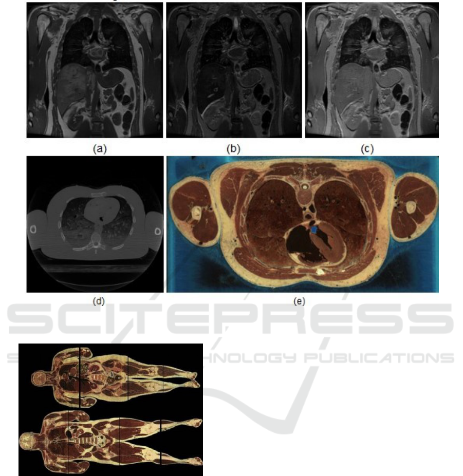
Figure 1: Images available on the website of The Visible Human Project: (a) MRI T1-; (b)MRI T2-; (c) proton density images;
(d) CT; (e) color axial image (U.S. National Library of Medicine, 2019).
Figure 2: Reconstruction of male and female bodies of The
Visible Human Project (Ackerman, 1998).
3.2 Interactive Simulator
The interactive dissection simulator for anatomy
study is consisted of two parts, hardware and soft-
ware. The hardware is formed by a computer with se-
lected components with the purpose of executing the
work satisfactorily. To attend the requirements of us-
ability in anatomy classes, it is presented dynamically
and it has a user-friendly interface. It also has an in-
teractive screen that supports resolutions of up to 4k
and about 1.70-m-human body, as shown in Figure 3.
Concerning the software, it is an application with
support for input by multiple touches on the screen,
enabling the dynamism and interactivity desired in a
computational application. The system has two main
areas, shown in Figure 4. In the first one the hu-
man body is visualized in a volumetric way and in
the second one the image in ultra-high definition of
the anatomical plane is selected by the user.
The interface between the user and the application
is made, mainly, with a dynamic dial, shown in Figure
5. It operates as a virtual mouse and can be positioned
anywhere on the screen to enable the mobility around
the high-resolution table.
The dial includes several tools. They allow the ex-
change of the visualization plane, manipulation and
selection of the volumetric model. Also the creation
of notes by means of a digital chart and content upload
to the study environment of the institution through In-
ternet.
Also in Figure 5, it is possible to observe that the
interaction with the images in an ultra-high-definition
is made through buttons and colored lines. They can
be dragged to apply anatomical cuts in the volumetric
model and to determine the cut displayed in the main
frame.
CSEDU 2020 - 12th International Conference on Computer Supported Education
398
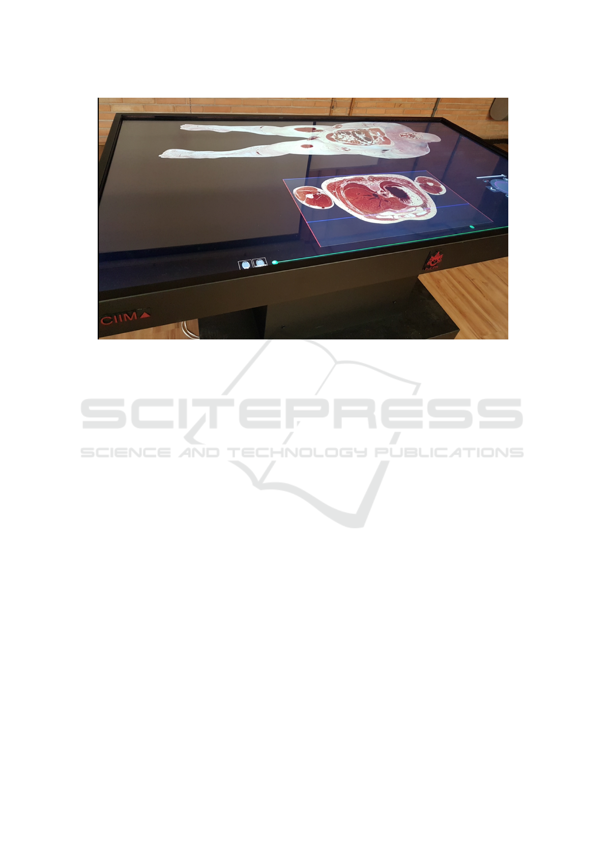
Figure 3: Visible Human Table.
For each anatomic plane, there are indexed win-
dows of MRI and CT scans which can be maximized
and positioned at any point on the screen creating the
desired corresponding association with the color im-
ages.
3.3 Active Learning
The Educational field is going through major changes
especially, concerning conceptions and teaching tech-
niques. So, new teaching-learning process under-
standing and alternative teaching operationalization
proposals have been elaborated. It is considered they
must admit an ethic, critic, reflexive and transform-
ing pedagogic practices, transposing merely technical
training limits to reach the man education as a historic
being (Mitre et al., 2008) and (Paiva et al., 2016).
Students must read, write, ask, discuss or engage
on problems solving and projects development in or-
der to make the active learning process happen. They
are encouraged to build the knowledge instead of re-
ceiving it from the teacher in a passive way. Thus, the
active learning strategies can be described as activi-
ties which keep students occupied in doing something
and, at the same time, help students think about what
is being done (Bonwell and Eison, 1991) and (Silber-
man, 1996). In an active learning environment, the
teacher acts as mentor, supervisor, learning process
facilitator and not just as the single source of infor-
mation and knowledge.
The active methodologies, serving to this educa-
tional revision, consist of alternatives to teaching-
learning process with benefits and challenges.
Flipped Classroom, Blended Learning, Peer Instruc-
tion, PBL (Problem Based Learning), GTD (Get-
ting Things Done), Think-pair-share, Just-in-time
teaching this is called “Customized Teaching” by
(Elm
ˆ
or Filho et al., 2019). They are some of the active
methodologies application possibilities. They aim to
put students in the middle of the learning process in
order to develop their own autonomy.
An active learning environment should create the
possibility to apply activities which will develop crit-
ical reflections through discussions to promote the
construction of meanings to an enduring learning.
Thus, students can understand the skills and compe-
tences acquisition needed to deal with professional
challenges of their training area. Apprenticeship
learning is built in action whose spontaneity is en-
hanced when it has meaning and it is noticed as neces-
sary (Piaget and Caixeiro, 1983). In the face of these
conditions, it is possible to raise questions to provoke
reflections because knowledge is built when students
appropriate of their thoughts.
In this respect, the interactive simulator fits per-
fectly because it allows the exploratory study of a
real human body anatomy. It is possible to visualize
image details with visual acuity, organs scale com-
pliance, texture, color and depth perception that are
not obtained at anatomy laboratories. Consequently,
students are stimulated to build knowledge instead of
receiving it from the teacher passively. The simula-
tor enables the teacher organize exploratory activities
Evaluation of Human Dissection in Anatomy Teaching using an Interactive Simulator
399
Figure 4: Interface of the Visible Human Table (Brongel. et al., 2019).
Figure 5: Dial visualization, volumetric section, and MRI
and CT exams.
into anatomy studies or problem resolution through
organs or human body systems observation whose re-
sults come from discussions involving all students.
Among many options which can also be con-
sidered in future works with the simulator usage,
the flipped classroom approach was chosen for this
work. Its application process is divided in three
stages: pre-assignment activities, pre-activities and
post-activities. These stages are applied to the pro-
posed experiment using the simulator. First, students
did previous research of anatomy contents. Second,
students had contact with the simulator under teacher
supervision, whereby it was possible to identify char-
acteristic such as color, depth, texture and anatomic
arrangement of previously studied organs. Third, they
were inquired about their perception of the simula-
tor’s contribution to the learning process. The experi-
ment’s results are presented in the following section.
4 METHOD
The simulator was evaluated during the anatomy
classes by students from different courses, such as
Biological Sciences, Dentistry, Physical Education,
Physiotherapy, Nursing and Nutrition. The flipped
classroom approach was chosen and the three stages
were carried out with teacher supervision. In the first
stage previously mentioned, students carried out stud-
ies on anatomy contents to recognize the organs, their
characteristics and spatial location in the human body
using a cadaver. On the stage, students explored the
resources of VHT visualizing images in the several
ways the environment allows. They applied cuts all
organs, checked colors and textures during the class.
The final stage, after all simulator functionalities were
presented to students, they were supposed to answer
an online questionnaire evaluating their experience
with the anatomy learning tool usage as after class
activity. Figure 6 shows in which course each student
respondent was.
The questionnaire above was composed by 7
close-ended questions and a five-point Likert scale
(Likert, 1932) with the following choices: I totally
agree (TA), partially agree (PA), indifferent (IN), par-
tially disagree (PD) and totally disagree (TD). The
questionnaire was applied through Google Forms in
order to obtain the degree of satisfaction and learn-
ing from students. According to (Wainerman, 1976),
the measurement instrument proposed by Likert aims
to verify the agreement level of the subject with sev-
CSEDU 2020 - 12th International Conference on Computer Supported Education
400
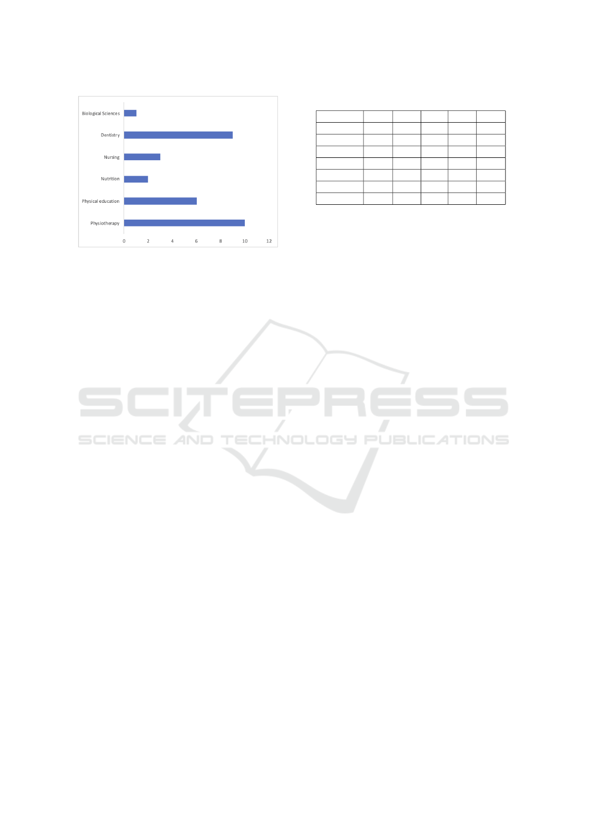
Figure 6: Distribution of students by course.
eral statements which express something favorable or
unfavorable in relation to an object. The questions
elaborated for the applied questionnaire were:
1. Would you use the digital table for further study
outside of school hours?
2. Do you consider that the corpses available on the
digital table contribute towards the proper assimi-
lation of organ positioning in the human body and
understanding of their structures, scale, color and
texture?
3. Do you believe that the possibility of visualizing
various axial, sagittal and coronal views using the
digital table contributes to your learning in your
anatomy studies?
4. Do you believe that the increased visual magni-
tude provided by the digital table provides a better
understanding of the structural details of an organ
and its relationship to others?
5. Do anatomical studies using only the digital table
meet your learning expectations?
6. Does the anatomy class using the digital table
combined with other resources available in the
anatomy lab contribute positively to your learn-
ing?
7. Does the use of manipulable volumetric digital
models of real bodies, such as the digital table
contribute positively to your learning?
Data collection was carried out on June, 2019 and
the online questions were answered by students vol-
untarily.
5 EVALUATION AND RESULTS
Table 1 shows the number of answers counted in each
proposition. The sum of the values should result in
the total of the participants.
Table 1: Survey Data.
Question T.A. P.A. I.N. P.D. T.D.
1 24 7 0 0 0
2 21 7 1 2 0
3 30 0 1 0 0
4 23 8 0 0 0
5 9 9 0 9 4
6 27 3 1 0 0
7 27 2 1 1 0
The result of the first question showed that the
virtual table is considered a very useful resource for
complementary studies outside of class hours by the
students. The answers highlighted an index of 77%
approval of the table use in this sense.
The results related to the second question indi-
cated that 67% of students totally agree that the bodies
available on the table contribute to the assimilation of
the structure, scale, color and texture of the human
body organs.
Third question showed that the visualization in the
axial, sagittal and coronal planes available in the tool
contributes to the learning in the anatomy studies with
96% of respondents totally agreeing with the affirma-
tion.
The possibility of increasing the visual magnitude
provided by the digital table is understood as another
benefit for learning. With this functionality it is easier
to visualize small structures. In this respect 74% of
the students completely approved this feature..
Students were asked if their learning expectations
of human anatomy would be met only when they used
the digital table. It was observed that 29% of them
partially disagree and 13% totally disagree, indicat-
ing how important the simulator is to complement the
anatomy learning. It was noticed that the real human
body is still needed and the table is not intended to
replace its usage. Although, it was also observed that
97% of all answers were in favor of using the table
as a complement. So, for students, the combined us-
ages of the table with the anatomy laboratories make
the learning more efficient, they also highlighted the
interactive simulator usage importance to the learning
process.
Finally, the results of question 7 attest that the ma-
nipulable volumetric models of the table assist in the
assimilation of the contents with 87% of the respon-
dents fully agreed. Figure 7 shows the percentages of
each answer per questions answered.
The answers indicated with TC and PC, that is to-
tal and partial agreement, when they were compared
to the other options, in general, demonstrate that the
data showed low disagreement in relation to the use
of the simulator, in other words most students agree
Evaluation of Human Dissection in Anatomy Teaching using an Interactive Simulator
401
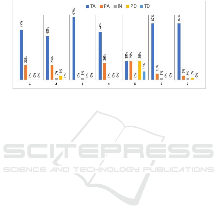
Figure 7: Percentages of each answer per question answered.
that the computer tool presented to them, the VHT is
promising for the learning process in human anatomy
studies.
6 CONCLUSIONS
This article presented the evaluation of an ultra-high-
definition simulator called VHT. Its purpose is for
dissection and teaching anatomy in classroom. The
difference from other anatomical tools and atlases is
the use of ultra-high-definition images and the use of
real human body images which provides visual acu-
ity, scale conformity with real human organs, texture,
color and depth perception.
The simulator complies with the precepts used in
the study of anatomy, that is, it presents the anatom-
ical planes of cut, visualization in the dissection pro-
cess. Another essential element of this learning tool
is the availability of imaging exams associated with
anatomical slices of images, the connectivity with
other online tools and the active blackboard.
The research also reported an experience with un-
dergraduate students which have the anatomy stud-
ies as a regular discipline. The flipped classroom
approach was used in order to evaluate the simu-
lator usage on the learning process. The results
were achieved through close-ended-questions analy-
sis based on the Likert scale. The questionnaire was
answered by students at the third stage of the method-
ology application.
The Flipped classroom use with the simulator re-
source in anatomy studies has created an appropri-
ate environment for knowledge construction in which
students identified organs studied in previous activ-
ities and raised questions, discussed and discovered
organs that were near each other on their own as it
was expected. It was possible just because it is a real
human body in size, shape, color and anatomical ar-
rangement like a living body. And, because of the
easy usage of the table provided by the interface gen-
erated by the dial whose usage was extremely sim-
ple e intuitive. The magnetic resonance and comput-
erized tomography images integration were also ele-
ments that draw attention due to the easy access to
perform anatomy studies.
The research has indicated the simulator is a
promising tool in active teaching-learning method-
ologies. It was also observed the tool is not sup-
posed to replace the human body dissection, but to
complement anatomic studies. It was also consid-
ered this resource can be extra-class used, outside of
school hours. It allows students to explore the human
body and do research. The answers received from the
questionnaire show that with the simulator usage in
a Flipped classroom, there was a contribution in the
anatomy teaching-learning process.
ACKNOWLEDGEMENTS
We thank to the US National Library of Medicine for
the acquisition and distribution of images The Visible
Human Body Project and The Voxel-Man Group for
granting segmented images which have contributed
to some studies and researches, the Financiadora de
Estudos e Projetos (FINEP) for project funding and
the Center of Innovation in Medical Imaging of the
PUCPR (CIIM) for project management.
CSEDU 2020 - 12th International Conference on Computer Supported Education
402

REFERENCES
Ackerman, M. J. (1998). The visible human project. In
Proceedings of the IEEE, volume 86, pages 504–511.
IEEE.
Anatomage Inc. (2018). Anatomage 3D
Anatomy — Anatomage Medical Home.
https://www.anatomage.com/anatomage-medical.
Last accessed 30 November 2019.
Baratz, G., Wilson-Delfosse, A. L., Singelyn, B. M., Allan,
K. C., Rieth, G. E., Ratnaparkhi, R., Jenks, B. P., Carl-
ton, C., Freeman, B. K., and Wish-Baratz, S. (2019).
Evaluating the anatomage table compared to cadaveric
dissection as a learning modality for gross anatomy.
Medical Science Educator, 29(2):499–506.
Bassete, F. (2009). ”doei meu corpo para o ensino da medic-
ina”, conta ex-taxista. https://www1.folha.uol.com.br/
ciencia/2009/04/553079-doei-meu-corpo-para-o-ensi
no-da-medicina-conta-ex-taxista.shtml. Last accessed
19 November 2019. Folha de S
˜
ao Paulo - Ci
ˆ
encia.
Beveridge, E., Ma, M., Rea, P., Bale, K., and Anderson, P.
(2013). 3d visualisation for education, diagnosis and
treatment of lliotibial band syndrome. In Proceedings
of the IEEE International Conference on Computer
Medical Applications - ICCMA, pages 1–6, Sousse,
Tunisia. IEEE.
Bonwell, C. C. and Eison, J. A. (1991). Active learning:
creating excitement in the classroom. ASHE-ERIC
Higher Education Reports, The George Washington
University, One Dupont Circle, Suite 630, Washing-
ton, DC 20036-1183.
Brongel., A. R., Brobouski., W. J. P., Pierin., L. M., Gomes.,
C., de Campos Almeida., M., and Justino., E. J. R.
(2019). An ultra-high definition and interactive sim-
ulator for human dissection in anatomic learning. In
Proceedings of the 11th International Conference on
Computer Supported Education - Volume 2: CSEDU,
pages 284–291. INSTICC, SciTePress.
Elm
ˆ
or Filho, G., Sauer, L. Z., de Almeida, N., and
Villas-Boas, V. (2019). Uma Nova Sala de Aula
´
e
Poss
´
ıvel: aprendizagem ativa na educac¸
˜
ao em engen-
haria. LTC, Rio de Janeiro.
Fyfe, G., Fyfe, S., Dye, D., and Crabb, H. (2013). Use
of anatomage tables in a large first year core unit.
In ASCILITE-Australian Society for Computers in
Learning in Tertiary Education Annual Conference,
pages 298–302. Australasian Society for Computers
in Learning in Tertiary Education.
Likert, R. (1932). A technique for the measurement of atti-
tudes. PhD thesis, Columbia University, New York.
Mitre, S. M., Siqueira-Batista, R., Girardi-de Mendonc¸a,
J. M., Morais-Pinto, N. M. d., Meirelles, C. d. A. B.,
Pinto-Porto, C., Moreira, T., and Hoffmann, L. M. A.
(2008). Metodologias ativas de ensino-aprendizagem
na formac¸
˜
ao profissional em sa
´
ude: debates atuais.
Ci
ˆ
encia & sa
´
ude coletiva, 13:2133–2144.
Olsen., D. R., de Almeida e Silva., F., Pierin., L. M.,
Moraes., A. H., and Justino., E. J. R. (2018). Genera-
tion of stereoscopic interactive learning objects true to
the original object. In Proceedings of the 10th Inter-
national Conference on Computer Supported Educa-
tion - Volume 1: CSEDU, pages 259–266. INSTICC,
SciTePress.
Paiva, M. R. F., Parente, J. R. F., Brand
˜
ao, I. R., and
Queiroz, A. H. B. (2016). Metodologias ativas de
ensino-aprendizagem: revis
˜
ao integrativa. SANARE-
Revista de Pol
´
ıticas P
´
ublicas, 15(2).
Piaget, J. and Caixeiro, N. (1983). A Epistemologia
gen
´
etica ; Sabedoria e ilus
˜
oes da filosofia ; Problemas
de psicologia gen
´
etica. Os Pensadores. Abril Cultural,
S
˜
ao Paulo, 2nd edition.
Silberman, M. L. (1996). Active Learning: 101 Strategies
To Teach Any Subject. Allyn and Bacon, Boston.
Silva, F. d. A., Olsen., D. R., Pierin., L. M., Bortolozzi., F.,
and Justino., E. J. R. (2019). Stereoscopic interactive
objects: Acquisition, generation and evaluation. In
Proceedings of the 11th International Conference on
Computer Supported Education - Volume 2: CSEDU,
pages 165–176. INSTICC, SciTePress.
Spitzer, V., Ackerman, M. J., Scherzinger, A. L., and Whit-
lock, D. (1996). The visible human male: a technical
report. Journal of the American Medical Informatics
Association, 3(2):118–130.
U.S. National Library of Medicine (2019). The visible hu-
man project - getting the data. https://www.nlm.nih.
gov/research/visible/getting data.html. Last accessed
01 December 2019.
Voxel-Man (2019). Segmented inner organs of the visi-
ble human. https://www.voxel-man.com/segmented-
inner-organs-of-the-visible-human/. Last accessed 01
December 2019.
Wainerman, C. (1976). Escalas de medici
´
on en ciencias
sociales. Ediciones Nueva Visi
´
on, Buenos Aires.
Evaluation of Human Dissection in Anatomy Teaching using an Interactive Simulator
403
