
Electroencephalography Registration of Laser Acupuncture Action
on Children with Autism Disorder
Anastasia I. Knyazkova
1,2 a
, Polina V. Shulmina
3
, Alice A. Samarinova
1
, Yury V. Kistenev
1,3 b
and Alexey V. Borisov
1,3 c
1
National Research Tomsk State University, Lenin Ave. 36, Tomsk, Russia
2
Institute of Strength Physics and Materials Science SB RAS, Academichesky Ave., 2/4, Tomsk, Russia
3
Siberian State Medical University, Moskovskiy trakt, 2, Tomsk, Russia
Keywords: EEG, Brain Activity, Laser Acupuncture, Autism Spectrum Disorders.
Abstract: Laser action on acupuncture points is an alternative to traditional acupuncture. We used red semiconductor
laser diodes with a wavelength of 650 nm. Three acupuncture points (GV20, LI4, P6) were selected to relieve
headaches and reduce anxiety in the study. EEG data had been obtained from two siblings, one of whom is
without pathologies of the nervous system (8 years), and the second has a diagnosis of autism (10 years). A
significant increase in the total activity of the brain for ASD patients up to a value close to the brain activity
of healthy patients was registered.
1 INTRODUCTION
Electroencephalography (EEG) is one of the most
informative methods of studying the human brain
from the standpoint of its holistic systemic activity.
This method is based on recording the total electrical
activity of brain neurons from the surface of the scalp.
EEG makes it possible to analyze qualitatively and
quantitatively the functional state of the brain and its
reactions under the influence of stimuli. EEG
recording is used widely in medical diagnostics and
treatment, in anesthesiology, as well as in the study of
brain activity related to the implementation of
functions such as perception, memory, and adaptation
(Louis et al. 2016).
Functional tests are of great importance in the
diagnosis of brain lesions: intermittent light irritation
(photostimulation), enhanced deep breathing for 2-3
minutes (hyperventilation), sound irritation, research
after a sleepless night (sleep deprivation), and others.
It is possible to identify changes in the EEG in
90% of patients with epilepsy using functional tests
(Dziadkowiak and Podemski 2019). The EEG allows
registering neoplasm transformation when the tumor
is located close to the surface of the brain and affects
a
https://orcid.org/ 0000-0002-1454-299X
b
https://orcid.org/ 0000-0001-5760-1462
c
https://orcid.org/ 0000-0003-1752-1649
mainly the cortex and subcortical structures. Local
pathological changes in the area of the projection of
the tumor are noted, such as inhibition of the alpha
rhythm, an increase in the amplitude of delta waves
(Roohi-Azizi et al. 2017). Intracerebral tumors cause
significant general changes in the EEG, masking
focal disorders of biopotentials.
Autism spectrum disorders (ASD) are a group of
lifelong disorders of the nervous system, and it is
believed to be the result of atypical neural
connections in the brain (Belmonte et al. 2004),
(Wang et al. 2013), (Assaf et al. 2010). Studies show
that ASD can be described as a dynamic disorder and
analyzed in terms of complex dynamic systems (Bosl
et al. 2011), (Megremi 2014). Changes in cortical
excitability may contribute to or be a manifestation of
disorders of connectivity (Boutros et al. 2015).
In (Duffy and Als 2019) showed that in patients
(N = 430 children) with a diagnosis of autism aged 2
to 12 years, the connections between different parts
of the brain are disrupted partially, which is reflected
in the EEG shape. To estimate the level of these
interactions, the authors have used the degree of
coordination (coherence) of the waves of electrical
activity in various areas of the brain. As a result of a
Knyazkova, A., Shulmina, P., Samarinova, A., Kistenev, Y. and Borisov, A.
Electroencephalography Registration of Laser Acupuncture Action on Children with Autism Disorder.
DOI: 10.5220/0009370503870394
In Proceedings of the 13th International Joint Conference on Biomedical Engineering Systems and Technologies (BIOSTEC 2020) - Volume 1: BIODEVICES, pages 387-394
ISBN: 978-989-758-398-8; ISSN: 2184-4305
Copyright
c
2022 by SCITEPRESS – Science and Technology Publications, Lda. All rights reserved
387

computer analysis of EEG signals, the authors
managed to identify 33 areas of wave combinations
characteristic only for autists, statistically different
from the EEG signals of children from the control
group, and this applies to all age categories.
Studies show that individual rehabilitation of a
person with autism has the most positive effect on his
abilities. There are various ways of influencing the
brain to rehabilitate the patient, including transcranial
magnetic stimulation (TMS), transcranial micro
polarisation (TCMP) and reflexotherapy.
The TMS, as a method for treating ASD, is based
on the stimulation of brain neurons with an
alternating magnetic field and recording responses to
stimulation using electromyography (Eldaief et al.
2013). The essence of this approach is the occurrence
of depolarization of the membranes of nerve cells
under the influence of a strong magnetic field. TMS
helps to regenerate neural connections in the cerebral
cortex, allowing purposeful stimulation non-
invasively individual structures of the cerebral cortex.
Depending on the regimen chosen by the specialist,
the effect on the central nervous system can be either
exciting or inhibitory. Regardless of the type of
influence, in the tissues of the cerebral cortex, there is
an improvement in the intercellular interaction and all
types of metabolism, and the blood microcirculation
is normalized. Some of the clinical trials of the
effectiveness of the TMS method suggested that its
use can help alleviate symptoms such as irritability
and stereotyped behavior, as well as reduce the
manifestation of autism symptoms associated with
deficiencies in areas of functioning and connections
such as coordination of vision and arm movement,
development of social skills (Oberman et al. 2016).
The TCMP of the brain consists of exposure to
certain parts of the brain with an electric current of
low intensity and is used in the comprehensive
rehabilitation of children with various forms of
cerebral palsy and other central nervous system
disorders, speech disorders, hearing loss, stuttering,
and so on. With the appointment of TCMP, the EEG
is recorded, based on the data of which the areas for
applying electrodes of the micro polarisation circuit
are determined. Thus, selective stimulation by
microcurrents of the weakened areas of the brain
responsible for the formation of speech, motor
activity, and mental development, allows achieving a
significant restoration of their functionality.
Reflexotherapy, as a method of rehabilitation in
autism, is based on exposure on biologically active
points located on the skin. As a result of exposure, a
local, regional or general reaction of the body is
caused, which leads to a restoration of balance in the
nervous, immune, endocrine systems, the production
of biologically active substances that block nerve
impulses and lead to pain relief, muscle relaxation,
stress relief, normalization of motor, autonomic and
emotional reactions in the body, regulation of blood
pressure.
One of the classic methods of reflexotherapy is
acupuncture, and one of the most innovative methods
is laser exposure to bioactive points. The main
objective of reflexotherapy in children with autism is
to strengthen the neuroendocrine links in the
regulation of autonomic tone and activation of the
subcortical formations of the brain. As a result of the
treatment, a decrease in stereotyped hyperkinesis and
phobias is observed, which helps to compensate for
the condition of children and their adaptation in the
family and children's team (Cheuk et al. 2011).
Laser action on acupuncture points is an
alternative to traditional acupuncture. Low-level laser
stimulation causes biological and physiological
changes. Using various frequencies of laser
stimulation was showed to cause activation of
different areas of the brain (Hsieh et al. 2011).
In the previous study (Knyazkova et al. 2019), we
showed that laser stimulation at the Hegu (LI4)
acupuncture point with low-level laser exposure (λ =
532 nm) entails a redistribution of brain activity
between its regions, without changing the average
total brain activity.
The aim of this work is to test the laser
reflexotherapy as a way of individual rehabilitation of
a patient with autism.
2 MATERIALS AND METHODS
Acupoint Selection and Application
Protocol in Our Study
Three acupuncture points were selected to relieve
headaches and reduce anxiety in the study.
BaiHui (GV20) is one of the most important
points of the Du (the government vessel) meridian
and is commonly used in neurology and psychiatry. It
is located on the crown, at the intersection of the line
connecting the tops of the two auricles and the
midline of the head, behind the front line of hair
growth (see Figure 1a). The main therapeutic effects
of the GV20 are usually relief from headache, stroke,
dizziness, tinnitus, and anxiety (Satoh et al. 2009),
(Zhao et al. 2007). In addition, this point is used to
activate the area of association (associated with
emotions, memory, and behavior).
NDNSNT 2020 - Special Session on Non-invasive Diagnosis and Neuro-stimulation in Neurorehabilitation Tasks
388
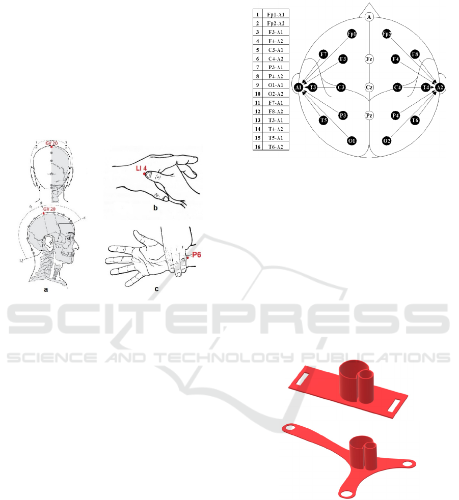
The Hegu point (LI4), also known as large
intestine 4, is located in the meridian of the colon in
the middle of the 2nd metacarpal bone on the radial
side (see Figure 1b). LI4 is considered one of the most
effective acupuncture points for general pain control,
especially headaches (Luong et al. 2018).
Nei Guan (P6) is commonly used to relieve
nausea, motion sickness, and headaches (Ezzo et al.
2006), (Lee and Fan 2009). P6 is located three fingers
below the wrist on the inner forearm between the two
tendons (see Figure 1c).
Figure 1: a - BaiHui (GV20), b - Hegu (LI4), and c - Nei
Guan (P6) acupoint.
EEG data had been obtained from two siblings,
one of whom is without pathologies of the nervous
system (8 years), and the second one has a diagnosis
of autism (10 years). Participation in the study of one
child with autism and the selection of the necessary
individual parameters is explained as an approach to
personalized medicine and individual rehabilitation.
The study protocol was approved by the local ethics
committee of TSU. Adult participants and official
representatives of the minors signed "the informed
consent" to the manipulation.
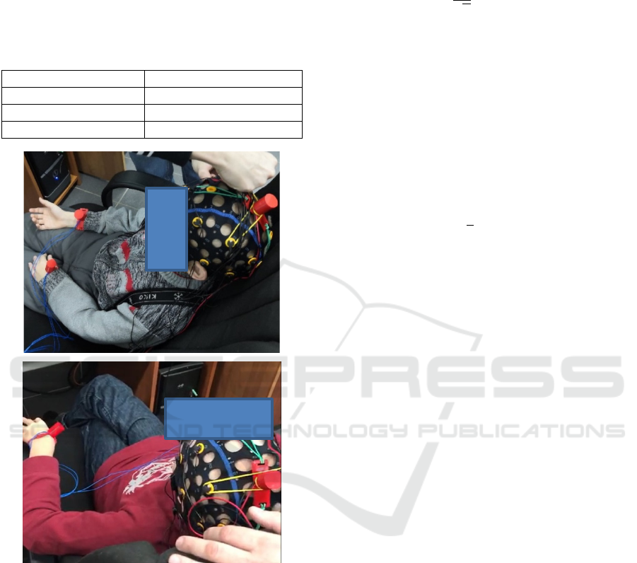
The subject was supposed to take a comfortable
position in the chair, which allowed him to relax the
muscles of the head, neck, and belt of the upper
extremities as much as possible, the left hand and
forearm of the right hand were released to attach the
lasers. It was forbidden to talk, chew anything,
completely close my eyes, remove the mounts for
lasers. In the case of a child with autism, hand
movements and slight head movements were allowed.
A helmet was put on a participant's head with
electrodes located according to the standard
international scheme 10-20 (Figure 2). The electrodes
were pre-lubricated with a special conductive gel for
EEG studies.
Figure 2: Scheme electrode overlay 10-20.
Data were recorded in a state of calm wakefulness
with open eyes when watching a video clip (cartoon).
Note that to avoid the occurrence of random artifacts
during EEG recording, all objects that could distract
children from the cartoon were out of sight.
We used a CONTEC KT88-1016 digital 16-
channel EEG analyzer (China) with the
EEG18V5.0.3 software. The recording time was
reduced to 5 minutes because there was no purpose to
track the temporal dynamics of any pathology, it was
only necessary to capture the moment when the laser
was turned on and to determine whether there was a
laser effect on the functioning of the brain in a state
of calm wakefulness with open eyes.
Figure 3: Holders for points GV20, P6 (top), and for point
LI4 (bottom).
We used red semiconductor laser diodes with a
wavelength of 650 nm. Special holders have been
developed to fix the lasers at selected acupuncture
points (see Figure 3). Holders printed on a 3D printer
made of PLA plastic.
Laser diodes were fixed in the holders and
connected to a YIHUA 305D power supply with an
adjustable output voltage in the range from 0 to 30V,
Electroencephalography Registration of Laser Acupuncture Action on Children with Autism Disorder
389
an output power of 150W and an adjustable output
current from 0 to 5A. The measurements were carried
out at the following power values: 3 mW, 4 mW, and
5 mW. Table 1 shows the output voltage values and
the corresponding current power value.
Table 1: The output voltage values for the corresponding
current power value.
Powe
r
Volta
g
e
3mW 2V
4mW 2.5V
5mW 3V
Figure 4: Disposition of lasers in selected acupuncture
points for a healthy child (top) and a child with autism
(bottom).
Lasers were placed on the hands and head of a
subject at the selected acupuncture points and were
fixed (see Figure 4). The diodes were mounted at a
distance of approximately 30 mm from the skin (spot
area 0.31 cm
2
) and perpendicular to the selected
point. Each of the selected points was stimulated by
the laser for 2.5 minutes.
We used power spectral density (PSD) to describe
the EEG signal. The PSD describes a signal spectral
power distribution as a function of frequency. To
calculate PSD, the truncated Fourier transform 𝑈
(𝜔)
in a finite interval [ t
, t
] of a raw signal 𝑈
(
𝑡
)
was calculated:
𝑈
(
𝜔
)
=
1
√
𝑇
𝑈
(
𝑡
)
𝑒
𝑑𝑡.
We used T=t
−t
= 20s.
Then the power spectral density (Rieke and
Warland 1999), (Millers and Childers 2012) can be
calculated
S
(
𝜔
)
=lim
→
𝐄𝑈
(
𝜔
)
,
where 𝐄 denotes an expected value.
The PSD used to compute a variance of a process
(net power) by integrating over frequency (Storch and
Zwiers 2001)
Var=
S
(
𝜔
)
𝑑𝜔
.
3 RESULTS
A preliminary study had been conducted with a group
of healthy adults. Low-level laser stimulation was
shown to be able to change the power of rhythms in
the head region, which corresponds to the stimulation
of various brain regions (Knyazkova et al. 2019).
Similarly (Knyazkova et al. 2019), an EEG signal
in a 300 s interval was divided into two parts, where
the first 150 seconds are the signal without any laser
action, and the second 150 seconds are the signal
when exposed to lasers. To minimize the factors
associated with the beginning and end of signal
acquisition and the inclusion of lasers, a time interval
of 90 s was cut from each part of the interval center.
Significant noise in the form of emissions was
observed in this area (Figure 5) associated with
patient movements.
Note that patients diagnosed with ASD have much
more such noise. Noise removal (filtering) in the form
of emissions was carried out by means of a
combination of gradient methods (Kistenev et al.
2019), taking into account the threshold value of the
absolute value of 80 μV. The limitation of 80 μV
obtained by us in preliminary studies characterizes
the maximum value of the signal generated on the
surface of the head during negligible movements of
patients. Figure 5 bottom shows the result of the
filtering of the signal depicted in Figure 5 top.
The minimum time interval of the signal after
filtering during the study in all cases was more than
50 s, therefore, when analyzing the data, intervals of
50 s were used (the rest of the signal was discarded).
NDNSNT 2020 - Special Session on Non-invasive Diagnosis and Neuro-stimulation in Neurorehabilitation Tasks
390
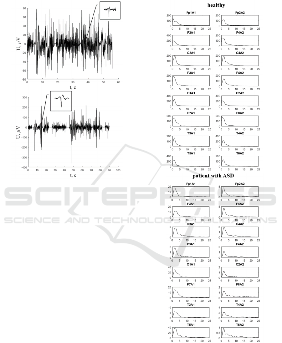
Figure 5: Example of an EEG signal from a healthy child
(top image) and a patient with ASD (bottom image) in
which there are noise components in the form of ejections.
A discrete window Fourier transform (Sherlock
1999) was applied to the filtered signal (Figure 5,
bottom), and many different spectrograms were
constructed with the Hamming weight function
(Harris 1978), the length of which was varied so that
it was possible to analyze time intervals from 0.1 s to
50 s in duration. When constructing spectrograms, the
overlap of adjacent signal segments varied from 0 to
50%.
Figure 6 shows an example of the dependence of
the PSD (Maral 2003)of the EEG signals averaged
over the window of the spectrograms for a healthy
child and a patient with ASD. It can be concluded that
the energy density signal from all electrodes for the
patient with ASD is significantly lower than for the
healthy child
.
Figure 7 shows an example of a dependence of
PSD averaged over the spectrogram window of EEG
signal signals for a healthy child and a patient with
ASD when exposed to laser radiation with a
wavelength of 650 nm at various power levels.
It is obvious that when exposed to lasers, the
energy density in a patient with ASD significantly
increases at all leads with increasing radiation power.
Figure 6: The dependence of the PSD on the frequency (Hz)
averaged over the windows of the spectrograms for a
healthy child (top) and a patient with ASD (bottom).
Electroencephalography Registration of Laser Acupuncture Action on Children with Autism Disorder
391
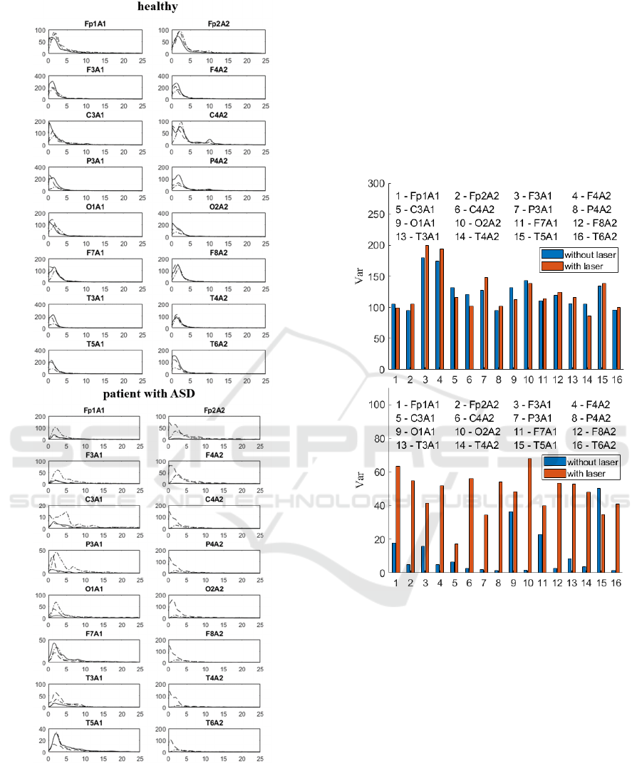
Figure 7: Dependence of the PSD on the frequency (Hz)
averaged over the window of the spectrograms for a healthy
child (top) and a patient with ASD (bottom) when exposed
to laser radiation with a wavelength of 650 nm at a power:
3mW - solid line, 4mW - dash - point line, 5mW - dotted
line.
The energy density in a healthy child does not change
significantly.
It was shown, that when exposed to Hego's point,
the total brain activity does not change on average for
healthy participants, but a redistribution of brain
activity between its spatial regions takes place
(Knyazkova et al. 2019). A significant increase in the
total activity of the brain for ASD patients up to a
value close to the brain activity of healthy child was
registered.
Figure 8:
The total value of PSD for all temporal
windows
inside 90s interval for the healthy child (top) and
the patient with ASD (bottom) before and after laser action.
Note that visual observation of the behavior of
patients with ASD showed that when exposed to laser
radiation, there were changes in the patient's
behavior, expressed in a decrease in anxiety. For
example, the patient with ASD was obsessively
played with a laser mount mounted on his arm
without laser exposure. When the laser was turned on
at a power of 5 mW, the patient with ASD practically
stopped paying attention to this mount.
Figure 8 shows the total value of PSD for all
temporal windows inside 90s interval for the healthy
child (top) and the child with ASD (bottom) before
and after laser action. The latter presented averaged
results for three levels of laser power: 3, 4, and 5 mW.
NDNSNT 2020 - Special Session on Non-invasive Diagnosis and Neuro-stimulation in Neurorehabilitation Tasks
392

4 CONCLUSIONS
Reflexotherapy, as a method of rehabilitation in
autism, is based on the exposure of biologically active
points located on the skin. Laser action on
acupuncture points is an alternative to traditional
acupuncture. We used red semiconductor laser diodes
with a wavelength of 650 nm. Three acupuncture
points (GV20, LI4, P6) were selected to relieve
headaches and reduce anxiety in the study. The
measurements were carried out at the following
power values: 3, 4, and 5 mW. EEG data had been
obtained from two siblings, one of whom is without
pathologies of the nervous system (8 years), and the
second one has a diagnosis of autism (10 years). In
the previous study (Knyazkova et al. 2019), we
investigate a group of healthy volunteers (N = 10),
adult men (the average age was 25 years old). It was
shown, that when exposed to Hego's point, the total
brain activity does not change on average for healthy
participants, but a redistribution of brain activity
between its regions takes place. In this study, it was
confirmed for a healthy child of 8 years old, and the
ASD child. A significant increase in the total activity
of the brain of the ASD child up to a value close to
the brain activity of healthy participants was
registered after the laser action.
Note that visual observation of the behavior of
patients with ASD showed that when exposed to laser
radiation, there were changes in the patient's
behavior, expressed in a decrease in anxiety.
ACKNOWLEDGEMENTS
This work was performed within the frame of the
Fundamental Research Program of the Russian
Academy of Sciences for 2013-2020, line of research
III.23.
REFERENCES
Louis, E.K., et al. 2016. Electroencephalography (EEG):
An Introductory Text and Atlas of Normal and
Abnormal Findings in Adults, Children, and Infants.
Chicago, IL: American Epilepsy Society, 96pp.
doi:10.5698/978-0-9979756-0-4.
Dziadkowiak, E. and Podemski, R., 2019. Impact of
Hyperventilation and Sleep Deprivation Upon Visual
Evoked Potentials in Patients with Epilepsy. Neurol
India, 67, pp. 1027-1032.
Roohi-Azizi, M., et al., 2017. Changes of the brain's
bioelectrical activity in cognition, consciousness, and
some mental disorders. Medical journal of the Islamic
Republic of Iran, 31, pp. 53. doi:10.14196/mjiri.31.53
Belmonte, M.K., et al., 2004. Autism and abnormal
development of brain connectivity. J Neurosci, 24(42),
pp. 9228–9231
Wang, J., et al., 2013. Resting state EEG abnormalities in
autism spectrum disorders. J Neurodev Disord., 5(1),
pp. 24.
Assaf, M., et al., 2010. Abnormal functional connectivity of
default mode sub-networks in autism spectrum disorder
patients. Neuroimage, 53(1), pp. 247-256
Bosl, W.J., et al., 2011. EEG complexity as a biomarker for
autism spectrum disorder. BMC Med, 9(18).
Megremi, A., 2014. Autism spectrum disorders through the
lens of complex-dynamic systems theory. Open Access
Autism, 22(2), pp.1–10.
Boutros, N.N., et al., 2015. EEG changes associated with
autistic spectrum disorders. Neuropsychiatr
Electrophysiol, 1(1), pp.1–20.
Duffy, F.H. and Als, H., 2019. Autism, spectrum or
clusters? An EEG coherence study. BMC Neurol,
19(27).
Eldaief, M.C., et al., 2013. Transcranial magnetic
stimulation in neurology: A review of established and
prospective applications. Neurology. Clinical practice,
3(6), pp.519–526.
Oberman, L.M., et al., 2016. Transcranial magnetic
stimulation in autism spectrum disorder: Challenges,
promise, and roadmap for future research. Autism
research: official journal of the International Society
for Autism Research, 9(2), pp. 184–203.
doi:10.1002/aur.1567.
Cheuk, D.K., et al., 2011. Acupuncture for autism spectrum
disorders (ASD). Cochrane Database Syst Rev. 7(9),
pp. CD007849
Hsieh, C.W., et al., 2011. Different brain network
activations induced by modulation and nonmodulation
laser acupuncture. Evidence-Based Complementary
and Alternative Medicine, 2011(951258).
Knyazkova, A.I., et al.,2019. Influence of laser
acupuncture on EEG characteristics. Proc. SPIE,
11322, pp. 113222O. doi 10.1117/12.2550757.
Satoh, H., et al., 2009. Acute effects of acupuncture
treatment with Baihui (GV20) on human arterial
stiffness and wave reflection. J. Acupunct Meridian
Stud. 2(2), pp. 130-134. doi 10.1016/S2005-
2901(09)60045-5.
Zhao, N.X., et al., 2007. Influence of moxibustion of bai hui
(GV 20) on hemodynamics of common carotid artery in
healthy subjects.
Zhen Ci Yan Jiu. 32(4), pp. 252-254.
Luong, T.G., et al., 2018. Electroencephalogram
Measurement: An Investigation into the Effects of
Laser Acupuncture at Acupoints on Brain. Conference
Paper in IFMBE proceedings. doi 10.1007/978-981-
10-4361-1_127.
Ezzo, J., et al., 2006. Cochrane systematic reviews examine
P6 acupuncture-point stimulation for nausea and
vomiting. J. Altern Complement Med. 12(5), pp. 489-
95. doi 10.1089/acm.2006.12.489
Electroencephalography Registration of Laser Acupuncture Action on Children with Autism Disorder
393

Lee, A. and Fan, L.T.Y., 2009. Stimulation of the wrist
acupuncture point P6 for preventing postoperative
nausea and vomiting. Cochrane Database of Systematic
Reviews, 2, CD003281. doi 10.1002/14651858.
CD003281.pub3.
Rieke, F. and Warland, D., 1999. Spikes: Exploring the
Neural Code (Computational Neuroscience).
Cambridge, MA: MIT Press, 416 pp.
Millers, S. and Childers, D., 2012. Probability and random
processes. Academic Press. 522 pp.
Storch, H. Von and Zwiers, F.W., 2001. Statistical analysis
in climate research. Cambridge University Press, 496
pp.
Kistenev, Y.V., et al., 2019. Application of multiphoton
imaging and machine learning to lymphedema tissue
analysis. Biomed. Opt. Express, 10(7), pp. 3353-3368.
Sherlock, B.G., 1999. Windowed discrete Fourier
transform for shifting data. Signal Processing, 74(2),
pp. 169-177.
Harris, F.J., 1978. On the use of windows for harmonic
analysis with the discrete Fourier transform. Proc. of the
IEEE, 66, pp. 51-83.
Maral, G., 2003. VSAT Networks. John Wiley & Sons, Ltd,
296 pp.
.
NDNSNT 2020 - Special Session on Non-invasive Diagnosis and Neuro-stimulation in Neurorehabilitation Tasks
394
