
Rheoophthalmography Used for the Analysis of Blood Flow in the
Posterior Part of the Eye
P. V. Luzhnov
1a
, A. A. Kiseleva
1
, E. N. Iomdina
2
, L. V. Vasilenkova
2
and O. A. Kiseleva
2
1
Bauman Moscow State Technical University, 5, 2-nd Baumanskaya St., Moscow, Russian Federation
2
Hemlholtz National Medical Research Center of Eye Diseases, 14/19, Sadovaya-Chernogryazskaya St.,
Moscow, Russian Federation
Keywords: Rheoophthalmography, Ocular Blood Flow, Primary Open-angle Glaucoma.
Abstract: The paper is aimed at developing a new rheoophthalmography (ROG) technique able to measure electrical
impedance signals of patients with primary open-angle glaucoma (POAG), which could facilitate early
diagnostics of the disease and the differentiation of its stages. We used a technique of electrode positioning
differing from the transpalpebral ROG technique used previously. Specifically, we changed the distance
between the measuring electrodes and their location pattern: the axis of symmetry was rotated 90° relative to
that used in the transpalpebral technique and located vertically rather than horizontally. This technique was
applied to ROG signals of 32 patients (42 eyes) aged 67.7 years averagely. Of these, 6 patients (10 eyes)
average aged 62.7 years had stage I (early) POAG or suspected POAG; 13 patients (17 eyes) average aged
69.4 years had stage II (developed) POAG; 10 patients (12 eyes) average aged 70.0 years had stage III
(advanced) POAG; 3 patients (3 eyes) average aged 69.3 years had stage IV (terminal) POAG. The results of
this study confirmed the feasibility of the above technique for ROG signal registration. The recorded signals
are informative for quantitative assessment of blood flow in the posterior part of the eye, which enables early
POAG diagnostics and differentiation between POAG stages.
1 INTRODUCTION
The normal functioning of the eye is essentially
determined by the blood flow level in its tissues. For
ocular pathologies such as progressive myopia,
diabetic retinopathy, and glaucoma, eye
hemodynamic examination provides the
ophthalmologist with additional information about
their pathogenesis, gives the opportunity for early
diagnosis, prognostication of the disease
development, and assessment of treatment
effectiveness. The research in this area is important
due to the growing incidence of these ophthalmic
diseases.
The electrical impedance technique allows non-
invasive assessment of blood flow in diverse
segments of the human body (Patterson, 2005; Bodo,
2010; Lazarenko, 2004). It extracts information about
the pulsatile blood supply of the studied segment.
Furthermore, electrical impedance provides
information about the biomechanical properties of
a
https://orcid.org/0000-0003-1111-7063
blood vessels. As far as eye electrical impedance is
concerned, a special technique, called
rheoophthalmography (ROG), was developed
(Avetisov, 1967; Lazarenko, 1999; Lazarenko, 2004).
The authors of subsequent research in the field,
including those of the present study (Luzhnov et al,
2015; Luzhnov et al., 2017) described one of the
varieties of ROG, the transpalpebral
rheoophthalmography (TP ROG), in which the
electrodes are superimposed on a closed eyelid. The
results of mathematical modeling were used as part of
the development of this technique, taking into
account the anatomical structure of the vascular bed
of the eyeball (Shamaev, 2017; Shamaev, 2018). Eye
blood flow during the development of myopia was
studied, which showed the applicability of this
technique forearly diagnosis of myopia progression in
children (Iomdina, 2014; Luzhnov, 2015). It should
be emphasised that low and moderate myopia affects
most of all the blood flow of the anterior eyeball
structures.
Luzhnov, P., Kiseleva, A., Iomdina, E., Vasilenkova, L. and Kiseleva, O.
Rheoophthalmography Used for the Analysis of Blood Flow in the Posterior Part of the Eye.
DOI: 10.5220/0009165902630267
In Proceedings of the 13th International Joint Conference on Biomedical Engineering Systems and Technologies (BIOSTEC 2020) - Volume 1: BIODEVICES, pages 263-267
ISBN: 978-989-758-398-8; ISSN: 2184-4305
Copyright
c
2022 by SCITEPRESS – Science and Technology Publications, Lda. All rights reserved
263

In the case of primary open-angle glaucoma
(POAG), even in its early stages, changes of blood
flow are observed in all parts of the eye
(Cherecheanu, 2013). As is well known, this disease
is one of the leading causes of blindness (Quigley,
2006). Currently, the pathogenesis of POAG is
mostly associated with increased individual level of
intraocular pressure (IOP). Increased IOP damages
the optic nerve fibers and cells of the retina, which
gradually leads to an irreversible loss of vision.
However, not only an into lerable IOP level but also
other factors can lead to the development and
progression of POAG. One of the risk factors of
progressive visual impairment due this disease is a
decrease in the blood supply level in the vessels of the
brain and the eye (Schmetterer, 2015). Eye
hemodynamics monitoring in POAG patients may
give useful diagnostic information for glaucoma
clinicians.
A number of studies, described in (Luzhnov,
2018), were carried out on the analysis of TP ROG
signals in patients with POAG. It was shown that the
estimation of amplitude parameters became more
informative if waveform analysis is used. So, the
analysis of electrical impedance signals in POAG
must include a qualitative and quantitative estimation.
A qualitative analysis of signals includes determining
the type of the pulse wave. It is affected by
biophysical, biomechanical and hydrodynamic
factors, which subsequently determine the diagnostic
result in the qualitative analysis of signals. At the
moment, the analysis of the pulse wave shape in TP
ROG is carried out using attractors (Luzhnov, 2018).
This analysis allows indirect evaluation of blood flow
in different parts of the eye, as well as their interaction
with each other. On the whole, however, the issue of
quantitative determination of blood flow indices in all
(not only the anterior) parts of the eye using non-
invasive electrical impedance methods remains
unresolved. It is especially vital for early diagnosis of
POAG and differentiation of the POAG stages.
The aim of this work, therefore, is to develop the
ROG technique, which could ensure quantitative
analysis of electrical impedance signals of the
posterior part of the eye in patients with POAG,
including the possibility of early diagnosis of the
disease and differentiation between its stages.
2 MATERIALS AND METHODS
Currently, various methods are used to study the
blood supply of eye structures (Kurysheva, 2017).
The electrical impedance diagnostic method allows a
comprehensive assessment of blood flow state in the
eye vessels as a whole, in contrast to research
methods that determine the blood supply of each
vessel individually. The technique of TP ROG
involves a quantitative assessment of blood supply at
the depth of sounding corresponding to the anterior
part of the eye (Luzhnov, 2015). Therefore, a new
technique for applying electrodes for the assessment
of posterior eye pole hemodynamic parameters of
POAG patients was used in the present work.
To reach the posterior eye pole vascular bed
(Roebuck, 2015), the estimated sounding depth
should be increased. Accordingly, we used increased
distance between the ROG measuring electrodes, and
their location was changed: the axis of symmetry was
rotated 90° relative to that used in the previous ROG
technique and was located vertically rather than
horizontally (see Fig.1).
The vertical orientation with the arrangement of
four electrodes (a pair of current electrodes and a pair
of measuring ones) in the tetrapolar system of leads
allowed us to provide the desired interelectrode
distance during its superimposition. The distance
between the electrodes was controlled by a rigid
fastening system, made similarly to the method
described in (Kiseleva, 2020).
To register the TP ROG signal, a semi-rigid
substrate was used for attaching the electrodes. A pair
of measuring electrodes was located at a distance of
13 mm from each other. This enables measuring the
blood flow at a distance corresponding to the anterior
part of the eye. To register the ROG PP signal, we use
an ABS plastic plate with holes for electrodes. A pair
of ROG PP measuring electrodes is spaced 60 mm
apart. Thanks to this arrangement we are able to
measure blood flow at the depth corresponding to the
posterior pole of the eye. Such system parameters
enable the registration of an electrical impedance
signal produced by blood supply of both the posterior
and the anterior part of the eye.
The distance between the round electrodes was
considered the distance between their centres, or
attachment points. In preliminary studies, the selected
methods of applying electrodes to adult patients and
children aged 8 years or older were tested. The
accuracy of positioning and the quality of electrode
placement made it possible to register ROG signals
for subsequent analysis of ocular blood flow.
To register the signals, an impedance measuring
transducer with a probe current frequency of 100 kHz
was used. The signal analysis of the ROG pulse blood
supply was carried out after filtering the obtained
electrical impedance signal with a Butterworth
fourth-order band-pass filter with a lower cut-off
BIODEVICES 2020 - 13th International Conference on Biomedical Electronics and Devices
264
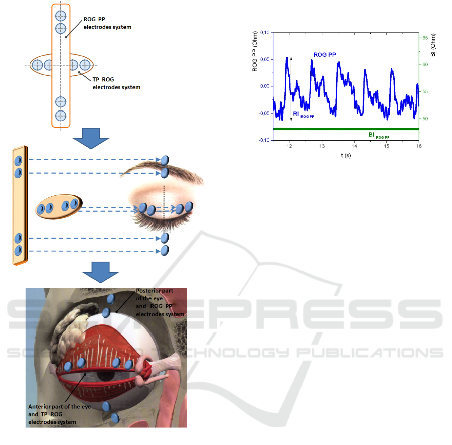
Figure 1: The location of two tetrapolar electrodes systems:
transpalpebral for anterior part of the eye (TP ROG) and
rotated for posterior part of the eye (ROG PP).
frequency of 0.15 Hz and an upper cut-off frequency
of 100 Hz. The signal was recorded for 20 seconds.
The amplitude of the ROG signal was determined as
the arithmetic mean of the amplitudes of all pulse
waves included in the 20-second recording period. A
frequency band below 0.15 Hz determined the level
of blood flow by the amplitude of the base impedance
signal. To achieve this, the initial signal was filtered
with a second-order Chebyshev filter. As a result,
when analysing the ROG signals, two indicators were
calculated: the amplitude of the pulse wave, or the
rheographic index (RI), and the value of the base
impedance (BI). An example of electrical impedance
signal registration for the posterior part of the eye
(ROG PP) is shown in Fig.2.
Figure 2: An example of ROG PP signal in a patient with
stage I POAG.
The study was conducted in the glaucoma
department of the Hemlholtz National Medical
Research Center of Eye Diseases. In total, ROG PP
signals of 32 patients (42 eyes) averagely aged 67.7
years were analysed. Of these, 6 patients (10 eyes,
average age 62.7 years) had stage I (early) POAG or
suspected POAG; 13 patients (17 eyes, average age
69.4 years) had stage II (developed) POAG; 10
patients (12 eyes, average age 70.0 years) had stage
III (advanced) POAG; 3 patients (3 eyes, average age
69.3 years) had stage IV (terminal) POAG.
This study was performed in accordance with the
Declaration of Helsinki and was approved by the
Local Committee of Biomedical Ethics of the
Moscow Hemlholtz National Medical Research
Center of Eye Diseases. A written informed consent
was obtained from all participants.
3 RESULTS
It was found that in the examined groups of patients,
the average value of the RI indicator increases with
the severity of the disease (from stage I to III, see
Table 1). In stage IV, the average RI indicator fell
slightly. An increase was also established in the
average value of the BI indicator with POAG
progression from stage I to stages II and III. The
increase was even more significant at stage IV of the
disease.
The increase of BI indicator reflects a drop in the
total blood supply in eye tissues. Our study showed
two characteristic changes of this indicator. The first
was observed when POAG progressed from stage I to
more advanced stages whilst the second change took
place with the onset of stage IV POAG.
Rheoophthalmography Used for the Analysis of Blood Flow in the Posterior Part of the Eye
265

Table 1: RI and BI indicators in the examined groups
(M±SD).
Stage of POAG RI, Ohm BI, Ohm
I 0.11±0.02 46.1±9.7
II 0.13±0.03 52.7±10.4
III 0.14±0.03 51.2±10.9
IV 0.10±0.04 57.3±10.3
In addition to averaging the diagnostic indices by
study groups, they were also compared for patients
with different stages of POAG in the right and the left
eye. In total, ROG PP signals of five patients were
analysed; five pairs of ROG records were examined.
In this case, similar proportions were observed but the
values were higher. For example, the RI of the eye
after antiglaucoma surgery and the non-operated eye
of the same patient was approximately 2.6 times
different.
4 CONCLUSIONS
The results obtained in the study confirm the
feasibility of the method of applying electrodes
proposed in the work for ROG signals registration.
The signals recorded in such a way are informative
for hemodynamics assessment in POAG patients,
which can be used for early diagnostics of POAG and
differentiation between its stages. The difference in
hemodynamic parameters observed between the
fellow eyes with different POAG stages of the same
patient confirms the efficiency of the new technique.
As a further development of this work, the authors
consider a joint study of ROG signals recorded
simultaneously using different leads, which will
allow us to differentiate the changes in the level of
blood flow depending on the depth of sounding, and,
consequently, on the part of the eye.
When using a device in which the ROG signal is
recorded transpalpebrally through one channel with a
tetrapolar lead system installed on the eyelid, and
through a second channel with a tetrapolar lead
system installed symmetrically with respect to the
anteroposterior axis of the eye on the face in the area
of the eye socket, it becomes possible to expand the
existing facilities of the electric impedance
diagnostics of the ocular blood flow.
A simultaneous analysis of electrical impedance
signals through two channels, TP ROG and ROG PP,
makes it possible to estimate the level of blood flow
in the anterior part of the eye by the signal of the first
channel and in the posterior part of the eye by the
signal received from the second channel.
A research into the relationship between blood
flow in the anterior and posterior parts of the eye at
different stages of glaucoma is also interesting for
understanding the pathophysiological processes of
the development of this disease.
CONFLICT OF INTEREST
The authors declare that they have no conflict of
interest. The paper was supported by a grant from
RFBR (No.18-08-01192).
REFERENCES
Avetisov, E.S., Katsnel'son, L.A., Savitskaia, N.F., 1967.
Rheocyclographic examinations in myopia. Vestnik
Oftal’mologii 80(3): 3–7.
Bodo, M. (2010) Studies in Rheoencephalography. Journal
of Electrical Bioimpedance 1, 18–40.
Cherecheanu A.P., Garhofer G., Schmidl D., Werkmeister
R., Schmetterer L., 2013. Ocular perfusion pressure and
ocular blood flow in glaucoma. Curr Opin Pharmacol
13: 36–42. DOI:10.1016/j.coph.2012. 09.003
Iomdina, E.N., Luzhnov, P.V., Shamaev, D.M., et al., 2014.
An evaluation of transpalpebral rheoophthalmography
as a new method of studying the blood supply to the eye
in myopia. Russian ophthalmological journal 7(4): 20–
24. (in Russian)
Kiseleva, A.A., Luzhnov, P.V., Shamaev, D.M., 2020.
Verification of mathematical model for bioimpedance
diagnostics of the blood flow in cerebral vessels.
Advances in Intelligent Systems and Computing 902:
251–259. DOI: 10.1007/978-3-030-12082-5_23
Kurysheva, N.I., Parshunina, O.A., Shatalova, E.O., et al.,
2017. Value of structural and hemodynamic parameters
for the early detection of primary open-angle glaucoma.
Curr. Eye Res. 42(3): 411–417.
Lazarenko, V.I., Komarovskikh, E.N., 2004. Results of the
examination of hemodynamics of the eye and brain in
patients with primary open-angle glaucoma. Vestnik
oftalmologii 120(1): 32–36. DOI:10.21687/0233-
528X-2017-51-3-22-30
Lazarenko, V.I., Kornilovsky, I.M., Ilenkov, S.S. et al.,
1999. Our method of functional rheography of eye.
Vestnik oftalmologii 115(4): 33–37.
Luzhnov, P.V., Shamaev, D.M., Iomdina, E.N. et al., 2015.
Transpalpebral tetrapolar rheoophtalmography in the
assessment of parameters of the eye blood circulatory
system. Vestn Ross Akad Med Nauk 70(3): 372–377.
DOI: 10.15690/vramn.v70i3.1336
Luzhnov, P.V., Shamaev, D.M., Iomdina, E.N., et al., 2017.
Using quantitative parameters of ocular blood filling
with transpalpebral rheoophthalmography. IFMBE
Proceedings 65: 37–40. DOI:10.1007/978-981-10-
5122-7_10
BIODEVICES 2020 - 13th International Conference on Biomedical Electronics and Devices
266

Luzhnov, P.V., Shamaev, D.M., Kiseleva, A.A., et al.,
2018. Using nonlinear dynamics for signal analysis in
transpalpebral rheoophthalmography. Sovremennye
tehnologii v medicine 10(3): 160–167. DOI:
10.17691/stm2018.10.3.20
Patterson, R. (2005) Electrical Impedance Tomography:
Methods, History, and Applications (Institute of
Physics Medical Physics Series), Physics in Medicine
and Biology 10: 2427–2428.
Quigley, H.A., Broman, A.T., 2006. The number of people
with glaucoma worldwide in 2010 and 2020. Br J
Ophthalmol 90(3): 262–267.
Roebuck, J., 2015. Anatomy 360: The Ultimate Visual
Guide to the Human Body. Thunder Bay Press, San
Diego.
Schmetterer L., 2015. Ocular perfusion abnormalities in
glaucoma. Russian Ophthalmological Journal 4: 100–
109.
Shamaev, D. M., Luzhnov, P. V., Iomdina, E. N., 2018.
Mathematical modeling of ocular pulse blood filling in
rheoophthalmography. IFMBE Proceedings 68(1):
495–498. DOI: 10.1007/978-981-10-9035-6_91
Shamaev, D.M., Luzhnov, P.V., Iomdina, E.N., 2017.
Modeling of ocular and eyelid pulse blood filling in
diagnosing using transpalpebral rheoophthalmography.
IFMBE Proceedings 65: 1000–1003.
DOI:10.1007/978-981-10-5122-7_250
Rheoophthalmography Used for the Analysis of Blood Flow in the Posterior Part of the Eye
267
