
Enhancing Vibroarthrography by using Sensor Fusion
Dimitri Kraft
1 a
, Rainer Bader
2
and Gerald Bieber
3 b
1
University of Rostock, Rostock, Germany
2
Unimedizin, Rostock, Germany
3
Fraunhofer IGD, Rostock, Germany
Keywords:
Endoprosthesis, VAG, Sensors, Mobile Device, Implants, Wear, Accelerometer, Microphone, Vibration.
Abstract:
Natural and artificial joints of a human body are emitting vibration and sound during the movement. The
sound and vibration pattern of a joint is characteristic and changes due to damage, uneven tread wear, injuries,
or other influences. Hence, the vibration and sound analysis enables an estimation of the joint condition. This
kind of analysis, vibroarthrography (VAG), allows the analysis of diseases like arthritis or osteoporosis and
might determine trauma, inflammation, or misalignment. The classification of the vibration and sound data is
very challenging and needs a comprehensive annotated data base. Current existing data bases are very limited
and insufficient for deep learning or artificial intelligent approaches. In this paper, we describe a new concept
of the design of a vibroarthrography system using a sensor network. We discuss the possible improvements
and we give an outlook for the future work and application fields of VAG.
1 INTRODUCTION
A joint is the connection between bones in the body,
which link the skeletal system into a functional whole.
They are constructed to allow for different degrees
and types of movement (Kim et al., 2009). Joint dis-
order and diseases have a huge impact on the life of
people and are the main cause for disability of el-
derly people. Disorders and diseases such as arthri-
tis causing huge economic costs for nations. In the
US, the costs for treating arthritis were $303.5 bil-
lion or 1% of the 2013 US Gross Domestic Product
(GDP). One of the most common disease of joints
is osteoarthritis (OA), a degenerative disease where
changes in bones, articular cartilages and soft tissues
occur. OA affecting nearly 10% of the population
worldwide and is frequently observed in elderly peo-
ple (44% in people ¿ 80 years). OA was the second
most costly health condition treated at US hospitals
in 2013. In that year, OA accounted for $16.5 bil-
lion, or 4.3%, of the combined costs for all hospital-
izations (Murphy et al., 2017). Tools and technologies
for quantify the health status of human joints outside
of the clinical setting are investigated by researchers
throughout the past decade include the analysis of vi-
bration caused by human joints, the range of motion
a
https://orcid.org/0000-0002-0604-5854
b
https://orcid.org/0000-0003-2496-6232
and gait analysis. The analysis of vibration is called
Vibroarthrography (VAG) (McCoy et al., 1987) or Vi-
bration arthrometry (Kernohan et al., 1990). VAG is
a non-invasive screening tool for vibration and sound
analysis of natural joints which was first introduced
by McCoy et al. in 1985 (McCoy et al., 1987) (see
Figure 1). The origin concept is older and referred to
Carl Hueter in 1883 (Hueter, 1883).
2 MOTIVATION
Because of the demographic development, joint dis-
eases are an increasing problem. Within the lifetime,
approx. 90 % of the population will suffer on seri-
ous knee or hip problems, hereby women are more
affected than men. In Germany, the most surgery is
knee joint operation by more than 150,000 times per
year. But often, a knee replacement is not needed and
an artificial knee joint wound last forever. More than
60 % of joints that had to be replaced are younger
than 7 years. Therefore, an easy to use measurement
system is necessary. The usage of a sensor network
that combines also the capability to measure mobility,
strength, sound, weight, acceleration, and light reflec-
tion of the veins enables the assessment of a compre-
hensive representation of the joint and body condi-
tion. A measurement over time provides the determi-
Kraft, D., Bader, R. and Bieber, G.
Enhancing Vibroarthrography by using Sensor Fusion.
DOI: 10.5220/0009098701290135
In Proceedings of the 9th International Conference on Sensor Networks (SENSORNETS 2020), pages 129-135
ISBN: 978-989-758-403-9; ISSN: 2184-4380
Copyright
c
2022 by SCITEPRESS – Science and Technology Publications, Lda. All rights reserved
129

nation of a trend and allows a prognosis beside the
diagnosis. To achieve this goal, the concept and de-
sign of a sensor system is necessary.
3 RELATED WORK
3.1 Sensor Selection
The analysis of knee joint sound experience a long
history of investigation since 1976. Chu et al. pub-
lished a series of papers investigating the acoustic
pattern of knee joints. The researchers found that
cartilage damage around the knee may be classified
with acoustic sensors (Chu et al., 1978). Further they
found, that pattern recognition in acoustic signals may
be used to distinguish between normal, rheumatoid
and degenerative knees (Chu et al., 1976). Later in
1985, a research team evaluated the diagnostic po-
tential of vibration arthrography by examining 250
subjects who were undergoing diagnostic arthroscopy
and suggested that VAG will become a significant di-
agnostic aid in the clinical evaluation of the locomo-
tive system. In 86% of these cases, a characteristic
signal was obtained. Further, it was possible to iden-
tify the affected side and determine how far posteri-
orly the mensical injury lay (Kernohan et al., 1986).
Figure 1: Subject attached to the recordings apparatus of
VAG (McCoy et al., 1987).
Acceleration sensors show the advantage against mi-
crophones that microphones are sensing ambient
noise from the environment too much and provide
a limited frequency response in the audible range
(Frank et al., 1990). Technologically, the accelera-
tion of the skin is a mechanical wave. Hence, mi-
crophones are sensing airwaves that are generated by
swinging material. The advantage of microphones is
that they are able to detect higher frequencies and that
no direct contact to the body is necessary. In rela-
tion to industrial inspections for mechanical machine
components of wear by vibration analysis (Tandon
and Choudhury, 1999; Orhan et al., 2006), the vibra-
tion and sound analysis of natural and artificial joints
seems also to be manageable.
Recently, Klemm et al. studied the effects of
sensor placement in their recent study and suggest,
that mechanical extensions to the sensors, like 3D-
printed caps may improve the data acquisition pro-
cess. This ensures a amplification and a good trans-
mission of lower frequency sounds (up to 125Hz)
without attenuation of higher bands. During measure-
ment, the sensors, off-the-shelf airborne audio sensors
(SPH0645LM4H-B), were attached to the medial and
lateral side of the knee. The measurement protocol
consists of 20 sit to stand movements cycles (Klemm
et al., 2019).
3.2 Feature Assessments
Several authors carried out research to investigate
acoustic emission to assessing the dynamic integrity
of joints (Prior et al., 2010). To detect acoustic emis-
sion, it is necessary to attach piezoelectric transducers
to the surface of a structure under load. Shark et al.
developed a joint angle based acoustic emission sys-
tem. After the data acquisition process, the authors
applied feature extraction techniques and dimension-
ality reduction to discriminate between healthy and
unhealthy knees (Shark et al., 2010). Shark et
al. found that the OA knees produce consistently
and substantially more acoustic emission events with
higher peak and mean magnitude than healthy knees.
Teague et al. used piezoelectric transducer, elec-
tret amd MEMS microphone, and IMU to assess the
click location of knees during movement. During
their study the researchers evaluated different types
of microphone and carried out a comparison between
on skin ad off skin joint sound signals. Further, they
found that acoustic events occur at consistent joint
angles during repetitive motions of healthy subjects.
They suggest that joint sound measurements are re-
peatable with sensing technology that can be imple-
mented in an inexpensive and wearable form factor
(Teague et al., 2016). In 2018, Andersen et al. ex-
amined the methodical aspects of VAG assessment
by investigating sensor placement and flexion-exten-
sion movements under load. Their study showed, that
VAG parameters are affected by load level, movement
type and the location of the accelerometer over the
knee joint in asymptomatic subjects (Andersen et al.,
2018).
SENSORNETS 2020 - 9th International Conference on Sensor Networks
130

3.3 Classification Techniques for VAG
The current research in Vibroarthrography underlie
different problems. Most research in vibroarthrog-
raphy focus on the classification process (Rangayyan
et al., 1997; Wu and Krishnan, 2011; Tai et al., 2015;
Nalband et al., 2016), not considering different meth-
ods to acquire the data. It is not clear, which sen-
sors (IMU, MEMS-microphone, piezoelectric trans-
ducer, Ultrasound) should be used to assess the cur-
rent health state. Further, only little research is done
evaluating the optimal sensor placement and envi-
ronmental setting during the measurement. Several
studies showed, that crepitus of the knee is depen-
dent on the angular velocity (Kernohan et al., 1990)
and the load affecting the knee joint during move-
ment(Andersen et al., 2018). The usage of multi-
ple sensors is needed due to the fact, that differ-
ent states of pathology are found in different fre-
quency bands (Frank et al., 1990) and may enable
the early prevention of certain diseases. Peat et al.
suggest that the American College of Rheumatology
(ACR) clinical criteria to classify osteoarthritis seem
to reflect later signs in advanced disease. They sus-
pect that other approaches may be needed to iden-
tify early, mild osteoarthritis in the general population
(Peat et al., 2006). The gold standard for diagnosing
certain joint disorder is still X-ray or MRI . Those
techniques are either radiating or expensive, render-
ing the constant monitoring impossible and not use-
ful for early prevention. Abbot et al. concluded that
the intense variability within signals is caused by con-
tacting joint surfaces and forces during motion. This
produce an understandable scepticism in the clinical
community as to the reliability of vibration arthrome-
try and therefore making an adaption in medicine hard
to accomplish (Abbott and Cole, 2013). The perfor-
mance and capabilities of Vibroarthrography in de-
tection of knee disorders are slight behind those of
MRI/MRT. McCauly et al. reported 86% sensitivity
and 74% specificity in detection of chondromalacia
patellae using MRI (McCauley et al., 1992). Pihlaja-
maki et al. reported 83% sensitivity and 84% speci-
ficity for MRI images for stage III chondromalacia
(Pihlajam
¨
aki et al., 2010).
3.4 Mobile VAG Systems
The need for constant monitoring of health status is
not a new concept, considering the newest technolo-
gies in the Internet of Things area. Several devices
such as smartwatches and fitness tracker measuring
heart rate, respiration rate or general activity of hu-
man beings since a decade. The concept of constant
monitoring of the knee health status, on the other
hand, was first introduced a by a research group in
2016. T
¨
oreyin et al. (Toreyin et al., 2016) provided a
prototype and framework to measure the sound made
by joints during daily activities such as sit to stand
transitions, walking or others. Another approach pro-
posed by Msayib et al. in (Msayib et al., 2017) de-
veloped an intelligent monitoring system to measure
the rehabilitation of total knee arthroplasty patients
by assessing the range of motion of the knee. A
recently proposed framework by Athavale and Kris-
han in (Athavale and Krishnan, 2019) is especially
designed for the IoT era. By encoding the signals,
the researchers enabling a efficient way for a con-
stant monitoring of the condition of knee joint with
vibroarthrography (or actigraphy). Unfortunately,
Athavale and Krishan did not propose a effective sen-
sor alignment to measure the knee joint sounds and
vibrations. This work try to outline a concept based
on the aforementioned research.
3.5 Challenges in VAG Systems
Although several researches reported high accuracy,
sensitivity and specificity (¿95%) in classification of
pathological knees via VAG (Kim et al., 2009; Nal-
band et al., 2017; Rangayyan et al., 2013), those re-
sults were obtained on a fairly dated dataset, consist-
ing of 38 abnormal and 51 normal VAG signals ob-
tained from a single axis accelerometer. Scalograms
of abnormal and normal VAG signals are depicted in
Figure 2. Rangayyan et al. suspect in (Rangayyan
et al., 2013) that those remarkable results are obtained
by overfitting on a specific dataset, regardless of the
leave-one-out validation method used. This overfit-
ting on an collected dataset may apply for other re-
search in VAG as well. The acceptance of VAG in
medicine is still far behind the established methods
in this field (Shieh et al., 2016). Reasons might be
the lack of resolution, the measurement principle, and
that the sensor attachment on the skin varies by each
individual. Further, the signal transportation is in-
fluenced by various parameters. This indicates that
further research is needed, e.g. optimal sensor place-
ment (Ota et al., 2016), number and type of sensors
(Klemm et al., 2019), weight load during examination
(Andersen et al., 2018) or state of the art feature ex-
traction and classification techniques. Even today, a
realistic recognition rate of certain joint diseases like
knee, hip or hand osteoarthritis is not sufficient to ob-
tain a precise result (Altman et al., 1986). Therefore,
we need an enhanced VAG assessment and analysis
system as follows.
Enhancing Vibroarthrography by using Sensor Fusion
131

(a) Scalogram of VAG signal without pathology
(b) Scalogram of VAG signal with pathology
Figure 2: VAG signal comparison.
4 ENHANCED CONCEPT OF THE
VAG SYSTEM
As we worked out in the related work section, the ex-
isting concept of the assessment of a VAG Sensing
System is insufficient. Therefore, we designed a con-
cept that is more advanced, fuses heterogeneous sen-
sors, and compensate the lacks and deficiencies of the
existing VAG procedure.
Figure 3: General Architecture of the enhanced VAG sys-
tem.
The major characteristic of the VAG joint analysis is
the trait of dynamic measurement, against still captur-
ing like MRT or X-ray. The VAG measurement can be
performed only in motion. Therefore, we propose and
distinguish between two types of assessment, a fully
controlled movement and the free movement.
The controlled movement is a setting like sitting
in a chair and moving the limbs (Figure 4). The free
movement describes the assessment during activities
like walking, stair climbing, repetitive activities (e.g.
cycling), or other activities of the daily living. The
classification of free movement activities and their pa-
rameters is much more difficult that under controlled
conditions. That is the reason we emphasis on the
controlled assessment and will consider the mobile
assessments subsequently in the future. The station-
ary concept (Figure 3) includes the usage of estab-
lished sensors and additional sensors to complete the
setting for a comprehensive assessment. The manda-
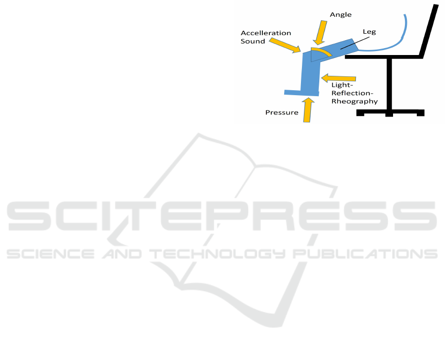
tory sensors for an enhanced vibroarthrography are:
• Ultrasound:
Acoustic emission from human knee joints
indicates, that healthy and unhealthy knees can be
successfully distinguished during the sit to stand
movement (Shark et al., 2010).
• Accelerometer:
Assessing the ROM of the joint, detection of
crepitus and other click and crack sounds (Toreyin
et al., 2016).
These sensors will be extended by a net of additional
sensors to improve the signal quality, to reduce the
noise, to enrich the information, and to combine val-
ues with other parameters, e.g. vibration in relation to
joint angle and angular velocity.
4.1 Improvements
The proposed concept includes several improvements
as follows:
• Frequency Band
The movements of the joint causes oscillations
in various frequencies and intensities. Slow os-
cillations can be easily measured with accelera-
tion sensors, higher frequencies are accessible by
microphones. The proposed concept combines
the two sensor types to be able to record the full
range of frequencies. To ensure a reliable over-
lap, we propose to use the acceleration sensor in
full range of sampling rate (e.g. up to 3.2 kHz
by AXDL345) and the microphone (e.g. 20Hz to
44kHz).
SENSORNETS 2020 - 9th International Conference on Sensor Networks
132
• 3D Assessment
Referring to the existing VAG database by Krish-
nan et al., our sensor system consists of multiple
sensors with multiple axes, in comparison to only
a one dimensional acceleration sensor.
Furthermore, we will include the trajectory of the
angle, to recognize a straight movement, torn liga-
ment, or misalignment. Therefore, the integration
of a radar sensor will be an option if the single use
of acceleration sensors is not sufficient.
• Sensor Placement
The coupling of the sensors to the joint is a chal-
lenge. In relation to ultrasonic assessment, a gel
and cuff can be used to enhance the vibration sig-
nal flow. Furthermore, the vibration of a joint are
transmitted also to the bones of the limb. There-
fore, we will examine the signal transmission to
the end of the related bones. We expect a leverage
effect, that the vibrations of the e.g. knee are am-
plified at the foot angle. Similar to (Klemm et al.,
2019) we may use extensions to enhance the vi-
bration signals.
• Additional Sensors
The concept of an enhances sensor network for
VAG measurements includes additional sensors
to examine supplementary effects, e.g. the mo-
bility and strength of legs with unhealthy and
healthy knees. To distinguish between joint dis-
ease and skeletal muscle disorder, the concept in-
cludes electromyography (EMG) for the measure-
ment of muscle activity (Hollander et al., 2018)
and PPG sensors to determine veins insufficiency
by the light reflection rheography (LRR).
• Correlation to the Movement Angle
The VAG database by Krishnan et al. contains
only vibration data over time without any correla-
tion of the angle of the joint. Our concept includes
the simultaneously assessment of the joint angle.
• Joint Load
We assume that the load free movement of the
joint generates other vibration than a joint under
stress, as proposed by (Andersen et al., 2018).
Therefore, we designed a load measuring of the
joint to receive a multi-parameter dataset.
• Execution Speed and Repetition
The vibration of the joint differs by the execution
speed of the joint motion (Kernohan et al., 1990).
Therefore, we assess the vibration during certain
motion speed. Currently, we are not aware if vi-
bration vary in repetitions but this is a hypothesis
that has to be examined in future work.
• Machine Learning and Artificial Intelligence
The core of the VAG Sensing System bases on a
classifier that consists of a neural network. Cur-
rently we propose a convolutional neural network
(CNN) that enables an analysis of complex mo-
tions even during daily activities.
The improvements are combined into a sensor net-
work with a multidimensional signal assessing and
analysis platform.
Figure 4: Heterogeneous sensors as a Sensor Network.
5 DISCUSSION
Signal analysis of mechanical bearings are state of the
art for wear and abrasion estimation. The condition of
human joints are currently only by interest when they
hurt or if a major decline of mobility occurs. Since
now, MRT, CT or ultrasonic examinations are pro-
cedures that are performed whenever a joint diagno-
sis is needed. These methods are expensive and pro-
vide only information recorded in a motionless state.
In contrast, a dynamic assessment is an option to be
used for further diagnostic. The VAG is a dynamic
assessment tool that has the potential to become a
very cheap diagnosis tool because the sensor data as-
sessment is easy to perform, except the data analysis.
Therefore, newest technological concepts in Machine
Learning may be used for a powerful and reliable di-
agnostic. Unfortunately, classifiers based on convo-
lutional or recurrent neural networks require a large
dataset that does not exist so far. Existing data bases
do not contain radiation related acceleration data and
mixing all kinds of knee diseases together. For fur-
ther research, we developed the concept for a com-
prehensive data assessment of VAG under the usage
of a sensor network. With this concept, we are able
to establish a comprehensive database that gives the
basis for neural network classifiers.
We assume that VAG still has some limitations
but we expect that the advantages outweigh the disad-
vantages. On one side, we are facing technical chal-
lenges like synchronization, easy handling for the pa-
tient with in a setting of self-assessment in the home
Enhancing Vibroarthrography by using Sensor Fusion
133

environment. On the other side, VAG is an indirect
measuring technique. We are not able to determine
the thickness of a cartilage, but we measure the crepi-
tus intensity. Therefore, it will be difficult to achieve
a reliable relation between thickness and sound or de-
gree of disorder or injury. VAG can be used as a gate-
keeper technology and it is convenient to be used for
a long term usage to obtain trends and progress states.
6 CONCLUSION AND FUTURE
WORK
In the paper, we describe a new concept of using the
technology of vibroarthrography (VAG) by using sta-
tionary and mobile assessment of vibrations of human
joints during motion. Hereby, we describe the gen-
eral concept of a stationary assessment system and
outline the improvements. The analysis of the vibra-
tion pattern assessed with the enhanced VAG system
enables a high sophisticate classification with neu-
ral nets and the discrimination of healthy or injured
joints. Therefore, we propose to build up a compre-
hensive database, consisting of heterogeneous sensor
data assessed by the enhanced VAG sensor network.
The future work will be the application of the con-
cept and the implementation of a database. Further-
more, we will investigate the relevance of trajectories
of the leg and the interplay of muscle strength, ve-
nous insufficiency, and joint disease. VAG did not
found the respected dissemination or usage as a diag-
nosis tool so far, but we assume that the advantage of
a harmless, easy to perform and cheap analysis leads
to its establishment. We propose that not only injured
but also artificial joints can be analyzed.
ACKNOWLEDGMENTS
This work receives funding from the German Federal
Ministry for Economic Affairs and Energy by ZIM-
16KN04913, related to the project MOREBA.
REFERENCES
Abbott, S. C. and Cole, M. D. (2013). Vibration arthrom-
etry: a critical review. Critical reviews in biomedical
engineering, 41(3):223–242.
Altman, R., Asch, E., Bloch, D., Bole, G., Borenstein, D.,
Brandt, K., Christy, W., Cooke, T., Greenwald, R.,
Hochberg, M., et al. (1986). Development of crite-
ria for the classification and reporting of osteoarthri-
tis: classification of osteoarthritis of the knee. Arthri-
tis & Rheumatism: Official Journal of the American
College of Rheumatology, 29(8):1039–1049.
Andersen, R. E., Arendt-Nielsen, L., and Madeleine, P.
(2018). Knee joint vibroarthrography of asymp-
tomatic subjects during loaded flexion-extension
movements. Medical & biological engineering &
computing, 56(12):2301–2312.
Athavale, Y. and Krishnan, S. (2019). A telehealth system
framework for assessing knee-joint conditions using
vibroarthrographic signals. Biomedical Signal Pro-
cessing and Control.
Chu, M., Gradisar, I., and Mostardi, R. (1978). A nonin-
vasive electroacoustical evaluation technique of carti-
lage damage in pathological knee joint. Electronics
Letters, 16(4):437–442.
Chu, M. L., Gradisar, I. A., Railey, M. R., and Bowling,
G. F. (1976). Detection of knee joint diseases using
acoustical pattern recognition technique. Journal of
Biomechanics, 9(3):111–114.
Frank, C. B., Rangayyan, R. M., and Bell, G. D. (1990).
Analysis of knee joint sound signals for non-invasive
diagnosis of cartilage pathology. IEEE Engineering in
Medicine and Biology Magazine, 9(1):65–68.
Hollander, D. B., Yoshida, S., Tiwari, U., Saladino, A.,
Nguyen, M., Boudreaux, B., and Hadley, B. (2018).
Dynamic analysis of vibration, muscle firing, and
force as a novel model for non-invasive assessment
of joint disruption in the knee: A multiple case report.
The Open Neuroimaging Journal, 12(1).
Hueter, C. (1883). Grundriss der chirurgie. FCW Vogel.
Kernohan, W. G., Beverland, D. E., McCoy, G. F., Hamil-
ton, A., Watson, P., and Mollan, R. (1990). Vibration
arthrometry. a preview. Acta orthopaedica Scandinav-
ica, 61(1):70–79.
Kernohan, W. G., Beverland, D. E., McCoy, G. F., Shaw,
S. N., Wallace, R. G., McCullagh, G. C., and Mollan,
R. A. (1986). The diagnostic potential of vibration
arthrography. Clinical orthopaedics and related re-
search, (210):106–112.
Kim, K. S., Seo, J. H., Kang, J. U., and Song, C. G.
(2009). An enhanced algorithm for knee joint sound
classification using feature extraction based on time-
frequency analysis. Computer methods and programs
in biomedicine, 94(2):198–206.
Klemm, L., S
¨
uhn, T., Spiller, M., Illanes, A., Boese, A.,
and Friebe, M. (2019). Improved acquisition of vi-
broarthrographic signals of the knee joint.
McCauley, T. R., Kier, R., Lynch, K. J., and Jokl, P. (1992).
Chondromalacia patellae: diagnosis with mr imaging.
AJR. American journal of roentgenology, 158(1):101–
105.
McCoy, G. F., McCrea, J. D., Beverland, D. E., Kernohan,
W. G., and Mollan, R. (1987). Vibration arthrography
as a diagnostic aid in diseases of the knee. a prelim-
inary report. The Journal of bone and joint surgery.
British volume, 69(2):288–293.
Msayib, Y., Gaydecki, P., Callaghan, M., Dale, N., and Is-
mail, S. (2017). An intelligent remote monitoring sys-
tem for total knee arthroplasty patients. Journal of
medical systems, 41(6):90.
Murphy, L., Cisternas, M., Pasta, D., Helmick, C., and
Yelin, E. (2017). Medical expenditures and earnings
SENSORNETS 2020 - 9th International Conference on Sensor Networks
134

losses among us adults with arthritis in 2013. Arthritis
Care Res.
Nalband, S., Prince, A., and Agrawal, A. (2017). Entropy-
based feature extraction and classification of vi-
broarthographic signal using complete ensemble em-
pirical mode decomposition with adaptive noise. IET
Science, Measurement & Technology, 12(3):350–359.
Nalband, S., Sreekrishna, R., and Prince, A. A. (2016).
Analysis of knee joint vibration signals using en-
semble empirical mode decomposition. Procedia
Computer Science, 89:820 – 827. Twelfth Inter-
national Conference on Communication Networks,
ICCN 2016, August 19– 21, 2016, Bangalore, India
Twelfth International Conference on Data Mining and
Warehousing, ICDMW 2016, August 19-21, 2016,
Bangalore, India Twelfth International Conference on
Image and Signal Processing, ICISP 2016, August 19-
21, 2016, Bangalore, India.
Orhan, S., Akt
¨
urk, N., and Celik, V. (2006). Vibration mon-
itoring for defect diagnosis of rolling element bearings
as a predictive maintenance tool: Comprehensive case
studies. Ndt & E International, 39(4):293–298.
Ota, S., Ando, A., Tozawa, Y., Nakamura, T., Okamoto, S.,
Sakai, T., and Hase, K. (2016). Preliminary study of
optimal measurement location on vibroarthrography
for classification of patients with knee osteoarthritis.
Journal of Physical Therapy Science, 28:2904–2908.
Peat, G., Thomas, E., Duncan, R., Wood, L., Hay, E., and
Croft, P. (2006). Clinical classification criteria for
knee osteoarthritis: performance in the general pop-
ulation and primary care. Annals of the rheumatic dis-
eases, 65(10):1363–1367.
Pihlajam
¨
aki, H. K., Kuikka, P.-I., Lepp
¨
anen, V.-V., Kiuru,
M. J., and Mattila, V. M. (2010). Reliability of clinical
findings and magnetic resonance imaging for the di-
agnosis of chondromalacia patellae. JBJS, 92(4):927–
934.
Prior, J., Mascaro, B., Shark, L.-K., Stockdale, J., Selfe, J.,
Bury, R., Cole, P., and Goodacre, J. A. (2010). Anal-
ysis of high frequency acoustic emission signals as a
new approach for assessing knee osteoarthritis. An-
nals of the rheumatic diseases, 69(5):929–930.
Rangayyan, R. M., Krishnan, S., Bell, G. D., Frank, C. B.,
and Ladly, K. O. (1997). Parametric representation
and screening of knee joint vibroarthrographic sig-
nals. IEEE Transactions on Biomedical Engineering,
44(11):1068–1074.
Rangayyan, R. M., Oloumi, F., Wu, Y., and Cai, S. (2013).
Fractal analysis of knee-joint vibroarthrographic sig-
nals via power spectral analysis. Biomedical Signal
Processing and Control, 8(1):23–29.
Shark, L.-K., Chen, H., and Goodacre, J. (2010). Discover-
ing differences in acoustic emission between healthy
and osteoarthritic knees using a four-phase model of
sit-stand-sit movements. The open medical informat-
ics journal, 4:116–125.
Shieh, C.-S., Tseng, C.-D., Chang, L.-Y., Lin, W.-C., Wu,
L.-F., Wang, H.-Y., Chao, P.-J., Chiu, C.-L., and Lee,
T.-F. (2016). Synthesis of vibroarthrographic signals
in knee osteoarthritis diagnosis training. BMC re-
search notes, 9(1):352.
Tai, S. M., Munir, S., Walter, W. L., Pearce, S. J., Wal-
ter, W. K., and Zicat, B. A. (2015). Squeaking
in large diameter ceramic-on-ceramic bearings in to-
tal hip arthroplasty. The Journal of arthroplasty,
30(2):282–285.
Tandon, N. and Choudhury, A. (1999). A review of vibra-
tion and acoustic measurement methods for the detec-
tion of defects in rolling element bearings. Tribology
international, 32(8):469–480.
Teague, C. N., Hersek, S., Toreyin, H., Millard-Stafford,
M. L., Jones, M. L., Kogler, G. F., Sawka, M. N., and
Inan, O. T. (2016). Novel methods for sensing acous-
tical emissions from the knee for wearable joint health
assessment. IEEE transactions on bio-medical engi-
neering, 63(8):1581–1590.
Toreyin, H., Jeong, H. K., Hersek, S., Teague, C. N., and
Inan, O. T. (2016). Quantifying the consistency of
wearable knee acoustical emission measurements dur-
ing complex motions. IEEE journal of biomedical and
health informatics, 20(5):1265–1272.
Wu, Y. and Krishnan, S. (2011). Combining least-squares
support vector machines for classification of biomed-
ical signals: a case study with knee-joint vibroarthro-
graphic signals. Journal of Experimental & Theoreti-
cal Artificial Intelligence, 23(1):63–77.
Enhancing Vibroarthrography by using Sensor Fusion
135
