
Development of a Smartphone-based Pupillometer for
Neuro-ophthalmological Diseases Screening
Ana Isabel Sousa
1 a
, Rui Valente Almeida
1 b
, Maria Narciso
1 c
, Fernando Sacilotto Crivellaro
1 d
,
Carlos Marques Neves
2 e
, Lu
´
ıs Abeg
˜
ao Pinto
2 f
and Pedro Vieira
1 g
1
Department of Physics, Faculty of Science and Technology, NOVA University of Lisbon, Caparica Campus,
2829-516 Caparica, Portugal
2
Faculty of Medicine, University of Lisbon, 1649-028 Lisbon, Portugal
Keywords:
Pupil, Pupillometry, Smartphone, Neuro-ophthalmological Diseases.
Abstract:
Over the last two decades pupillometry gained a renewed interest, due to the discovery of intrinsically pho-
tosensitive retinal ganglion cells (ipRGCs) and their function in pupil light reflex (PLR). This technique is
usually used to assess patient’s neurological state and has been researched as a screening tool for neuro-
ophthalmological diseases. Several automated pupillometers have been developed, as they allow a quanti-
tative measure of PLR, but most of them are expensive and not portable, which reduces their possibility to
be a widespread screening tool. Taking advantage of low price and accessible smartphone technology, a
smartphone-based pupillometer was developed in this work. An Android application was developed that al-
lows pupil’s dynamic video recording and its processing for pupil detection. The preliminary tests made to
validate the application and the algorithms have shown that the proposed system is a promising tool for a
simple, inexpensive and portable pupillometry.
1 INTRODUCTION
Pupil light reflex (PLR) has been widely used to as-
sess the patient’s consciousness in both qualitative
and quantitative ways. Over the last 20 years, due
to the discovery of intrinsically photosensitive retinal
ganglion cells (ipRGCs) and their function in pupil re-
sponse to light (Hattar et al., 2002; Lucas et al., 2001),
pupillometry gained a new interest. Particularly be-
cause these cells discovery and research showed that
pupil light reflex is not only pursued by rods and
cones, but also by ipRGCs, as they are sensitive to the
absorption of blue light (Gamlin et al., 2007). This
renewed interest in pupillometry research also lead
to an increase in its potential to be applied to neuro-
ophthalmological diseases screening and detection,
such as Parkinson (Giza et al., 2011; Wang et al.,
a
https://orcid.org/0000-0003-2980-4742
b
https://orcid.org/0000-0002-2269-7094
c
https://orcid.org/0000-0001-5079-9381
d
https://orcid.org/0000-0002-7534-9149
e
https://orcid.org/0000-0002-3842-2466
f
https://orcid.org/0000-0002-9960-7579
g
https://orcid.org/0000-0002-3823-1184
2016), Alzheimer (Granholm et al., 2017) or Glau-
coma (Rukmini et al., 2019; Rukmini et al., 2015;
Gracitelli et al., 2014)
Usually known as pupillometry, this technique al-
lows an objective measurement of pupil’s dynamic to
a certain stimulus when automated. Pupillometry is
non-invasive and allows a functional assessment of
the pupil light reflex. With ipRGCs discovery, chro-
matic pupillometry also gained an important role as
it allows to study different types of damage to rod,
cones and ipRGCs, measuring pupil responses to red
or blue light stimuli (Rukmini et al., 2019). This
technique using red or blue stimuli has been stud-
ied and applied for the previously mentioned neuro-
ophthalmological diseases screening.
Several types of pupillometers have been devel-
oped over the years based in infrared video acquisi-
tion, first established by Loewenstein et al. (Lowen-
stein and Loewenfel, 1958) with the construction of a
photoelectric pupillograph in 1947. Technology im-
provements over the last decades allowed continuous
video recording of the pupil and automatic computer
data analysis, leading to a large upgrade in pupil-
lometry technique. Since Loewenstein et al. (Lowen-
stein and Loewenfel, 1958) work, pupillometry has
50
Sousa, A., Almeida, R., Narciso, M., Crivellaro, F., Neves, C., Pinto, L. and Vieira, P.
Development of a Smartphone-based Pupillometer for Neuro-ophthalmological Diseases Screening.
DOI: 10.5220/0008962600500056
In Proceedings of the 13th International Joint Conference on Biomedical Engineering Systems and Technologies (BIOSTEC 2020) - Volume 1: BIODEVICES, pages 50-56
ISBN: 978-989-758-398-8; ISSN: 2184-4305
Copyright
c
2022 by SCITEPRESS – Science and Technology Publications, Lda. All rights reserved
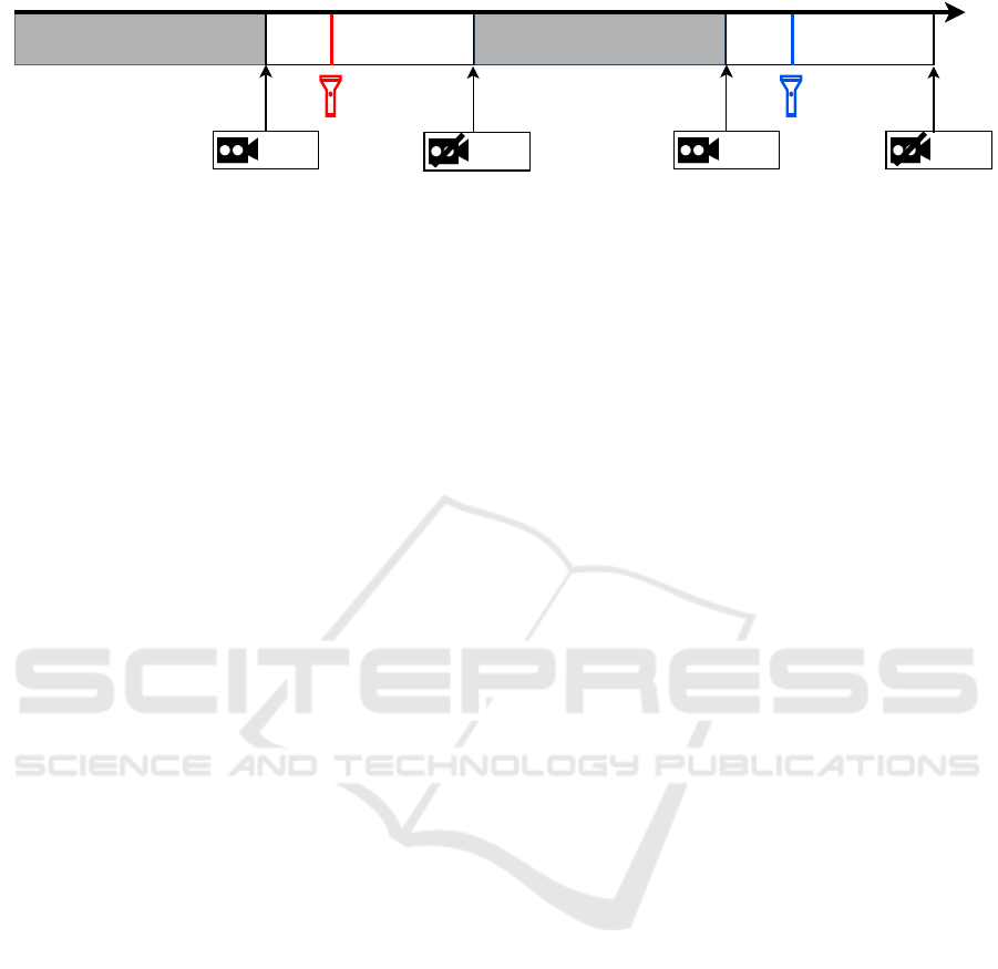
Initial light adaptation post-stimulusbaseline Pupil recovery
Start
Stop
post-stimulusbaseline
Start Stop
time
Figure 1: Chromatic pupillometry protocol schema.
been infrared camera based, which allows a proper
image contrast and the environment light does not
have an impact in pupil size. These type of systems,
with infrared cameras, are highly effective and pro-
vide precise measurements of the pupil. However,
they are also expensive, not portable and usually re-
quire some trained operator, which is a restriction for
its widespread use.
Mobile smartphones industry exponential increase
has brought a new interest for medicine applications,
as they overcome those previously referred limita-
tions, such as price, portability and accessibility. As
smartphones are present in everyones daily routines,
they could lead, in the limit, to a self-diagnosis tool
easily widespread over the population. The usage of
smartphones for pupillometry has started in 2013, by
Kim et al. (Kim and Youn, 2013), with the devel-
opment of a smartphone-based infrared video pupil-
lometer. This device included an optical apparatus at-
tached to the camera, with four infrared light emitting
diodes (LEDs), one white LED to work as the stim-
uli, one infrared cut-off filter and one microcontroller.
Tight occlusion around the eye was also taken care
to protect the eye from environment light, to reduce
its influence in PLR. All the acquired data was trans-
fered to a laptop and the analysis was made using a
proposed algorithm processed in MATLAB
R
(Math-
works Inc., Natick, MA).
Another smartphone app has been developed in
2016 by Shin et al. (Shin et al., 2016) used only to ac-
quire five steady images of the eye in different stages:
one before the flash, another one during the light stim-
uli and the remaining three photos after the flash. The
acquired images were then analyzed by a clinician
and compared the measurements with a penlight mea-
surement. In this research study, no automated neither
computerized algorithm was used to analyze pupil’s
size variations with the stimuli. The results showed
that pupil size measurements with a smartphone ap-
plication were similar to the ones made by a trained
clinician.
More recently, McAnany et al. (McAnany et al.,
2018) have developed an iPhone-based pupillometer,
which uses the rear camera to capture a high speed
video, its flash as a white stimulus and processes real
time measurements. The major difference from this
research study to the other two mentioned is the com-
pletely software based system, without the need to
any external optic apparatus, and the full process be-
ing taken care by the iPhone. The algorithm used to
analyze the acquired data uses a ratio between pupil
diameter and iris diameter. The measurements with
this system were then compared to ones acquired with
an infrared pupillometer and the results were in agree-
ment.
Although these studies show an increased po-
tential in smartphone-based pupillometry, apart from
McAnany et al. (McAnany et al., 2018) system, they
do not provide a low-cost, real-time, accessible and
portable device for pupillometry. The iPhone project
can be considered low cost when comparing to the in-
dustrial pupillometers, but comparing to other smart-
phones is a high range one. It is also important to
notice that the referred studies using smartphones do
not specifically target chromatic pupillometry, which
has been studied as a proper neuro-ophthalmological
diseases screening method (Rukmini et al., 2019).
The present study aims to describe an all-in-
one smartphone-based chromatic pupillometer using
a medium range Android device. All-in-one indicates
that the smartphone is used to acquire and process the
pupillometry data, running image processing algo-
rithms through the Android application, without the
need of high computing machines. The main goal of
this study was to show Android capability to perform
chromatic pupillometry measures and to run pupil de-
tection algorithms in real-time.
2 METHODS
Given the state-of-the-art of mobile pupillometers,
it is intended to develop a low cost system using a
medium range Android smartphone, with a camera to
allow pupil recording, a flash to work as a stimulus
and with enough capability to run image processing
algorithms. The system should also allow chromatic
pupillometry, using both blue and red stimuli in order
to be used to screen neuro-ophthalmological diseases
according to the recent findings and protocols.
Development of a Smartphone-based Pupillometer for Neuro-ophthalmological Diseases Screening
51
The acquisition protocol should start with a period
for the eye to adapt to the environmental light condi-
tions, then the recording period with some initial time
to acquire pupil’s baseline, then a short colored light
stimulus flash followed by a post-stimulus period. Be-
fore a new acquisition a pause should be made for
pupil recovery and the process should then be re-
peated with change of light stimulus color. A repre-
sentation schema of this type of protocol is shown in
Figure 1. Each of the protocol periods duration should
be tested and optimized in future work, particularly
to be applied to neuro-ophthalmological pathologies
such as Alzheimer, Dementia, Glaucoma or Parkin-
son.
Essentially, the main goal is to develop a system
with all these characteristics, that could be, in the
future, used in any Android device as a tool spread
through the population for neuro-ophthalmological
diseases early screening.
2.1 System Architecture
The system proposed in this work consists only in a
smartphone that allows acquiring and processing the
pupillometric data. Development was made using a
Nokia 7 Plus (Nokia Corporation, HMD Global, Fin-
land) which is an Android One device, with Android
9 Pie operating system (Android sdk 28). The appli-
cation was developed in Java programming language
using Android Studio (IntelliJ
R
Platform). Video and
image processing was made using OpenCV library
(Open Source Computer Vision Library) with Java
Native Interface (JNI) framework, which allows Java
to run C or C++ code, being then incorporated in the
Java Android application.
Acquisition
- Camera 2 API
- Rear facing flash control
Image Processing
- Video and Image Processing
- ElSe Algorithm
JAVA
C++
JNI
Figure 2: Proposed system architecture.
For the acquisition part of the application An-
droid’s Camera2 API was used, which provides an in-
terface to individual camera devices available in the
smartphone and proper adjustments of the recording
and image characteristics. In this case, Nokia 7 Plus
has one front camera and two rear cameras. The
rear-cameras are considered as one logical camera by
Camera2 API, which means that it is not possible to
access each of the physical rear cameras, they work as
one, so when changing recording characteristics the
result comes from both physical cameras combined
in one image.
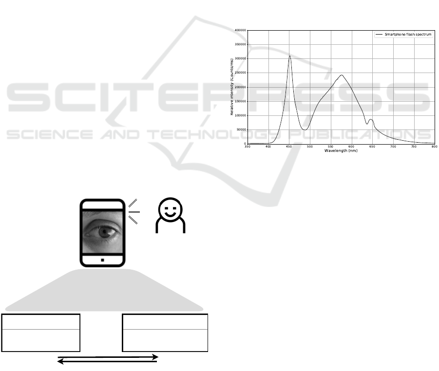
Camera2 API also allows to control rear-facing
flash light, which was used as light stimuli for pupil-
lometry measures. The spectral emission of the
rear-facing flash of the Nokia smartphone was mea-
sured using a spectrometer (AvaSpec - Mini2048CL
- UVI25 by Avantes, Netherlands) and the average
resultant of three acquisitions made is shown in Fig-
ure 3.
Figure 3: Spectral emission characteristics of the rear-
facing camera flash of the Nokia 7 Plus.
To allow chromatic pupillometry, a simple filter
made with standard grade cellophane paper, with blue
or red colors, was placed in front of the rear-facing
flash. In this way the color of the flash gets filtered to
get blue or red flash lights, whose spectra, which was
also acquired with Avantes spectrometer, are shown
in Figure 4. This low cost and easy solution to get
colored stimuli allows to get the proper wavelength
light stimuli (red and blue), according to literature.
2.2 Video and Image Processing
After video acquisition, the second main part of the
proposed smartphone application is the video and im-
age processing algorithm. Using OpenCV for An-
droid and C++ language, as previously referred, the
acquired video is then processed, converted to frames
and pupil is detected in each frame through a proper
algorithm.
BIODEVICES 2020 - 13th International Conference on Biomedical Electronics and Devices
52
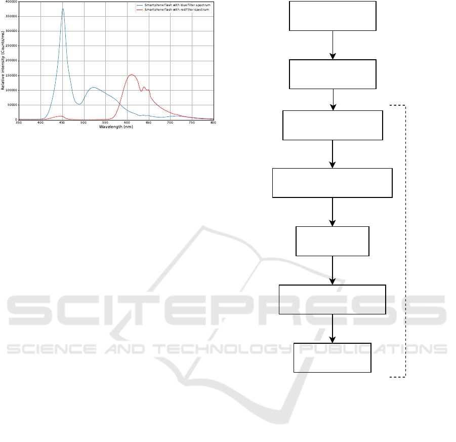
Figure 4: Spectral emission characteristics of the rear-
facing camera flash of the Nokia 7 Plus with red and blue
filters.
The acquired video does not contain only the eye,
but also some part of subject’s face, due to the dis-
tance to the smartphone to allow proper image focus.
To overcome this and to reduce non relevant informa-
tion in the image for the application of pupil detec-
tion algorithm, an eye detection algorithm is applied.
OpenCV offers a pre-trained Haar cascade algorithm
for face and eye detection, based in Viola and Jones
Haar cascade object detection algorithm (Viola and
Jones, 2001).
In this work, OpenCV Haar cascade eye detection
was applied to each frame,and was then cropped in the
obtained location. This algorithm sometimes fails and
considers some other part of the image to be an eye;
these detections with smaller size were automatically
discarded.
After eye detection, a process was made as sum-
marized in Figure 5 in order to detect the pupil.
Contrast Limited Adaptive Histogram and ElSe Al-
gorithm are going to be further explained in 2.2.1
and 2.2.2.
2.2.1 Contrast Limited Adaptive Histogram
One of the problems of images acquired with non in-
frared cameras or in non ideal lightning conditions is
the low contrast ratio between iris and pupil, particu-
larly in dark colored iris. To overcome this situation,
a contrast enhancement of the image increases pupil’s
visibility.
Histogram equalization distributes the intensities
on the histogram, leading to an increase in the global
contrast of an image. It is highly efficient and simple,
however it can produce ”washed out” effect or can
destroy the brightness of the image.
Adaptive Histogram Equalization allows locally
enhancement of the contrast, as it perform histogram
equalization in different sections of the image redis-
tributing the lightness values of the image. One of the
VideoAcquisition
GetVideoFrames
ConvertRGBtoGrayScale
ApplyCLAHE
ApplyElSeAlgorithm
Pupildetected
For each frame
EyeDetectionand
ImageCrop
Figure 5: Flowchart of the image processing algorithms.
main problems of this technique is the high compu-
tational complexity, not being favorable for real-time
applications.
An extended case of adaptive histogram equaliza-
tion is Contrast Limited Adaptive Histogram Equal-
ization (CLAHE), which performs an histogram clip-
ping at some threshold and redistributes the image us-
ing the maximum values. It has a lower computational
complexity and prevents over-amplification of noise
signals.
According to Hassan et al. (Hassan et al., 2017),
CLAHE algorithm outperforms a simple histogram
equalization or an adaptive histogram equalization for
iris recognition. Taking these results into considera-
tion, in this work CLAHE was tested and applied to
video frames before running the pupil detection algo-
rithm, using the OpenCV CLAHE function with cut
limit = 4.
Development of a Smartphone-based Pupillometer for Neuro-ophthalmological Diseases Screening
53
2.2.2 Pupil Detection Algorithm
After image acquisition and eye detection, the con-
cern rests in pupil detection algorithms as one of the
main parts in automated pupillometry systems. It is
important to clarify that pupil detection refers to find-
ing its center and size (area or diameter) in the image,
either in pixels or converted to some unit of measure.
There are some difficulties regarding achieving this,
that can go from low contrast images, blur, illumina-
tion issues and many others.
With the increasing interest in automated pupil-
lometry has also increased the need to have better and
more precise pupil detection algorithms. Particularly,
as the applications of pupillometry are being studied
to be in real-world environments and not only under
laboratory and controlled conditions.
One of those is ElSe algorithm, developed by Fuhl
et al. (Fuhl et al., 2015), based on ellipse evaluation
of a filtered edge image thought to be applied in real-
world scenarios, such as in-door environments or dur-
ing driving. In a state-of-the-art review published in
2016 (Fuhl et al., 2016) that compared several pupil
detection algorithms behavior has considered ElSe al-
gorithm as a gold standard for pupil detection. This is
an open source algorithm which is the one chosen to
be used in the system proposed in this work, due to
its easy access and it is targeted for real world envi-
ronments, which is what having a smartphone-based
pupillometer pursues.
The input is a gray scale image and in a very sum-
marized way the algorithm tries to find an ellipse that
could most likely be the pupil. First applies a Canny
filter to have the image edges, then they are filtered
using straightening patterns, the straight lines are re-
moved and the best ellipse is selected through least
square ellipse fitting. The final step is the ellipse eval-
uation, excluding those that are unlikely to be pupils.
If this first process fails there is a second approach that
the algorithm tries through coarse positioning. This
second analysis is made by downscaling and convolv-
ing the image with two different filters: a surface dif-
ference filter and a mean filter. The results of both
convolutions are multiplied and the maximum value
is the starting point to be refined. The surrounding
pixels of this point is verified, and the new pupil po-
sition is the center of mass of the pixels under this
threshold.
Even though ElSe algorithm was developed to
real-world scenarios it is important to notice that it
was tested and validated in datasets acquired with in-
frared cameras, which in most of the cases have a very
evident pupil. In the present work, this algorithm was
applied to images acquired using Nokia 7 Plus smart-
phone camera, without any optical apparatus, as a pre-
liminary test and validation of the proposed solution.
Some of Fuhl et al. (Fuhl et al., 2015) datasets pos-
sess eye images where the pupil is hidden, sideways
or even more far-fetched scenarios. In this prelimi-
nary study, ElSe algorithm was only applied to im-
ages where the pupil is normal, with subject looking
straight forward and the pupil is not occluded by eye-
lashes for example.
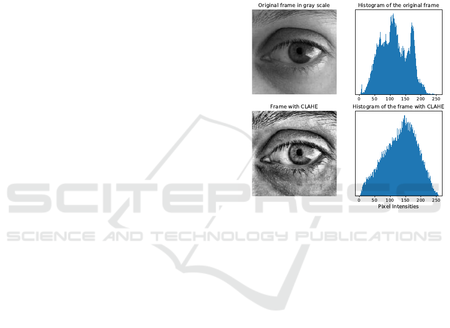
Figure 6: Original video frame in gray scale with and with-
out CLAHE with respective histograms.
3 PRELIMINARY RESULTS
First, pupil detection algorithm running in the An-
droid application was validate with eye images
datasets published by Fuhl et al. (Fuhl et al., 2015).
As expected, pupil center and size were the same as
Fuhl research group has labeled. This simple test
was just to guarantee that ElSe algorithm was prop-
erly running in the Android application, considering
the system architecture and the linkage between pro-
gramming languages.
Preliminary tests were made using the developed
smartphone application to acquire videos of the eye
and apply the processing algorithms to get the pupil.
ElSe algorithm was tested in frames with and with-
out CLAHE. An example of the same frame with and
without CLAHE is shown in Figure 6 with the re-
spective histograms.
The image processing algorithms proposed in this
work for each frame are exemplified in Figures 7
and 8 with a frame from a video acquired with the
developed smartphone application.
BIODEVICES 2020 - 13th International Conference on Biomedical Electronics and Devices
54
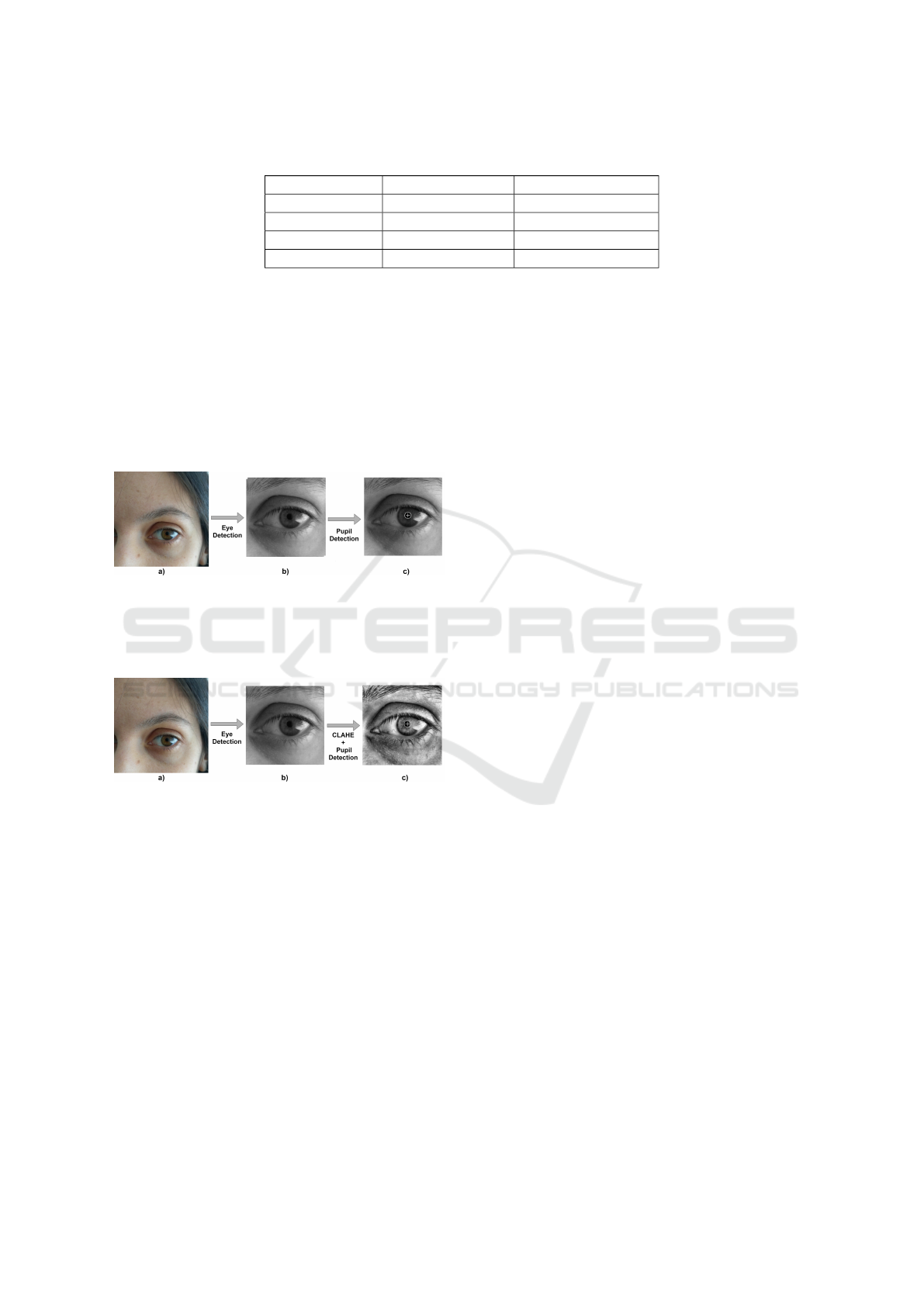
Table 1: Pupil parameters mean values obtained for 41 images with and without CLAHE. Unit of measure: pixels.
Pupil parameter Original Image Image with CLAHE
Center (124 ± 6, 106 ± 5) (124 ± 6, 106 ± 5)
Height 20 ± 8 21 ± 7
Width 19 ± 7 19 ± 6
Angle 108 ± 32 105 ± 36
The pupil detection algorithm with and without
CLAHE was also applied to 41 frames from the same
acquisition made with the same person. The average
results for these 41 images are summarized in Table 1
for both CLAHE and non CLAHE algorithms. From
this test it is possible to verify that the results are sim-
ilar in terms of center, height and width, being the
angle the most different value, in average, and with
higher standard deviation.
Figure 7: Image Process Schema. a) Frame from the
video acquired using the smartphone-bases pupillometer
with Nokia 7 Plus; b) Cropped image around the eye ob-
tained through eye detection algorithm; c) Eye image with
pupil detected.
Figure 8: Image Process Schema with CLAHE. a) Frame
from the video acquired using the smartphone-bases pupil-
lometer with Nokia 7 Plus; b) Cropped image around the
eye obtained through eye detection algorithm; c) Eye image
with CLAHE and pupil detected.
With the images acquired in these preliminary
tests this algorithm seems promising to deal with im-
ages acquired with this smartphone camera without
any optical apparatus and further tests should be made
to better validate the proposed algorithm.
4 CONCLUSION
Using low-cost, portable and accessible technology
for medical applications, particularly for screening
and monitoring diseases, is gaining interest and mar-
ket all over the world. The usage of a smartphone for
this purpose is a smart and easy way to spread early
screening and make it more available and accessible
to everyone and all over the world. This work pro-
poses a solution for pupillometry measurements with
a smartphone, which overcomes the main problems
with the existing pupillometers to spread this tech-
nique into medical and screening usage. The pre-
liminary tests made with the proposed prototype in-
dicate a great potential of this solution, particularly
due to its low price, easy accessibility and portability.
Another advantage of the proposed system is that it
only needs the smartphone, diminishing the requisites
of high range technology and apparatus. To perform
chromatic pupillometry it needs a standard grade cel-
lophane paper, which, in the future in a commercial
solution, can be available as a kit to complement the
smartphone application.
Further work is to improve the algorithms for
pupil detection and make more validation tests. It
is also relevant to test this solution in different light
conditions and make some adjustments in the camera
recording characteristics in order to get a more effi-
cient and precise pupillometer application.
After these improvements and algorithm val-
idations, the next step should be validating
this smartphone-based pupillometer for neuro-
ophthalmological diseases screening, using the
colored filters to apply colored stimuli allowing to do
chromatic pupillometry. With these colored stimuli,
should be tested different acquisition protocols
that could early screen pathologies as Alzheimer,
Dementia, Glaucoma or Parkinson in a portable and
accessible way.
In general, a smartphone-based pupillometer
seems to be the future of pupillometry to lower the
gap between academic research and clinical applica-
tion. The preliminary tests made with the proposed
system show its potential to be used as a pupillome-
ter and, in the future, to screen and monitor neuro-
ophthalmological diseases.
ACKNOWLEDGEMENTS
This work is funded by National Funds through FCT
- Portuguese Foundation for Science and Technology
Development of a Smartphone-based Pupillometer for Neuro-ophthalmological Diseases Screening
55

and Compta S.A. under the PhD grant with reference
PD/BDE/135002/2017. A special acknowledgment to
Compta S.A. team for all the support given.
REFERENCES
Fuhl, W., Santini, T. C., Kuebler, T., and Kasneci, E. (2015).
ElSe: Ellipse Selection for Robust Pupil Detection in
Real-World Environments.
Fuhl, W., Tonsen, M., Bulling, A., and Kasneci, E. (2016).
Pupil detection for head-mounted eye tracking in the
wild: an evaluation of the state of the art. Machine
Vision and Applications, 27(8):1275–1288.
Gamlin, P. D., McDougal, D. H., Pokorny, J., Smith, V. C.,
Yau, K. W., and Dacey, D. M. (2007). Human
and macaque pupil responses driven by melanopsin-
containing retinal ganglion cells. Vision Research,
47(7):946–954.
Giza, E., Fotiou, D., Bostantjopoulou, S., Katsarou, Z., and
Karlovasitou, A. (2011). Pupil light reflex in Parkin-
son’s disease: Evaluation with pupillometry. Interna-
tional Journal of Neuroscience, 121(1):37–43.
Gracitelli, C. P. B., Duque-Chica, G. L., Moura, A. L.,
Nagy, B. V., de Melo, G. R., Roizenblatt, M., Borba,
P. D., Teixeira, S. H., Ventura, D. F., and Paranhos, A.
(2014). A Positive Association Between Intrinsically
Photosensitive Retinal Ganglion Cells and Retinal
Nerve Fiber Layer Thinning in Glaucoma. Investiga-
tive Ophthalmology & Visual Science, 55(12):7997–
8005.
Granholm, E. L., Panizzon, M. S., Elman, J. A., Jak, A. J.,
Hauger, R. L., Bondi, M. W., Lyons, M. J., Franz,
C. E., and Kremen, W. S. (2017). Pupillary Responses
as a Biomarker of Early Risk for Alzheimer’s Disease.
Journal of Alzheimer’s disease : JAD, 56(4):1419–
1428.
Hassan, R., Kasim, S., Jafery, W. A. Z. W. C., and Shah,
Z. A. (2017). Image enhancement technique at differ-
ent distance for Iris recognition. International Journal
on Advanced Science, Engineering and Information
Technology, 7(4-2 Special Issue):1510–1515.
Hattar, S., Liao, H. W., Takao, M., Berson, D. M., and Yau,
K. W. (2002). Melanopsin-containing retinal ganglion
cells: Architecture, projections, and intrinsic photo-
sensitivity. Science, 295(5557):1065–1070.
Kim, T. H. and Youn, J. I. (2013). Development of a
smartphone-based pupillometer. Journal of the Op-
tical Society of Korea, 17(3):249–254.
Lowenstein, O. and Loewenfel, I. E. (1958). Electronic
Pupillography; A New Instrument and Some Clini-
cal Applications. A.M.A. Archives of Ophthalmology,
59(3):352.
Lucas, R. J., Douglas, R. H., and Foster, R. G. (2001).
Characterization of an ocular photopigment capable
of driving pupillary constriction in mice. Nature Neu-
roscience, 4(6):621–626.
McAnany, J. J., Smith, B. M., Garland, A., and Kagen, S. L.
(2018). IPhone-based pupillometry: A novel approach
for assessing the pupillary light reflex. Optometry and
Vision Science, 95(10):953–958.
Rukmini, A. V., Milea, D., Baskaran, M., How, A. C., Per-
era, S. A., Aung, T., and Gooley, J. J. (2015). Pupillary
Responses to High-Irradiance Blue Light Correlate
with Glaucoma Severity. Ophthalmology, 122(9[1]
A. V. Rukmini et al., “Pupillary Responses to High-
Irradiance Blue Light Correlate with Glaucoma Sever-
ity,” Ophthalmology, vol. 122, no. 9, pp. 1777–1785,
2015.):1777–1785.
Rukmini, A. V., Milea, D., and Gooley, J. J. (2019). Chro-
matic pupillometry methods for assessing photorecep-
tor health in retinal and optic nerve diseases. Frontiers
in Neurology, 10(FEB):1–20.
Shin, Y. D., Bae, J. H., Kwon, E. J., Kim, H. T., Lee, T.-S.,
and Choi, Y. J. (2016). Assessment of pupillary light
reflex using a smartphone application. Experimental
and Therapeutic Medicine, 12(2):720–724.
Viola, P. and Jones, M. (2001). Rapid object detection us-
ing a boosted cascade of simple features. In Proceed-
ings of the 2001 IEEE Computer Society Conference
on Computer Vision and Pattern Recognition. CVPR
2001, volume 1, pages I–511–I–518. IEEE Comput.
Soc.
Wang, C. A., McInnis, H., Brien, D. C., Pari, G., and
Munoz, D. P. (2016). Disruption of pupil size mod-
ulation correlates with voluntary motor preparation
deficits in Parkinson’s disease. Neuropsychologia,
80:176–184.
BIODEVICES 2020 - 13th International Conference on Biomedical Electronics and Devices
56
