
Thyroid Ultrasound Images Classification using the Shearlet Coefficients
and the Generic Fourier Descriptor
Noura Aboudi
a
and Nawres Khlifa
Laboratoire de Biophysique et Technologies M
´
edicales, Universit
´
e de Tunis El Manar, Tunis, Tunisia
Keywords:
Thyroid Nodule, Feature Extraction, Shearlet Transform, Generic Fourier Descriptor, Feature Selection,
Random Forest.
Abstract:
To ameliorate the classification accuracy of the thyroid ultrasound imaging computer-aided diagnosis (CAD)
system based on feature extraction, we used the Shearlet Transform (ST) to extract texture features, and the
Generic Fourier Descriptor (GFD) to extract shape feature descriptor based on contours information. The ST
supplies a rotation invariant descriptor at various scales. The GFD descriptor is autonomous, robust, and has
no redundant features. Then, we applied a feature selection method on the extracted shearlet descriptor to
build up the performance metrics. Finally, we used the objective metrics(sensitivity, specificity, and accuracy)
to validate the performance of the proposed method. Experimentally, we apply our novel methods on a public
dataset and we use the Support Vector Machine(SVM) and Random Forest (RF) as classifier. The obtained
results prove the superiority of the proposed method.
1 INTRODUCTION
Thyroid nodule is an abnormal growth of cancerous
lumps in the thyroid gland, it is the accumulation of
malignant cells in thyroid gland tissues. It is one of
the most leading cause of cancer deaths. In 2017,
56,870 new patients in the United States have been
reported to have involved nearly thyroid cancer (Ab-
basian Ardakani et al., 2018). Generally, the most of
thyroid nodules diseases are benign.
Currently, thyroid ultrasound imaging has been
the most used tools for early thyroid nodules de-
tection and diagnosis. It is inexpensive, radiation-
free imaging tool, and provides the benefits infor-
mation needed for medical diagnosis (Abbasian Ar-
dakani et al., 2018; Zhang and Lu, 2002). How-
ever, the diagnosis of thyroid ultrasound image de-
pends greatly on personal experience and skills. Thus,
many benign and malignant nodules have similar vi-
sual characteristics. Hence, experienced radiologists
have a high good diagnosis rate than beginner radi-
ologists. Thyroid ultrasound Computer Aided Diag-
nosis (CAD) system becoming progressively a cru-
cial tool, that assists to offers an objectivity evalu-
ation diagnostic and a better decision accuracy. In
general, the thyroid ultrasound CAD system is con-
stituted of four steps, containing image preprocess-
a
https://orcid.org/0000-0002-5101-853X
ing, image segmentation, feature extraction and se-
lection, and classification. Feature extraction is one
of the crucial stages in thyroid ultrasound CAD. The
extracted features are usually classified into textural
and shape (morphological) features. Latterly, many
researches are focused on the feature extraction and
selection steps. Usually, the thyroid nodules classi-
fication problems depend on the extracted features.
Also, the textural features are commonly used in thy-
roid ultrasound CAD.
Usually, the textural features descriptors are cal-
culated using a diversity of statistical and structural
approaches, such as Grey Level Co-occurrence Ma-
trix (GLCM), autocorrelation-based approaches, Lo-
cal Binary Pattern, and auto-covariance coefficients.
These methods can describe the statistical features of
grey level variation in a Region of Interest (ROI). The
popular advantage of these methods is they are easy to
implement, but the extracted textural features by these
methods are mostly from the special domain and ig-
nore the frequency features, which are very important
in the classification step. Moreover, the multiscale
properties of an image not evaluated in these methods.
The Multiscale Geometric Analysis (MGA) grants
complete features analysis using different scales.
Recently, image analysis based on transform ap-
proaches has been widely used in the image feature
extraction. The transform approaches is the represen-
292
Aboudi, N. and Khlifa, N.
Thyroid Ultrasound Images Classification using the Shearlet Coefficients and the Generic Fourier Descriptor.
DOI: 10.5220/0008956902920298
In Proceedings of the 15th International Joint Conference on Computer Vision, Imaging and Computer Graphics Theory and Applications (VISIGRAPP 2020) - Volume 4: VISAPP, pages
292-298
ISBN: 978-989-758-402-2; ISSN: 2184-4321
Copyright
c
2022 by SCITEPRESS – Science and Technology Publications, Lda. All rights reserved

tation of an image in the frequency and scale space;
when the features description and interpretation are
related on this special coordinate. The MGA is ap-
plied in different fields. One common field is the tex-
ture feature extraction in thyroid ultrasound images.
The shearlet transform is a powerful spatial frequency
analysis method. Shearlet provides sufficient tools
to exactly detect the orientations, the scales and the
positions of pixels (Easley et al., 2008a). Shearlet
transform has been fully utilized in image processing,
Edge and Ridge Detection and Analysis, image sep-
aration, and image denoising(?). Despite, a limited
research used the shearlet transform on the textural
features extraction.
Also, thyroid nodules analysis based on the shape
description is very important. Shape features can be
categorized into two principal groups: region-based
features and contour-based features. The contour-
based description method explores the boundary in-
formation and ignores the internal content of the
shape, so the versatility is not high. The region-
based description method uses the internal pixel in-
formation shape. Contour-based Descriptors can be
described by the fourier descriptor, the wavelet de-
scriptor, and the shapes signatures. In many exist-
ing shape feature descriptors, the Generic Fourier De-
scriptor (GFD) has several desirable features, such as
low computational complexity, sharpness to fine de-
scription, which makes it a popular descriptor. The
GFD is one of the boundary feature extraction tech-
niques. GFD is obtained by applying the 2D Fourier
Transform in polar image and it extracts the spectral
features in radial and circular direction.
In this paper, we use the ST to extract textural fea-
ture and the GFD to explore shape descriptor features.
To achieve better classification performance, we pro-
pose a hybrid approach combining ST and GFD for
ultrasound thyroid nodules classification. The rest of
paper is organized as follows. In section 2, we in-
terpret the related work. The proposed method is de-
scribed in section 3. The section 4 represents the ob-
tained results and discussion. Finally, conclusion with
some feature works idea is given in section5.
2 RELATED WORKS
The calcification and detection of thyroid nodules in
ultrasound images was evolved in many studies. Dif-
ferent Computer-Aided Diagnosis was developed us-
ing a variety of features and classifiers for the clas-
sification of thyroid nodules. The most studies has
proven the potentiality and the importance of textural
and morphological features on the diagnostic of nod-
ules. Many CAD has developed for thyroid diseases
classification. The neural networks has used by Ozy-
ilmaz et al.(Ozyilmaz and Yildirim, 2002) for the di-
agnosis of thyroid nodules, they are applied different
architectures on their database (13, 2017). The pro-
posed method attained 88.3% as maximal accuracy
value. Also, Keles et al.(Keles¸ and Keles¸, 2008), de-
veloped a CAD system based on neuro fuzzy clas-
sification testing on the similar dataset (13, 2017),
it attaining 95.33% accuracy value. Iakovidis et
al.(Iakovidis et al., 2010), proposed a method based
on textural and echogenicity features, focused on im-
age analysis. In this work, they used the fuzzy local
binary pattern to represent the texture feature. The
proposed method used 250 thyroid ultrasound im-
ages, achieving 97.5% as the best ROC AUC, utiliz-
ing polynomial kernel SVM as classifier. Acharya et
Al.(Acharya et al., 2011), proposed a system for the
diagnosis and classification of malignant thyroid nod-
ules using 20 contrast enhancement images(CEUS).
They used the Discrete Wavelet Transform (DWT)
and texture parameters for feature extraction. The
DWT detects the small variations in malignant and
benign nodules. The accuracy values achieve 98% us-
ing KNN classifier. In another study(Acharya et al.,
2012), the same author combined a Fourier Descrip-
tor (FD), local binary patterns, fractal dimensions and
Law’s texture energy to detect features from 20 im-
ages. The highest accuracy value is of 100% us-
ing SVM and fuzzy classifier. On other lately stud-
ies, in (Raghavendra et al., 2017) authors used the
Binary Stack Decomposition (BSD) algorithm and
Two-Threshold Binary Decomposition algorithm to
extract 120 features from 242 images. In this case,
a 97.52% accuracy value was attained using SVM
classifier. The higher number of extracted features
decreases the performance and the exactitude of the
impact of these features. So, they have applied the
fisher analysis (MFA) to select and reduce the fea-
tures sets. MFA based on the fuse of existing features
and the created features. Chi et al(Chi et al., 2017),
proposed a recently study published on 2017; they
used a deep learning features extraction method using
a GoogLeNet model. In this approach, 1024 features
was extracted and used to classify the nodules using
Random Forest classifier. They used two dataset in
the evaluation of their method, the accuracy value at-
tained 98.29% of the first dataset (357); and 96.34%
for the second dataset (164 images). The obtained re-
sults improves that deep learning offer a good results.
Thyroid Ultrasound Images Classification using the Shearlet Coefficients and the Generic Fourier Descriptor
293
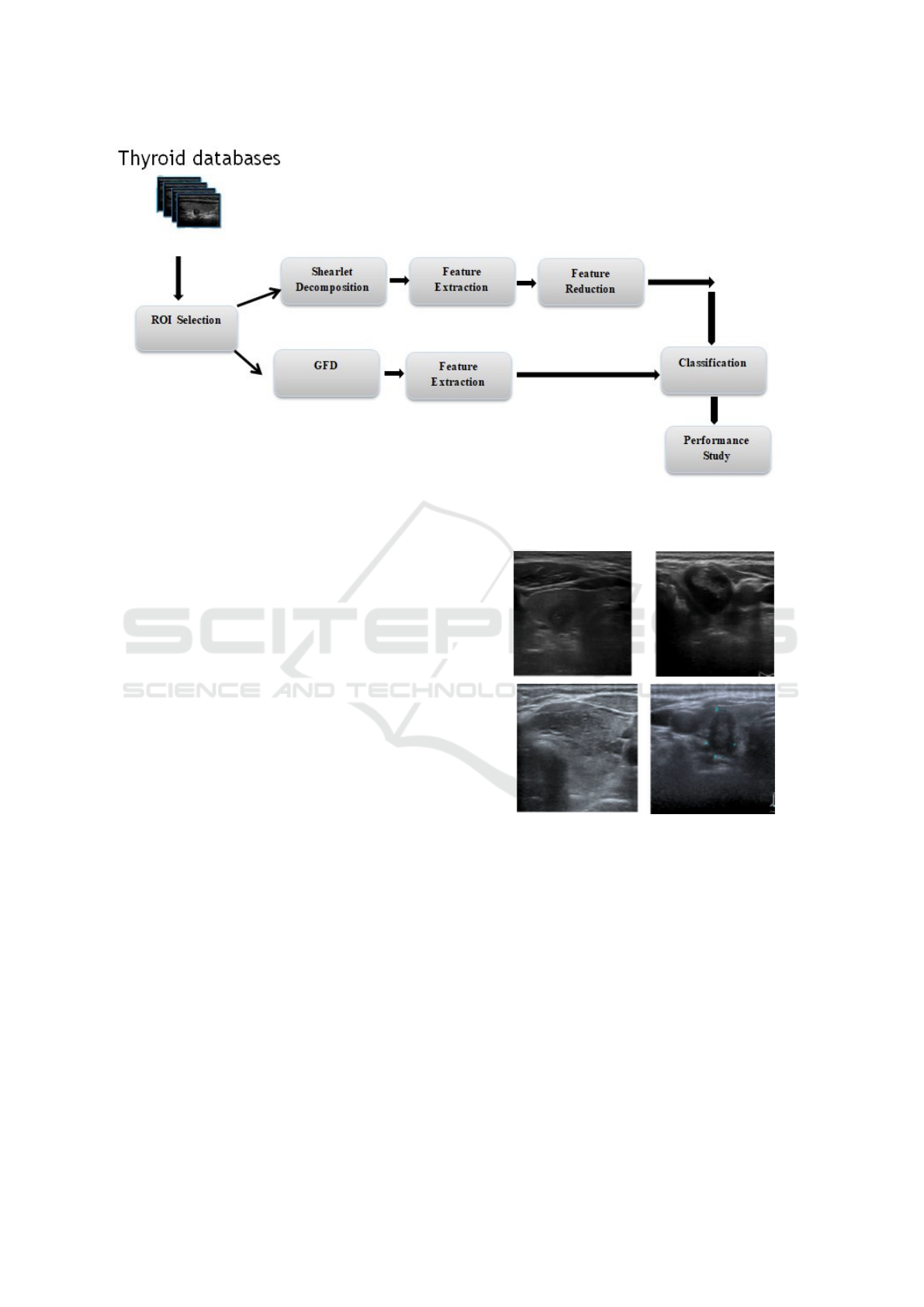
Figure 1: Diagram of the proposed method.
3 MATERIALS AND METHODS
The proposed method is composed of three steps:
ROI selection, Shearlet Transform and GFD decom-
position feature extraction, feature selection, and clas-
sification based on SVM and RF. The proposed ap-
proach was presented in figure 1.
3.1 Data Collection
In our experiment, a total of 447 thyroid ultrasound
images with benign and malignant thyroid nodules
were used to evaluate the performance of the pro-
posed method. Thyroid ultrasound images used in this
work are acquired from the laboratory CIM @ LAB of
the National University of Colombia and the medical
diagnostic institute. Each image involves one or more
nodules (attached with XML file ready by expert and
contain annotation and patient’s information). The
ROI was selected using the position denoted in the
XML file. The original image had a resolution of
546*410. These extracted nodules are grouped into
two classes: benign and malign. In this work, out of
447 thyroid nodules, 372 nodules are malignant and
75 nodules are benign. An example of thyroid benign
and malign is presented in figure2 respectively.
Figure 2: Example of benign and malign thyroid nodules.
3.2 Feature Extraction
3.2.1 Shearlet-based Texture Feature
Descriptors
The textural feature are extracted using the Shearlet
Transfrom (ST) , it is briefly defined as follows:
Shearlet systems were first introduced by K. Guo, G.
Kutyniok, D. Labate, W.-Q Lim and G. Weiss in (Guo
et al., 2006; Labate et al., 2005). ST is a multiscale
directional transform method which allows efficient
encoding of anisotropic features, based on directional
filter followed by Laplacian pyramid (LP). Shearlet
Transform provides an effective tool for combining
VISAPP 2020 - 15th International Conference on Computer Vision Theory and Applications
294

the multi-scale and the invariance notation. This
multi-scale decomposition improves the robustness
multi-directional and multi-scale analysis and the
representation of the data image. It represent the
image in the frequency space where the texture
description is closely related to this coordinate. ST
used to identify directional features in images (Easley
et al., 2008b; Guo et al., 2006). It is generated by
applying a set of operators to a single function. The
most important advantage of the ST is not sensitive
with scales and orientations variations, and it is
more powerful in understanding the geometry of
images. Recently, ST constitute one of the most
successful methods for the efficient representation
of multidimensional data and in the understanding
the geometry of images. Like that, it is only con-
nected with two parameters, the scaling parameter
a and the translation parameter t. For the input im-
age f , continuous Shearlet Transform is described by:
f −→ SH
ψ
f (a,s,t) = h f ,ψ
a,s,t
i (1)
Where:
ψ: is the generating function;
a : is the scaling parameter;
s ∈ R: shear parameter;
t ∈ R :translation parameter;
and ψ
a,s,t
:shearlet basis functions.
Important properties of shearlets are they are well lo-
calized, they follow parabolic scaling law, they have
high directional sensitivity, and they are optimally
sparse.
In the proposed method, ultrasound thyroid nod-
ule is decomposed by ST into three layers, therefore
the textural features descriptors were extracted from
these layers. The contrast, correlation, energy, homo-
geneity, entropy, skewness, variance, mean, standard-
deviation of each sub-bands are extracted from these
three layers for the horizontal and vertical cone and
are used as directional features. Shearlet can capture
directional features like orientations in images, which
are in fact one of the most discriminating features. By
practicing the ST to an image, a number of decompo-
sition levels and directional subbands are produced.
3.2.2 Fourier Feature Descriptors
In this study, we have utilized the generic Fourier
descriptor (GFD) for efficient shape representation.
Most of the actual shape descriptors are non-robust.
GFD is proposed to crucify the disadvantages of
the current shape representation methods that inde-
pendent, easy to implement, less sensitive to noise,
and robust. GFD introduced by Zhang(Zhang and
Lu, 2002). It is a contour-based shape descriptor for
image classification. To obtain invariance to rotation,
the image is first converted to polar coordinates then
we use a 2D Fourier transform on a polar image.
GFD uses the modified polar Fourier transformation
(MPFT) of a region shape to the polar coordinate
system. So, the coordinates of all pixels of the
initial images are converted into polar coordinates.
It detects spectral feature in both radial and circular
direction. The determination of the number T and
R for the description of the forms is physically
feasible, because the shape characteristics are usually
extracted by the low frequencies. Finally, the GFD
has the following expression:
For an shape image
I = { f (x,y);0 ≤ x < M,0 ≤ N} (2)
p f (ρ, ψ) =
R
∑
r=0
T
∑
i=0
f (r,θ
i
)
j2π(
r
R
ρ+
2πi
T
ψ)
(3)
Where:
T : is the angular resolution;
R : is the radial resolution;
The Fourier coefficients obtained are translation in-
variant. So to realize scaling and rotation invariance,
the following normalization is calculated:
GFD = {|
p f (0, 0)
aire
,|
p f (0, 1)
p f (0, 0)
,..., |
p f (0, n)
p f (0, 0)
,..., |
p f (m, n)
p f (0, 0)
}
(4)
Where:
area : symbolize the area of the border circle in which
the polar image exists;
andn= max (angular frequencies);
m = max (radial frequencies).
In this paper, we have used the Generic Fourier
Fescriptor (GFD) as shape descriptor. For efficient
shape description, only a small number of GFD fea-
tures are selected for shape representation. In our
implementation, 36 GFD features reflecting 4 radial
frequencies and 9 angular frequencies are selected to
index the shape. The selected GFD features form a
feature vector which is used for indexing the shape.
Therefore, the online matching is efficient and sim-
ple. The extracted features with GFD are no redun-
dant and it grants a multi-resolution feature analy-
sis in radial and angular directions. The GFD fea-
ture vector is introduced by the following expression:
GFD(0, 0),GFD(0, 1), . . . ,GFD(0, n), . . . ,GFD(m,
0). . . ,GFD(m, n).
3.3 Feature Selection
Feature selection is a dimensionality reduction
method which aims to select a subset of relevant and
Thyroid Ultrasound Images Classification using the Shearlet Coefficients and the Generic Fourier Descriptor
295

informative features from the initial features set by
eliminating irrelevant and redundant features. It aug-
ments the classification performance, the efficiency in
learning stage, and reducing the computation cost and
the complexity. The total number of extracted texture
feature was 207. Some of these are irrelevant and not
significant in the differentiation between benign and
malignant nodules and not appropriate for classifica-
tion. The feature selection method utilized should be
able to choose a subset of relevant and most repre-
sentative features. In this study, we have applied the
Reliable Attribute Selection Based on Random Forest
(RASER)(Noura et al., 2016) to eliminate the non-
informative features. The features results obtained af-
ter applying this feature selection method was used
in the classification, and for construct SVM and RF
models.
3.4 Classification Algorithm
In order to evaluate the performance of the proposed
method, the extracted features were fed to the classi-
fier for discriminating the malignant from the benign
nodules. Machine Vector Support (SVM) and Ran-
dom Forest (RF) were applied to measure the perfor-
mance of these features. Concise descriptions of these
two classifiers are given below.
3.4.1 Support Vector Machine
Support Vector Machine (SVM) is a supervised learn-
ing algorithm, SVM belongs to the class of linear
classifiers (that use a linear separation of data). It
is known for their strong theoretical guarantees, their
great flexibility and their ease of use even without
much knowledge of data. SVM is intended to sep-
arate data into classes using a boundary, so that the
distance between the different groups of data and the
boundary between them is maximum. SVM is based
on the generation of hyperplanes to discriminate fea-
tures categorizing to two different classes. In this
study, a weighted SVM algorithm is used to equili-
brate imbalance classes. Weighted SVM (W-SVM)
solves the problem of having two classes with unequal
training data. W-SVM sets the penalty parameter C
in proportion to the size of the class. With regard to
RF, the principal advantage of SVM is its simpler ge-
ometric interpretation and lower computational cost.
The main advantages of SVM are its simpler inter-
pretation and computation cost compared to Random
Forest (RF).
3.4.2 Random Forest
Random Forest (RF) is a one of the ensemble meth-
ods of classification. The ensemble methods type is
based on vote to predict the final decision. RF is con-
structs of a large number of decision trees based on
averaging random selection of variables. RF is based
on the idea of bagging and Random subspaces in the
construction of decision trees. The randomness no-
tion is in the subsampling of the training data and in
the selection of the node tests, each tree is build us-
ing different subset. RF uses the majority votes in the
classification case in the terminal leaf nodes. More
than, RF has the ability to measure the importance of
used the features.
3.4.3 Validation of Classifier
We have used the group 10 fold cross-validation on
the evaluation of the proposed method. K-fold cross
validation based on the random split of the dataset
into k equal samples, and it guarantee that the same
set of data not be selected in both testing and train-
ing sets. Between the k samples, one sample is used
as test dataset and the remaining k-1 sets are used as
training dataset. The accuracy, sensitivity, specificity
were chosen as critical measure performance of the
proposed method for both SVM and random forest
classifiers. Their definitions are as follows:
Sensitivity = T P/(T P + FN) (5)
Speci f icity = T N/(T N + FP) (6)
Accuracy =
T P + T N
T P + T N + FP + FN
(7)
where : T P represent the correct classification rate of
malignant instances;
FN is the misclassification rate of malignant in-
stances;
T N is the correct benign instances; and
FP indicates the misclassification rate of benign in-
stances.
4 RESULTS AND DISCUSSION
In this paper, the thyroid nodules are classified into
two classes: benign and malignant. The ROIs are
extracted initiallly from the ultrasound images and
then subjected to the shearlet transform and generic
fourier descriptor. All nodules are decomposed with
shearlet transform into three layers. The 9 textu-
ral features(contrast, correlation, energy, homogene-
ity, entropy, skewness, variance, mean,and standard-
deviation) are extracted from each sub bands. In total,
VISAPP 2020 - 15th International Conference on Computer Vision Theory and Applications
296
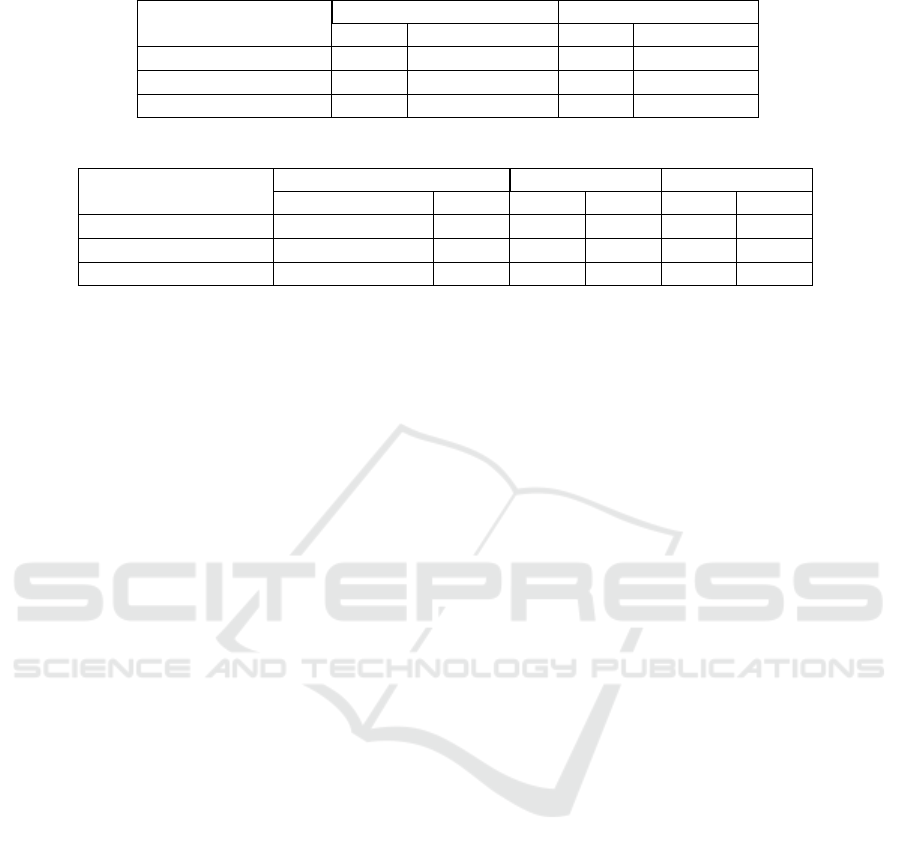
Table 1: Classification performance for Shearlet feature using SVM and RF(unit: %).
Index Without feature selection With feature selection
SVM RF SVM RF
Accuracy 93.3 94.65 94.59 96.64
Sensitivity 89.86 92.95 91.78 93.15
Specificity 91.6 95.79 93.11 95.98
Table 2: Classification performance for different features using SVM and RF (unit:%).
Index Shearlet GFD Fusion feature
SVM RF SVM RF SVM RF
Accuracy 94.59 96.64 97.74 95.9 96.55 98.51
Sensitivity 91.78 93.15 97.58 97.4 95.87 98.17
Specificity 93.11 95.98 98.11 89.2 97.11 96.08
207 features were extracted from the all sub bands.
Further, these features were subjected to Reliable At-
tribute Selection Based on Random Forest (RASER)
dimensionality reduction method to eliminite redun-
dant and irrelivant features. Finally, relevant features
were fed to SVM and RF classifier to test the pro-
posed method based on 10 cross validation. The table
1 represents a comparison of the classification per-
formance using SVM and RF classifier. The feature
selection method provides also a good classification
performance compared to the classification without
feature reduction method.
It can be note that best accuracy, sensitivity and
specificity values are 94.59%, 91.78%, and 93.11%
for SVM and 96.64%, 93.15%, and 95.98% for RF
after apply the feature selection. In rest of study,
only the relevant shearlet coefficient are used. Unlike
shearlet transform, GFD has no redundant features.
Table 2 represents the obtained evaluation metrics
value between different feature type using SVM
and RF classifiers, respectively. GFD achieves
better classification compared to the Shearlet-based
method. The accuracy value achieve 97.74%,96.64 %
respectively for GFD using SVM and RF classifier,
and 94.59%, 95.9% for the shearlet coefficient using
SVM and RF.
Therefore, the quantitative results of classification
accuracy, sensitivity, and specificity for different
combination of texture features and classifiers are
shown in table 2. They show that the contribution of
the new descriptors improves the overall accuracy,
sensitivity, and specifity. The classification accuracy
of the fusion features is 96.55% and 98.51% using
SVM and RF respectively, which are much higher
than those of other methods.
Recently, several researchers have encouraged to
propose new efficient method to diagnose thyroid
cancer using ultrasound images. Table 3 summarizes
the achieved results on thyroid nodule classification,
we introduce the obtained performances by present-
ing the accuracy value. The compared methods are:
Conic Section Function Neural Network (Ozyilmaz
and Yildirim, 2002); neuro fuzzy Classification(Keles¸
and Keles¸, 2008); fuzzy local binary pattern(Iakovidis
et al., 2010); Discrete Wavelet Transform (DWT)
and texture parameters(Acharya et al., 2011) that
is based in the texture feature and the wavelet
transform; Two-Threshold Binary Decomposition
algorithm(Acharya et al., 2012); and deep learning
features extraction(Chi et al., 2017). Really, we
produced the accuracy value for the proposed method
as well as for six relevant thyroid nodules classifi-
cation methods from the state of the art. It can be
clearly seen that the proposed method reaches a good
performances and still better to other methods.
In this paper, we have proposed an efficient
method for classification of thyroid nodules using the
shearet transform, generic fourier descriptor and in-
variant texture features. Shearleat transform has some
important properties, like multiresolution, multidirec-
tion and multiscale, which approve the uniqueness
above different levels. At present, textural feature is
commonly used in CAD system to classify the ultra-
sound thyroid nodules. The combination of statistical
and transform based features improve the classifica-
tion accuracy for thyroid nodules classification.
5 CONCLUSION
In conclusion, we proposed a new feature extrac-
tion method based on shearlet transform and generic
fourier descriptor for characterizing thyroid nodules
in ultrasound image. The comparative experiment re-
sults indicated that the combination of both shearlet-
based texture and fourier based edge features have
the best classification performance. The proposed
method was tested on public thyroid database re-
Thyroid Ultrasound Images Classification using the Shearlet Coefficients and the Generic Fourier Descriptor
297
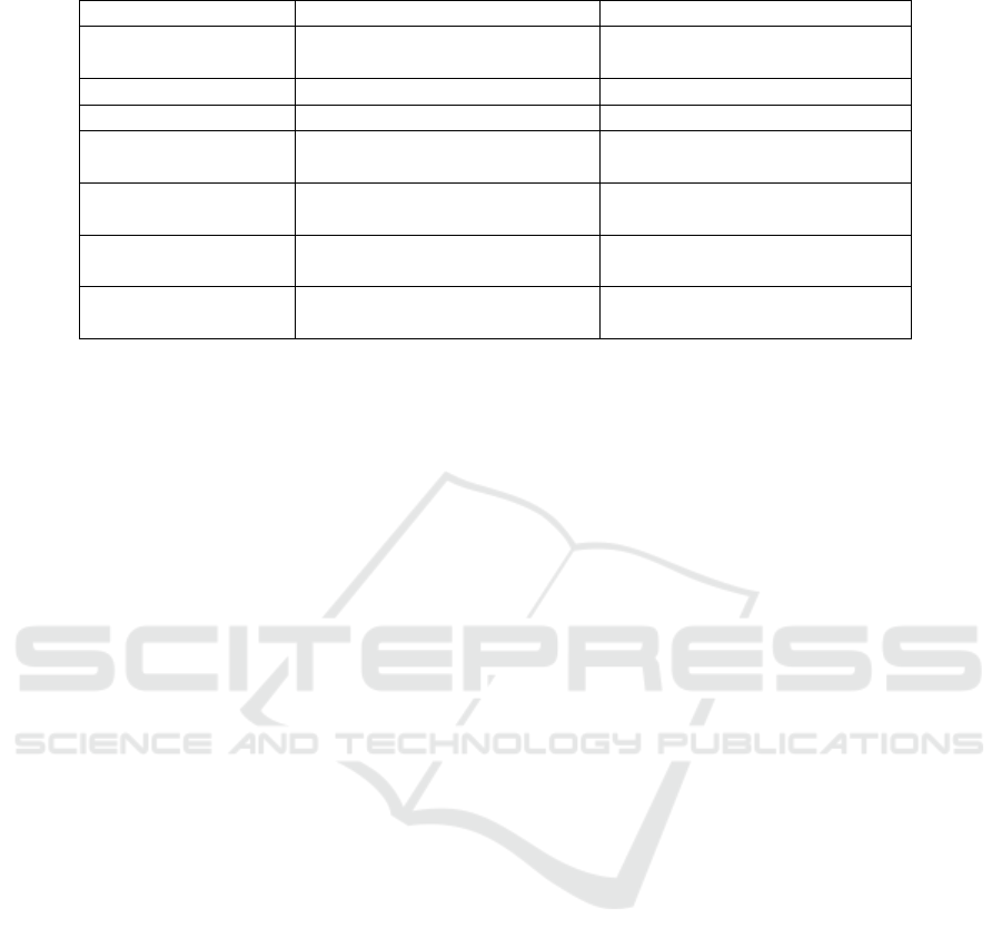
Table 3: Evaluation of the proposed method comparatively to the other methods (unit: %).
Methods Accuracy
Ozyilmaz et al(2002) Conic Section Function Neural
Network
88.3
Keles et al(2008) neuro fuzzy Classification 95.33
Iakovidis et al(2010) fuzzy local binary pattern 97.5
Acharya et Al(2011) Discrete Wavelet Transform
(DWT) and texture parameters
98
Acharya et Al(2017) Two-Threshold Binary Decom-
position algorithm
97.52
Chi et al(2017) deep learning features extrac-
tion
98.29
Proposed methods Shearlet Transform and
Generic Fourier Descriptor
98.51
quired from the laboratory CIM @ LAB. The classifi-
cation performance of textural feature has also been
optimized by the RASER dimensionality reduction
method. A comparative study shows that the metrics
performance is better with the application of feature
selection step. In addition, GFD was used to extract
the boundary information. Finally, the combination of
texture feature obtained with shearlet decomposition
and boundary information acquired using GFD give
the highest classification performance.
Thus, in our future work, we want to propose an
automated CAD system for detection and classifica-
tion of thyroid nodules.We aim also to study and pro-
pose other feature selection methods.
REFERENCES
(2017). Uci repository of machine learning databases, uni-
versity of columbia at irvine.
Abbasian Ardakani, A., Reiazi, R., and Mohammadi, A.
(2018). A clinical decision support system using ultra-
sound textures and radiologic features to distinguish
metastasis from tumor-free cervical lymph nodes in
patients with papillary thyroid carcinoma. Journal of
Ultrasound in Medicine, 37(11):2527–2535.
Acharya, U. R., Faust, O., Sree, S. V., Molinari, F., Gar-
beroglio, R., and Suri, J. (2011). Cost-effective and
non-invasive automated benign & malignant thyroid
lesion classification in 3d contrast-enhanced ultra-
sound using combination of wavelets and textures: a
class of thyroscan
TM
algorithms. Technology in can-
cer research & treatment, 10(4):371–380.
Acharya, U. R., Sree, S. V., Krishnan, M. M. R., Moli-
nari, F., Garberoglio, R., and Suri, J. S. (2012). Non-
invasive automated 3d thyroid lesion classification in
ultrasound: a class of thyroscan
TM
systems. Ultrason-
ics, 52(4):508–520.
Chi, J., Walia, E., Babyn, P., Wang, J., Groot, G., and
Eramian, M. (2017). Thyroid nodule classification
in ultrasound images by fine-tuning deep convolu-
tional neural network. Journal of digital imaging,
30(4):477–486.
Easley, G., Labate, D., and Lim, W.-Q. (2008a). Sparse
directional image representations using the discrete
shearlet transform. Applied and Computational Har-
monic Analysis, 25(1):25–46.
Easley, G., Labate, D., and Lim, W.-Q. (2008b). Sparse
directional image representations using the discrete
shearlet transform. Applied and Computational Har-
monic Analysis, 25(1):25–46.
Guo, K., Kutyniok, G., and Labate, D. (2006). Sparse multi-
dimensional representations using anisotropic dilation
and shear operators.
Iakovidis, D. K., Keramidas, E. G., and Maroulis, D.
(2010). Fusion of fuzzy statistical distributions for
classification of thyroid ultrasound patterns. Artificial
Intelligence in Medicine, 50(1):33–41.
Keles¸, A. and Keles¸, A. (2008). Estdd: Expert system for
thyroid diseases diagnosis. Expert Systems with Ap-
plications, 34(1):242–246.
Labate, D., Lim, W.-Q., Kutyniok, G., and Weiss, G.
(2005). Sparse multidimensional representation using
shearlets. In Wavelets XI, volume 5914, page 59140U.
International Society for Optics and Photonics.
Noura, A., Shili, H., and Romdhane, L. B. (2016). Reliable
attribute selection based on random forest (raser). In
International Conference on Intelligent Systems De-
sign and Applications, pages 11–24. Springer.
Ozyilmaz, L. and Yildirim, T. (2002). Diagnosis of thy-
roid disease using artificial neural network methods.
In Proceedings of the 9th International Conference on
Neural Information Processing, 2002. ICONIP’02.,
volume 4, pages 2033–2036. IEEE.
Raghavendra, U., Acharya, U. R., Gudigar, A., Tan, J. H.,
Fujita, H., Hagiwara, Y., Molinari, F., Kongmebhol,
P., and Ng, K. H. (2017). Fusion of spatial gray level
dependency and fractal texture features for the charac-
terization of thyroid lesions. Ultrasonics, 77:110–120.
Zhang, D. and Lu, G. (2002). Generic fourier descriptor for
shape-based image retrieval. In Proceedings. IEEE In-
ternational Conference on Multimedia and Expo, vol-
ume 1, pages 425–428. IEEE.
VISAPP 2020 - 15th International Conference on Computer Vision Theory and Applications
298
