
Analysis of Functional Connectivity When using Complementary
Methods of Treatment in Patients with Asymptomatic Carotid
Stenosis
A. S. Lepekhina
1
, M. L. Pospelova
2
, G. E. Trufanov
1
, T. M. Alekseeva
2
, D. N. Iskhakov
1
,
T. A. Bukkieva
1
, D. S. Chegina
1
, N. N. Semibratov
1
, B. S. Litvincev
3
and Y. N. Tsarevskaya
1
1
MRI Department, Almazov National Medical Research Centre, Akkuratova Str., 2, Saint-Petersburg, Russia
2
Neurology Department, Almazov National Medical Research Centre, Akkuratova Str., 2, Saint-Petersburg, Russia
3
Clinical Depertment, Institution of Toxicology, FMBA of Russia, Bekhtereva Str., 1, Saint-Petersburg, Russia
Keywords: Asymptomatic Carotid Stenosis, Leeching, Leech Therapy, Functional MRI, Connectome.
Abstract: Investigation of the mechanisms of neuroplasticity and detection of changes in the connectome in patients
with asymptomatic carotid stenosis is relevant for the development of new preventive, therapeutic strategies
and prediction of disease outcomes. Leech therapy (hirudotherapy, leeching) is one of the most well-known
and studied methods of complementary medicine, with proven pathogenetic mechanisms, widely and
justifiably used in patients with vascular diseases. Patients received a leech therapy course (10 sessions). All
patients underwent resting state functional MRI, complaints and neurological status were evaluated before
and after the course of leeching. After a leech therapy course, patients with asymptomatic carotid stenosis
noted a significant improvement in their condition (reduction of headaches, dizziness, noise in the head and
ears, optical and vestibular disorders, visual impairment). The connectivity of the main structures of the brain
increased, which is an important morphofunctional indicator of improved brain function. The connectome
study provides new approaches to understand the integrative brain function in health and disease, and to assess
the effectiveness of the treatment.
ABBREVIATIONS
TIA – transient ischemic attack
ACAS – asymptomatic carotid stenosis
FC – functional connectivity
ICA – internal carotid artery
1 INTRODUCTION
Stenosis of the internal carotid artery without
transient ischemic attack (TIA) or acute
cerebrovascular accident is asymptomatic. Cognitive
impairment has been described in patients with
clinically asymptomatic stenosis (Wang, 2017; Lin,
2012; Lin, 2014; Cheng, 2012). The pathogenetic
mechanism of the occurrence of these disorders in
patients with asymptomatic carotid stenosis (ACAS)
has not been fully studied. Patients with
asymptomatic carotid stenosis are at an increased risk
of developing acute vascular episodes, so the
frequency of stroke and TIA in patients with stenosis
of more than 70% reaches 40% over the next 2 years
NASCET (Streffer, 1992).
The proven fact is that the disorders in functional
connectivity (FC) are the basis of the most common
cerebral pathology. Currently, in the development of
pathophysiological models of cerebral pathology, not
only the pathology of specific parts of the brain is
evaluated, but also the characteristics of disorders of
neural networks (Seung, 2014; Fornito, 2015). The
basis of compensation of impaired functions of the
nervous system are the mechanisms of neuroplasticity
- the ability of nervous tissue to structural and
functional restructuring after its damage. Several
mechanisms of the functional connectivity
reconstruction, which underlie neuroplasticity, are
described: changes in the proportion of the
connections, recombination, reconnection, and
regeneration.
The use of multimodal neuroimaging techniques
makes it possible to identify structural and functional
disorders in patients with ACAS (Fornito 2015,
Lepekhina, A., Pospelova, M., Trufanov, G., Alekseeva, T., Iskhakov, D., Bukkieva, T., Chegina, D., Semibratov, N., Litvincev, B. and Tsarevskaya, Y.
Analysis of Functional Connectivity When using Complementary Methods of Treatment in Patients with Asymptomatic Carotid Stenosis.
DOI: 10.5220/0008953603730378
In Proceedings of the 13th International Joint Conference on Biomedical Engineering Systems and Technologies (BIOSTEC 2020) - Volume 1: BIODEVICES, pages 373-378
ISBN: 978-989-758-398-8; ISSN: 2184-4305
Copyright
c
2022 by SCITEPRESS – Science and Technology Publications, Lda. All rights reserved
373

Efimtsev A.Yu. et al. 2016). It is known that changes
in the connections between neurons occur within
three minutes after the onset of a stroke. Disruption
of the functional connectivity may be associated with
the loss of neurons or indirect effects in remote areas
of the brain. Therefore, structural and interregional
neuroimaging of the FC can demonstrate high clinical
potential for ischemic brain damage (Gulyaeva,
2016).
The study of the mechanisms of neuroplasticity in
patients with ACAS, the detection of changes of the
FC in cerebrovascular diseases and during treatment
is relevant for the development of new preventive and
therapeutic strategies, as well as predicting the
outcome (Bukkieva, 2015). As a result of the work, a
high potential for the restoration of the disturbed
connectome due to the activation of neuroplasticity
processes was noted (Kublanov 2018, Kublanov
2018, Petrenko, 2019).
There is a decrease in the severity or termination
of headaches, dizziness, noise in the head, flickering
flies before the eyes in more than 70% of cases in
patients with chronic cerebral ischemia and with
ACAS during leech therapy (Konyrtayeva, 2015;
Chernetsky 2003, Pospelova 2008
).
2 PURPOSE
Сonnectome study in patients with asymptomatic
carotid stenosis of more than 65% during leech
therapy course for develop therapeutic and preventive
strategies.
3 MATERIALS AND METHODS
3.1 Study Population
The study was conducted in accordance with the
principles of the Helsinki Declaration. Under our
supervision, there were 16 patients (10 women and 6
men, aged 61 to 81 years, average age 72.4 ± 3.4
years) with ACAS of one or both of the internal
carotid artery (ICA) 60-75%. 3 patients underwent
carotid enderectomy surgery from one internal
carotid artery, with persistent stenosis of more than
65% of the contralateral ICA. All patients suffered
from hypertension (disease duration from 3 to 20
years). 2 patients had diabetes (type II). The diagnosis
of ACAS was made on the basis of anamnesis,
complaints, and instrumental examination data.
All patients underwent ultrasound triplex
examination of the brachiocephalic arteries on a
Logiq Q7 Expert General Electric apparatus using B-
mode, color and energy Doppler mapping, and pulse-
wave Doppler. The study used a linear sensor with a
frequency of 8.5-10.0 mHZ, a standard protocol with
an assessment of the degree of stenosis of the lumen
of the common and internal carotid arteries by area
and diameter (European Carotid Surgery Trialists).
The study was approved by the ethics committee
of the Federal State Budgetary Institution «National
Medical Research Center V.A. Almazova» of the
Ministry of Health of Russia (extract from the
protocol No. 41 of 02/12/2018).
Criteria for exclusion of patients from the study
were: 1. The presence of a history of psycho-organic
pathology, epilepsy, brain tumors, injuries of the
brain and spinal cord. 2. The presence of severe
concomitant pathology (exacerbation of rheumatism,
acute infections, cirrhosis, alcoholism, drug
addiction, cardiomyopathy with thromboembolism in
the arteries of the brain, acute myocardial infarction,
heart failure 3-4 severity, blood diseases). 3. The
simultaneous administration of drugs that can distort
the results of treatment (anxiolytics, antidepressants,
barbiturates, lithium preparations, narcotic
analgesics, reserpine).
Patients complained of paroxysmal and / or
persistent headaches of one- or two-sided
localization, aching, pulsating; unsystematic and / or
systemic instant, short-term or prolonged dizziness;
noise in the head and / or in the ears; hearing loss; the
inability to look at moving objects; flashing flies
before the eyes.
In neurological status, the main symptoms were
manifested by lethargy of pupil reactions - in 6
patients, convergence failure - in 4 patients,
nystagmus with extreme leads - in 5, asymmetry of
tendon reflexes - in 7, vegetative instability - in 8,
tremor of fingers of extended arms - in 5, dynamic
elements in 6 patients and static-locomotor ataxia in
5 patients.
Patients underwent a leech therapy course
consisting of 10 sessions (2,5 months) according to
their own patent of the Russian Federation No.
2327494 (Pospelova M.L., 2008). Leech therapy was
carried out 1-2 times a week, once 2-6 leeches were
placed. The most common points of the prefix: the
occipital zone (along the edge of hair growth), the
cervical spine (paravertebral), mastoid processes,
lumbar, sacral spine (paravertebral), coccyx (in the
gluteal fold), the region of the liver, spleen, heart,
around the navel. Against the background of leech
NDNSNT 2020 - Special Session on Non-invasive Diagnosis and Neuro-stimulation in Neurorehabilitation Tasks
374

therapy, patients continued to take antihypertensive,
antiplatelet, and lipid-lowering drugs.
Complaints (scale: yes/no) of subjective
sensations and neurological status were evaluated
before the start of leech therapy and after the end of
the course of treatment (after 2-2.5 months).
Patients were instructed to lie with their eyes open
(do not sleep), without fixing the gaze. Thus, for
everyone there were identical conditions of a state of
rest, and this had a minimal effect on the visual and
auditory working networks of the brain.
3.2 MR Imaging Protocol
All patients underwent structural MRI with obtaining
T1 and T2 weighted images and FLAIR (Fluid
attenuated inversion) to exclude brain tumors, strokes
and other pronounced pathological changes. All
patients underwent functional resting state MRI at 2
time points - before and after the course of leech
therapy. Pulse sequence data of a T1-weighted
gradient echo (MP-RAGE – Magnetization Prepared
Rapid Acquired Gradient Echoes) was collected to
combine fMRI data with anatomical structures of the
brain, slice thickness – 4.5 mm, number of slices – 29,
the number of repetitions – 120, scan time – 6
minutes. The main feature of this sequence is its high
resolution and 0.8 mm isotropic voxel. BOLD (Blood
Oxygenation Level Dependent) were using with
repetition time (TR) = 3000 ms, echo time (TE) = 50
ms, field of view (FOV) = 230 mm and matrix size
128*128, slice thickness – 4.0 mm, the number of
repetitions – 120, scan time – 6 minutes.
3.3 Image Analyses
Analyzing the data of functional MRI, when
performing an intergroup statistical analysis (two-
sample t-test, comparing the resting state before
treatment and after a course of leech therapy) with the
choice of the medial prefrontal cortex (MPFC) as the
region of interest.
3.4 Statistical Analyses
For statistical analysis, the non-parametric McNemar
test for dependent binary indicators was used.
Statistical processing and evaluation of the results of
neuroimaging studies of each patient individually, as
well as their group totality (resting state fMRI data)
were carried out using the CONN v.18 software
package (Functional connectivity toolbox), designed
to determine the relationships between different parts
of the brain, statistical mapping of activation zones,
determining the structure of various resting state
networks and functional networks of the brain.
3.5 Results
Against the background of the course of leech therapy
in patients with asymptomatic carotid stenosis, a
significant improvement was noted in the assessment
of complaints. There was no deterioration in the
condition of patients, adverse and allergic reactions
during treatment. The improvement ranged from
16.67% to 100%, on average - 58.35%. Statistically
significant changes in the state were observed
according to the following indicators - aching,
paroxysmal and aching, unilateral, bilateral, and
overall headache; unsystematic, instantaneous, short-
term, and generally dizziness; opto-vestibular
syndrome and transient visual disturbances.
Improvement in 100% of cases was achieved by the
following indicators: throbbing, persistent headache;
systemic, systemic in combination with non-
systemic, prolonged dizziness.
Similar positive dynamics in evaluating
complaints of headaches and dizziness, noise in the
head and in the ears was noted by us and a number of
other authors in patients with chronic vertebral-
basilar insufficiency, hypertensive
angioencephalopathy, posthypoxic encephalopathy
(Portik O.A., Efimtsev A.Yu. et al, 2019; Pospelova
M.L, 2010; Frolov 2006).
Analyzing the functional MRI data, when
performing an intergroup statistical analysis (two-
sample t-test, comparing the state of rest before
treatment and after a course of leech therapy) with the
choice of the medial prefrontal cortex (MPFC) as the
ROI, we determined the enhancement of the positive
functional connections of MPFC with zone 10, right
cerebellum, with cerebellar vermis. Such changes of
connectom correlate with clinical data on a decrease
in the severity of vestibular disorders in this group of
patients.
Region of interest - cerebellar area. Demonstrated
positive FC with right and left hemisphere of the
cerebellum (8 area), the vermis cerebellum, the
precuneus, angular gyri and posterior divisions of the
cingulate gyrus (part of the default mode network)
(Figure 1).
When performing an analysis based on graph
theory with global efficiency assessment after a
course of leech therapy, stable functional
relationships between the middle temporal gyrus
(posterior, right), upper temporal gyrus (anterior,
right) and lower temporal gyrus were determined
comparing to the results of the study before the start
Analysis of Functional Connectivity When using Complementary Methods of Treatment in Patients with Asymptomatic Carotid Stenosis
375
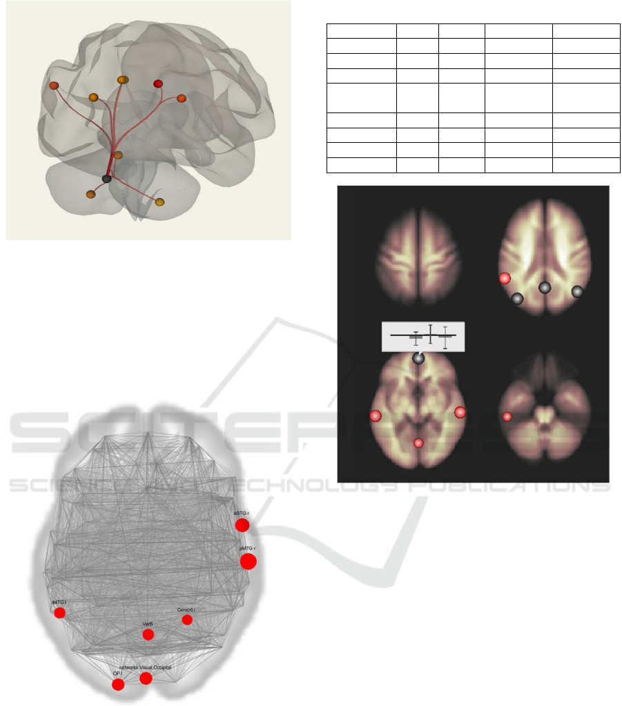
Figure 1: Cerebellar network. Intergroup comparison.
of treatment (temporal-occipital departments, left),
lingual network, visual network, cerebellar vermi,
zone 6 of the right hemisphere of the cerebellum, pole
of the occipital lobe. At the same time, the degree of
severity of activation of the lingual network (upper
and lower frontal gyrus on the right) was decreased
(Figure 2, Table 1).
Figure 2: Maps of functional connectivity. Graph theory
results.
There was an increase of the negative functional
connections of MPFC with the left middle frontal
gyrus and a weakening of the negative functional
connections of MPFC with the right parahippocampal
gyrus p<0.001 (Figure 3).
Table 1: The degree of activations severity.
ROI beta T
p
-unc
p
-FDR
Networ
k
0.00 -0.98 0.822939 0.931425
p
MTG r 0.05 2.95 0.008122 0.931425
aSTG
r
0.07 2.48 0.017570 0.931425
Visual
Occi
p
ital
0.05 2.24 0.026075 0.931425
OP l 0.03 2.19 0.028042 0.931425
Ve
r
mis 6 0.04 2.03 0.036683 0.931425
toITG l 0.06 1.98 0.039213 0.931425
Cereb6
r
0.05 1.85 0.048780 0.931425
Figure 3: Intragroup comparison of the subjects before and
after a course of treatment. The changes that occur after
treatment are shown. Activation sites combined with
anatomical images of the head — brain regions positively
functionally associated with MPFC are mapped in red, and
negatively functionally associated with MPFC are mapped
in blue (p<0,001).
The analysis of functional MRI showed that after
the course of leech therapy, the activation of the main
structures of the salience network and the executive
control network was noted. The functional
connections of MPFC — the area of the brain
responsible for controlling and making decisions with
the cerebellum — increased, which was clinically
manifested in a decrease in vestibular disorders, and
the functional connections with the left middle frontal
gyrus decreased, which may indirectly indicate a
decrease in the inhibitory component of the network.
In patients with chronic cerebrovascular accident,
cerebral microangiopathy, there is a gradual loss of
inter- and intrahemispheric functional connections
between the structures of the salience network and the
executive control network, which is the functional
NDNSNT 2020 - Special Session on Non-invasive Diagnosis and Neuro-stimulation in Neurorehabilitation Tasks
376

MRI equivalent of the disconnection phenomenon
(Dobrynina, 2018). After a course of leech therapy,
the connectivity of the leading structures of the brain
significantly increased, which is a morphological and
functional indicator of improving brain functioning
and improving cognitive, emotional and behavioral
disorders in patients with asymptomatic carotid
stenosis.
When assessing the colorability and safety of the
therapy, no hemorrhagic events during the treatment
(hemorrhagic stroke, retinal hemorrhage,
gastrointestinal bleeding, hemorrhoids, nose, uterine
bleeding) were noted.
As a result, we can talk about a significant positive
effect of the leech therapy course on the complex of
complaints and indicators of brain connectivity in
patients with asymptomatic carotid stenosis.
4 CONCLUSIONS
The study of connectome provides new approaches to
the analysis of integrative brain function, and to
assessment of the treatment effectiveness. A course
of leech therapy significantly altered the functional
connectivity of the brain in patients with
asymptomatic carotid stenosis.
CONFLICT OF INTERESTS
The authors declare no conflict of interest.
REFERENCES
Wang T, Xiao F, Wu G et al. Impairments in Brain
Perfusion, Metabolites, Functional Connectivity, and
Cognition in Severe Asymptomatic Carotid Stenosis
Patients: An Integrated MRI Study. Neural Plast. 2017.
doi: 10.1155/2017/8738714. Epub 2017 Feb 1.
Lin CJ, Chang FC, Chou KH et al. Intervention versus
Aggressive Medical Therapy for Cognition in Severe
Asymptomatic Carotid Stenosis. AJNR Am J
Neuroradiol. 2016; 37 (10): 1889-1897. doi:
10.3174/ajnr. A 4798. Epub 2016 Apr 28.
Lin CJ, Tu PC, Chern CM et al. Connectivity features for
identifying cognitive impairment in presymptomatic
carotid stenosis. PLoS One. 2014 Jan;15 ;9(1): e85441.
doi: 10.1371/journal.pone.0085441. eCollection 2014.
Cheng HL, Lin CJ, Soong BW et al. Impairments in
cognitive function and brain connectivity in severe
asymptomatic carotid stenosis. Stroke. 2012 Oct; 43
(10): 2567-73. Epub 2012 Aug 30.
Streffer JX, Benavente OR, Harbison JW et al. Prognostic
implications of retinal versus hemispheric TIA in
patients with high grade carotid stenosis: Observations
from NASCET. Stroke. 1992; (23): 159.
Efimtsev A.Yu., Trufanov G.E., Litvintsev B.S., Fokin
V.A. et al. Neuroimaging diagnosis of depressive and
addictive disorders. // Psychiatry, Psychotherapy and
Clinical Psychology. 7(1): 30-40, 2016.
Seung S. Connectom. Moscow: BINOM. Knowledge
laboratory; 2014.
Fornito A, Bullmore ET. Connectomics: a new paradigm
for understanding brain disease. European
Neuropsychopharmacology. 2015;25(5):733-748.
Fornito A, Zalesky A, Breakspear M. The connectomics of
brain disorders. Nature Reviews Neuroscience. 2015;
16(3): 159-172.
Gulyaeva NV. Plasticity of the brain and connectopathy:
the mechanisms of comorbidity of neurological
diseases and depression. // Korsakov journal of
Neurology and Psychiatry. 2016; 11: 157-162.
Chen M. Speech at the WHO Congress on Traditional
Medicine. Practical phytotherapy. 2008; 4: 43-48.
Nargiza EO, Mirdjuraev EM, Ergasheva NO. Leech therapy
to prevent ischemic stroke. European Journal of
Neurology. 2010;17(3):170.
Porshinsky BS, Saha S, Grossman MD et al. Clinical uses
of the medicinal leech: a practical review. Journal of
Postgraduate Medicine. 2011;57(1):65-71.
Konyrtayeva NN, Grzhibovsky AM, Kausova GK et al.
Leech therapy in diseases of the circulatory system.
Ecology of man. 2015; 6: 57-64.
Chernetsky VK, Pashkovsky VM, Madar GI et al.
Efficiency of leeching in the complex treatment of
chronic cerebrovascular insufficiency. Clinic and
examination of pathology. 2003;2(1):117-119.
Pospelova ML. Review of the pathogenetic mechanisms of
action of leeching and the rationale for its use in the
treatment of patients with cerebrovascular diseases.
Modern problems of science and education. 2012.
Patent RUS №2327494/ 27.06.2008. Byul. №18. Pospelova
ML, Barnaulov OD, Kadinskaya MI et al. The method
of treatment of patients with ischemic cerebrovascular
diseases.
Pospelova ML, Barnaulov OD. Doppler assessment of the
effectiveness of leeching in patients with chronic
vertebrobasilar insufficiency and circulatory
encephalopathy stage 1. Regional blood circulation and
microcirculation. 2010; 2(34): 40-43.
Frolov VA, Frolova EA. Leech therapy as the main method
in the complex rehabilitation of patients with the
consequences of ischemic stroke. Leeching: yesterday,
today, tomorrow. Materials of the IX Scientific
Practical Conference of the Association of leeching.
Russia, Balakovo; 2006; 2: 34–36.
Semikova TS, Semikova MV. Leech therapy in ophthalmic
practice. New technologies of eye microsurgery.
Materials of the XII Scientific Practical Conference.
Orenburg; 2001. p. 65–66.
Mukhanko IZh, Kulagin AN. The experience of complex
treatment of organic and functional disorders of vision
Analysis of Functional Connectivity When using Complementary Methods of Treatment in Patients with Asymptomatic Carotid Stenosis
377

in the sanatorium-resort conditions of the Caucasian
mineral waters with leeching. Materials of the VIII
Scientific Practical Conference of the Association of
leeching of Russia and CIS. 2003. p. 37-39.
Bukkieva Т.А., Chegina D.S., Еfimtsev А.Yu et. al. Resting
state functional MRI. General issues and clinical
application. REJR 2019; 9(2): 150-170.
DOI:10.21569/2222-7415-2019-9-2-150-170.
Kublanov, V., Dolganov, A., Aftanas, et. al, 2018
Investigation of the Neuroelectrostimulation
Mechanisms by Means of the Functional MRI: Case
Study. In: Proceedings of the 11th International Joint
Conference on Biomedical Engineering Systems and
Technologies. Volume 3: NENT (BIOSTEC 2018).
Presented at the Special Session on Neuro-
electrostimulation in Neurorehabilitation Tasks, pp.
319–324 doi.org/10.5220/0006712203190324
Kublanov V.S., Petrenko T.S., Efimcev A.A. Application
of Multichannel Electrical Stimulation of the Neck
Nervous Structures in Patients with Depressive
Disorders: An fMRI Case Study. BIOSTEC–2019:
Proceedings of the 12th International Joint Conference
on Biomedical Engineering Systems and Technologies,
Prague, Czech Republic, 22-24 February 2019 /
Portugal: SCITEPRESS, 2019, Vol.5: NNSNT. P. 564–
571. doi.org/10.5220/0007681705640571
T.S. Petrenko, V.S. Kublanov, K.Yu. Retyunskiy1, et al.
The effect of multichannel electrostimulation of neck
nervous structures on the brain connectivity of patients
with depressive disorders // S.S. Korsakov Journal of
Neurology and Psychiatry. 2019, 9 (119): 49-52.
doi.org/10.17116/jnevro201911909149]
Dobrynina LA, Gadzhiyeva Z.Sh., Morozova SN et al.
Control functions of the brain: functional magnetic
resonance tomography with the Stroop test and the
serial account test in healthy people. Korsakov journal
of neurology and psychiatry. 2018; 3: 64-71.
Portik O.A., Tsarevskaya Yu.N., Efimtsev A.Yu.,
Alekseeva T.M., Trufanov G.E. Posthypoxic
encephalopathy in patients undergoing coronary artery
bypass surgery: Clinical, neuropsychological, and
neuroimaging aspects. // Nevrologiya, Neiro
psikhiatriya, Psikhosomatika. 11(3):35-42. DOI:
10.14412/2074-2711-2019-3-35-42
NDNSNT 2020 - Special Session on Non-invasive Diagnosis and Neuro-stimulation in Neurorehabilitation Tasks
378
