Efficacy of Topical Binahong Leaf Ethanolic Extract Administration
on Serum TGF-β1 in Wistar Rats with Staphylococcus aureus-infected
Wounds
Rizki Andini Nawawi
11
, Muhammad Totong Kamaluddin
22
and Theodorus Theodorus
23
1
Biomedical Sciences Graduate Program, Faculty of Medicine, Universitas Sriwijaya, Palembang, Indonesia
2
Department of Pharmacology, Faculty of Medicine, Universitas Sriwijaya, Palembang, Indonesia
Keywords: Anredera Cordifolia, Wound Infection, Transforming Growth Factor Beta, Wound Healing, In Vivo
Abstract: Binahong (Anredera cordifolia (Ten.) Steenis) is a medicinal plant traditionally used as a topical wound
treatment. Saponin content in Binahong leaf extract has been thought to increase TGF-β secretion, which
plays an important role in accelerating wound healing. This study’s aim was to assess the efficacy of topical
Binahong leaf ethanolic extract administration on serum TGF-β1 in infected wounds. An experimental study,
in vivo, was conducted in the Biotechnology Laboratory and Animal House, Faculty of Medicine, Universitas
Sriwijaya, Palembang, from July to September 2020. Thirty male Wistar rats aged 10-12 weeks with
excisional wounds were infected with Staphylococcus aureus ATCC 25923. The rats were divided into five
groups and received three concentrations of Binahong leaf extracts (2.5%, 5%, and 10%), salve base, and
povidone iodine 10% topically twice daily for 14 days. Serum was obtained before treatment and after day 14
of treatment. Wound area was also recorded. After 14 days of topical administration of Binahong leaf extract
on rats with Staphylococcus aureus-infected wounds, a decrease in wound size was most significantly
observed in rats receiving 10% Binahong leaf extract (p = 0.02), but no significant serum TGF-β1 increase
was observed in all treatment groups.
1 INTRODUCTION
The presence of wounds poses a risk of colonization
and infection by pathogenic bacteria on the wounded
site, which might spread and cause systemic infection
if it is not adequately managed (Negut, Grumezescu,
and Grumezescu 2018). One of the most common
causative pathogens in wound and skin infections is
Staphylococcus aureus (Roy et al. 2018). S. aureus
disrupts normal wound healing process through the
release of extracellular adherence protein (Eap). Eap
release prolongs inflammation and prevents
angiogenesis, especially in the proliferative phase of
wound healing (Pereira-franchi et al. 2017; Wong,
Manikam, and Muniandy 2015). In Indonesia, a
multicenter study reported S. aureus isolation from
45.3% patients with wound infections and SSTI
(Santosaningsih et al. 2018).
1
https://orcid.org/0000-0002-5079-3016
2
https://orcid.org/0000-0002-8670-9867
3
https://orcid.org/0000-0003-1106-8396
The wound healing process involves a complex
interaction between the skin’s cellular components,
such as keratinocytes, fibroblasts, vascular endothelia,
immune system cells, and extracellular matrix (Martin
and Nunan 2015). These cells interact by secreting
various mediators and growth factors, one of which is
transforming growth factor-beta (TGF-β). TGF-β is a
pleiotropic growth factor secreted by platelets,
fibroblasts, and proinflammatory cells (Lichtman,
Otero-vinas, and Falanga 2016; Sutrisno et al. 2018).
TGF-β has an extensive role in wound healing, such
as stimulating collagen synthesis, angiogenesis, and
keratinocyte migration (Lichtman, Otero-vinas, and
Falanga 2016; Tejiram et al. 2016). TGF-β may also
induce epithelial-mesenchymal transition, which is an
important morphogenetic event in the formation of
scar tissue and regeneration (Martin and Nunan 2015;
Qi et al. 2018). TGF-β has three known isoforms,
Nawawi, R., Kamaluddin, M. and Theodorus, .
Efficacy of Topical Binahong Leaf Ethanolic Extract Administration on Serum TGF-1 in Wistar Rats with Staphylococcus aureus-infected Wounds.
DOI: 10.5220/0010491202670272
In Proceedings of the 1st Jenderal Soedirman International Medical Conference in conjunction with the 5th Annual Scientific Meeting (Temilnas) Consortium of Biomedical Science Indonesia
(JIMC 2020), pages 267-272
ISBN: 978-989-758-499-2
Copyright
c
2021 by SCITEPRESS – Science and Technology Publications, Lda. All rights reserved
267

TGF-β1 to TGF-β3, in which TGF-β1 is the most
abundant (Wang et al. 2017).
Recently, while topical therapy remains an
important modality in wound management, it has
become a concern that most available topical therapies
do not give additional benefits in speeding up the
wound healing process (Powers et al. 2019). In case of
infected wounds, rising tolerance and resistance
towards common topical antibiotics and antiseptic
agents also impose a challenge in choosing the
appropriate treatment (Hardy et al. 2018).
Natural products and their active compounds are
starting to be considered in the development of novel
products for wound management (Istyastono and
Yuliani 2016). Anredera cordifolia (Ten.) Steenis
(Binahong) is a plant from the Basellaceae family,
which has long been used in traditional medicine
around the world (Astuti et al. 2011; Leliqia,
Sukandar, and Fidrianny 2017). In Indonesia,
Binahong has been traditionally used for various
diseases, including in the treatment of wounds and
bacterial infections (Astuti et al. 2011; Sukandar and
Kurniati 2014).
A number of studies have been conducted on the
efficacy of A. cordifolia extract in wounds, and so far
there have been no data yet on the in vivo efficacy of
A. cordifolia extract on TGF-β1 concentrations in
infected wounds. This study aimed to determine the
efficacy of A. cordifolia ethanolic extract on
increasing serum TGF-β1 in rats with Staphylococcus
aureus-infected wounds.
2 MATERIALS AND METHODS
An experimental study, in vivo, was conducted at the
Biotechnology Laboratory and Animal House,
Faculty of Medicine, Universitas Sriwijaya,
Palembang, in July to September 2020. The study
population was male Wistar rats. There were 30 male
Wistar rats aged 10-12 weeks, weighing 150-200
grams which fulfilled the inclusion criteria.
Rats showing signs of infection in the first 24
hours after S. aureus inoculation were included in
this study. Ethical clearance had been approved by
Health Research Ethics Committee, Faculty of
Medicine, Universitas Sriwijaya, prior to the
commencement of the study (Certificate No.
024/kepkrsmhfkunsri/2020).
2.1
Extract Preparation
Four hundred grams of dried Binahong leaves
obtained from Karangpandan, Tawangmangu,
Central Java (elevation of 800 m above sea level),
was extracted by maceration with 96% ethanol which
is in accordance with the Indonesian Herbal
Pharmacopoeia and concentrated through rotary
evaporation (Kementerian Kesehatan Republik
Indonesia 2009). The obtained concentrated extract
was then formulated into salve with vaseline album
and adeps lanae base. Three concentrations of salves
were formulated, each containing 2.5%, 5%, and
10% Binahong leaf ethanolic extract, respectively.
Salve base was used as negative control and
povidone iodine 10% (Betadine®, PT Mahakam
Beta Farma, Jakarta, Indonesia, Batch No. GB20045)
was used as positive control.
2.2
In Vivo Efficacy Test
Rats were anesthetized by using ketamine, and their
dorsal skin was depilated with scissors and depilatory
cream before wounding. A 2 cm
2
circular excision
wound was made with scalpels and surgical scissors,
followed by inoculation with Staphylococcus aureus
ATCC 25923 suspension containing 2 x 10
7
cfu and
a 24-hour incubation period. The rats were then
divided into 5 treatment groups, each receiving salve
base (negative control), three concentrations of
Binahong leaf ethanolic extract salves, and povidone
iodine 10% (positive control). All groups received
treatments twice daily for 14 days.
Wounds were photographed on days 4, 7, 10, and
14 of treatment, and wound area was measured with
image processing software. Serum samples were
obtained during the 24-hour incubation period before
treatment started and on the 14
th
day of treatment.
TGF-β1 assay was performed by ELISA
(MyBioSource, San Diego, CA, USA), following
protocols specified by the manufacturer.
2.3
Statistical Analysis
Homogenity and normality of data was assessed prior
to further analysis. Efficacy of each treatment was
assessed by using paired T test. Efficacy comparison
between treatment groups and controls was
performed by using unpaired T test, and significance
test was performed with Post Hoc test. Significance
is assumed at p < 0.05.
JIMC 2020 - 1’s t Jenderal Soedirman International Medical Conference (JIMC) in conjunction with the Annual Scientific Meeting
(Temilnas) Consortium of Biomedical Science Indonesia (KIBI )
268
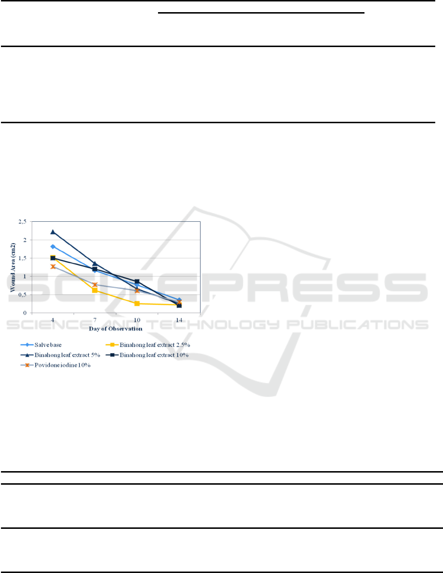
Table 1: Efficacy of topical Binahong leaf ethanolic extract on decreasing wound area.
Treatment Group Wound area (cm
2
) p
Pre-
treatment
Mean
Pos
t
-
treatment
Mean
Salve base
Binahong leaf ethanolic
extract
2.5%
5%
10%
Povidone iodine 10%
2.403
2.368
1.427
2.069
2.208
0.356
0.225
0.259
0.198
0.302
0.001
0.001
0.001
0.001
0.001
Paired T test, p = 0.05
3 RESULTS
Wound area was significantly decreased in all
treatment groups (Table 1).
Figure 1: Wound area contraction in all treatment groups
during the course of the experiment.
The most significant decrease in wound area was
found in treatment groups receiving 10% Binahong
leaf extract, where a significant difference in efficacy
was found in comparison to negative control group (p
= 0.02), but there was no significant efficacy
difference in comparison to povidone iodine 10%
(Table 2).
After 14 days of topical Binahong leaf ethanolic
extract administration, there was no significant
increase in serum TGF-β1 concentration in all
treatment groups (Table 3).
4 DISCUSSION
This study aimed to assess the efficacy of topical
Binahong leaf ethanolic extract administration on
serum TGF-β1 in infected wounds, while its efficacy
in decreasing wound area was also examined for
baseline data. Topical administration of Binahong
leaf ethanolic extract did not increase TGF-β1 levels,
but effectively decreased wound area. Treatment
with 10% Binahong leaf ethanolic extract was shown
to be most efficacious in decreasing wound area.
Table 2: Efficacy comparison between different doses of topical Binahong leaf ethanolic extract and controls on decreasing
wound area after 14 days of administration.
Comparison Group Treatment Group p
Salve base
(negative control)
2.5% Binahong leaf extrac
t
5% Binahong leaf extract
10% Binahong leaf extract
Povidone iodine 10%
0.129
0.109
0.022
0.427
Povidone iodine 10%
(positive control)
Salve base
2.5% Binahong leaf extract
5% Binahong leaf extract
10% Binahon
g
leaf extrac
t
0.427
0.323
0.360
0.063
Unpaired T test, p = 0.05
Efficacy of Topical Binahong Leaf Ethanolic Extract Administration on Serum TGF-1 in Wistar Rats with Staphylococcus aureus-infected
Wounds
269
Table 3: Efficacy of topical Binahong leaf ethanolic extract on decreasing wound area.
Treatment Group Serum TGF-β1 (p
g
/ml) p
Pre-treatment
Mean
Pos
t
-treatment
Mean
Salve base
Binahong leaf ethanolic extract
2.5%
5%
10%
Povidone iodine 10%
1,036.736
1,034.923
1,018.898
1,065.949
1,072.551
1,076.681
952.615
998.705
969.090
1,006.461
0.128
0.225
0.753
0.463
0.249
Paired T test, p = 0.05
Presently, studies conducted on the efficacy of
Binahong leaf extract in wound healing have
reported various findings. A study by Paju, Yamlean
and Kojong (2013) in rabbits with incisional wounds
reported the efficacy of 10% Binahong leaf ethanolic
extract in wound healing, which is similar to our
findings (Paju, Yamlean, and Kojong 2013).
Meanwhile, Sukrama et al. (2017) reported that
concentrated Binahong leaf ethanolic extract
effectively decreased the area of burn wounds in
murine models (Sukrama, Wihandani, and Manuaba
2017). A study on excisional wounds in guinea pigs
by Miladiyah and Prabowo (2012) showed that 40%
Binahong leaf ethanolic extract in distilled water was
efficacious in decreasing wound area (Miladiyah and
Prabowo 2012). Histopathological studies on rats
receiving 5% Binahong leaf extract showed a greater
decrease in polymorphonuclear (PMN) infiltration
and increase in collagen deposition, angiogenesis,
and fibrosis in comparison to silver sulfadiazine
(Yuniarti and Lukiswanto 2017).
Three main bioactive compound classes in
Binahong leaves are known to play important roles in
the wound healing process, namely saponin, tannin,
and flavonoid (Leliqia, Sukandar, and Fidrianny
2017; Yuniarti and Lukiswanto 2017). Saponin
enhances wound healing process through stimulation
of procollagen synthesis. Saponin also enhances the
proliferation of monocytes, which will differentiate
into macrophages and secrete various growth factors.
In the reepithelialization process, saponin stimulates
fibroblast proliferation and keratinocyte migration
(Yuniarti and Lukiswanto 2017). Fibroblasts will also
secrete growth factors, such as VEGF, interleukins,
and TGF-β (Sukrama, Wihandani, and Manuaba
2017; Sutrisno et al. 2018). Flavonoid and tannin are
aromatic compound classes known for their
astringent properties. The astringent properties of
flavonoid and tannin compounds cause the skin pores
to contract, hence stopping capillary bleeding and
exudation and stimulates wound contraction (Ibrahim
et al. 2018). In addition, Flavonoid enhances wound
healing through stimulation of collagen matrix
rearrangement, while tannin stimulates wound
contraction through its role in fibroblast migration
and proliferation (Budovsky, Yarmolinsky, and Ben-
shabat 2015; Ibrahim et al. 2018; Yuniarti and
Lukiswanto 2017).
No significant increase in serum TGF-β1 was
observed in all treatment groups. A previous study
suggested that low concentration povidone iodine
(0.5%) administered topically on clean wounds could
stimulate TGF-β secretion (Wang et al. 2017).
Therefore, higher concentration of povidone iodine
administered and wound infection might have
significantly impaired povidone iodine’s effects on
TGF-β. Saponin content in Binahong leaves has been
thought to indirectly increase TGF-β secretion
through stimulating fibroblast and monocyte
proliferation (Sukrama, Wihandani, and Manuaba
2017; Sutrisno et al. 2018). Harvesting conditions
might have influenced the saponin content of
Binahong leaves prior to extraction. A previous study
quantified more saponin content in older Binahong
leaves than younger ones (2.36 µg/mg and 1.37
µg/mg, respectively) (Hasbullah 2016).
Most previous studies on the role of saponin
compounds in increasing TGF-β release had not
specified the exact TGF-β isoform studied. Among
all TGF-β isoforms, TGF-β1 has so far been
considered more important as it is also the most
abundant. TGF-β1 has been known to induce integrin
expression from keratinocytes in the skin epidermis,
which facilitates the migratory components of
reepithelialization (Ibrahim et al. 2018). Recently,
TGF-β3, another isoform of TGF-β, has also been
reported to play an important role in the later stages
of wound healing. A study in murine models reported
that TGF-β1 and TGF-β3 affected cell cycle
progression and cell migration differently, which
showed clinically in the formation of scar tissues.
Higher TGF-β1 concentration tended to cause scar
tissue formation, while higher TGF-β3 concentration
JIMC 2020 - 1’s t Jenderal Soedirman International Medical Conference (JIMC) in conjunction with the Annual Scientific Meeting
(Temilnas) Consortium of Biomedical Science Indonesia (KIBI )
270

tended to promote scarless wound healing
(Lichtman, Otero-vinas, and Falanga 2016).
Binahong leaf extract’s effect on fibroblast and
macrophage proliferation might have enhanced
secretions of other growth factors and mediators as
well. Fibroblasts and macrophages secrete a plethora
of growth factors and cytokines, such as interleukins
(IL-1, IL-6, IL-11, IL-17, IL-18), TNF-α, IFN-γ,
VEGF, PDGF, and granulocyte-macrophage colony-
stimulating factor (GM-CSF), which have known
roles in the wound healing process (Gonzalez et al.
2016; Zeinali, Rezaee, and Hosseinzadeh 2017).
Previously, a study on rats with Pseudomonas
aeruginosa-infected burn wounds showed an
increase of IL-6 and VEGF concentrations after
administration of Binahong leaf concentrated extract
(Sukrama, Wihandani, and Manuaba 2017).
Our findings have reinforced existing evidence
on the efficacy of Binahong leaf ethanolic extract
administration in enhancing wound healing, hence
further suggesting its possible clinical application
both as a single wound management product and in
combination with other agents. However, our current
study’s findings were limited in that serum TGF-β1
was assayed only before treatment and after day 14
of treatment, which corresponded with later phases
of wound healing. Considering the vast influence of
TGF-β in all phases of wound healing, possible
increases in TGF-β1 during the earlier phases of
wound healing still needs to be determined in order
to better comprehend the effects of Binahong leaf
ethanolic extract administration on TGF-β1
secretion. Another limitation was that this study
focused on a single isoform of TGF-β, and the
possible effects of Binahong leaf ethanolic extract on
other TGF-β isoforms remains to be elucidated.
5 CONCLUSION
In rats with Staphylococcus aureus-infected wounds,
topical administration of Binahong (Anredera
cordifolia (Ten.) Steenis) leaf extract for 14 days was
efficacious in decreasing wound area but did not
increase serum TGF-β1 levels. Further studies need
to assess the effects of Binahong leaf ethanolic
extract administration on TGF-β1 levels in
accordance with each phase of wound healing and
investigate the effects of Binahong leaf ethanolic
extract on other isoforms of TGF-β.
ACKNOWLEDGEMENTS
The authors wish to thank Dr. dr. M. Irsan Saleh,
M.Biomed, dr. Iqmal Perlianta, SpBP-RE, and dr.
Ella Amalia, M.Kes for their valuable inputs in this
study.
The authors also thank Maisa Pusrita and Laila
Wardani Hasanah for their assistance with
interpreting the ELISA results.
REFERENCES
Astuti, Sri Murni, Mimi Sakinah A.M., Retno Andayani
B.M., and Awalludin Risch. 2011. “Determination of
Saponin Compound from Anredera Cordifolia (Ten)
Steenis Plant (Binahong) to Potential Treatment for
Several Diseases.” Journal of Agricultural Science
3(4): 224–32.
Budovsky, Arie, Ludmila Yarmolinsky, and Shimon Ben-
shabat. 2015. “Effect of Medicinal Plants on Wound
Healing.” Wound Repair and Regeneration 23: 171–83.
Gonzalez, Ana Cristina de Oliveira, Zilton de Araujo
Andrade, Tila Fortuna Costa, and Alena Ribeiro Alves
Peixoto Medrado. 2016. “Wound Healing - A Literature
Review.” Anais Brasileiros de Dermatologia 91(5):
614–20.
Hardy, Katherine et al. 2018. “Increased Usage of
Antiseptics Is Associated with Reduced Susceptibility
in Clinical Isolates of Staphylococcus Aureus.” mBio
9(3): 1–10.
Hasbullah, Umar Hafidz Asy’ari. 2016. “Kandungan
Senyawa Saponin Pada Daun , Batang Dan Umbi
Tanaman Binahong (Anredera Cordifolia (Ten)
Steenis).” Planta Tropika Journal of Agro Science 4(1):
20–24.
Ibrahim, Nurul Izzah, Sok Kuan Wong, Isa Naina
Mohamed, and Norazlina Mohamed. 2018. “Wound
Healing Properties of Selected Natural Products.”
International Journal of Environmental Research and
Public Health 15: 2360.
Istyastono, Enade Perdana, and Sri Hartati Yuliani. 2016.
“Scarless Wound Healing Gel with Binahong
(Anredera Cordifolia (Ten) Steenis) Leaves Extract and
Celecoxib as the Active Ingredients.” In AIP
Conference Proceedings 1755, 160001, AIP
Publishing, 1–6.
Kementerian Kesehatan Republik Indonesia. 2009.
Farmakope Herbal Indonesia. I. Jakarta: Kemenkes RI.
Leliqia, Ni Putu Eka, Elin Yulinah Sukandar, and Irda
Fidrianny. 2017. “Overview Of Efficacy , Safety And
Phytochemical Study Of Anredera Cordifolia (Ten.)
Steenis.” Pharmacologyonline 1: 124–31.
Lichtman, Michael K, Marta Otero-vinas, and Vincent
Falanga. 2016. “Transforming Growth Factor Beta
(TGF-b) Isoforms in Wound Healing and Fibrosis.”
Wound Repair and Regeneration 24: 215–22.
Efficacy of Topical Binahong Leaf Ethanolic Extract Administration on Serum TGF-1 in Wistar Rats with Staphylococcus aureus-infected
Wounds
271

Martin, P, and R Nunan. 2015. “Cellular and Molecular
Mechanisms of Repair in Acute and Chronic Wound
Healing.” British Journal of Dermatology 173: 370–78.
Miladiyah, Isnatin, and Bayu Rizky Prabowo. 2012.
“Ethanolic Extract of Anredera Cordifolia ( Ten .)
Steenis Leaves Improved Wound Healing in Guinea
Pigs.” Universa Medicina 31(1): 4–11.
Negut, Irina, Valentina Grumezescu, and Alexandru Mihai
Grumezescu. 2018. “Treatment Strategies for Infected
Wounds.” Molecules 23: 2392.
Paju, Niswah, Paulina V Y Yamlean, and Novel Kojong.
2013. “Uji Efektivitas Salep Ekstrak Daun Binahong
(Anredera Cordifolia (Ten.) Steenis) Pada Kelinci
(Oryctolagus Cuniculus) Yang Terinfeksi Bakteri
Staphylococcus Aureus.” Pharmacon 2(01): 51–62.
Pereira-franchi, Eliane Patricia Lino et al. 2017.
“Prevalence of and Risk Factors Associated with the
Presence of Staphylococcus Aureus in the Chronic
Wounds of Patients Treated in Primary Health Care
Settings in Brazil.” Revista Sociedad Brasileira de
Medicina Tropica 50(6): 833–38.
Powers, Jennifer G, Catherine Higham, Karen Broussard,
and Tania J Phillips. 2019. “Chronic Wound Care and
Management.” Journal of American Dermatology
74(4): 607–25.
http://dx.doi.org/10.1016/j.jaad.2015.08.070.
Qi, Min et al. 2018. “Growth Factors in the Pathogenesis of
Diabetic Foot Ulcers.” Frontiers in Bioscience 23: 310–
17.
Roy, Sushmita et al. 2018. “Evaluation of Antibiotic
Susceptibility in Wound Infections : A Pilot Study from
Bangladesh.” F1000Research 6: 2103.
Santosaningsih, Dewi et al. 2018. “Prevalence and
Characterisation of Staphylococcus Aureus Causing
Community-Acquired Skin and Soft Tissue Infections
on Java and Bali , Indonesia.” Tropical Medicine and
International Health 23(1): 34–44.
Sukandar, Elin Yulinah, and Neng Fisheri Kurniati. 2014.
“Evaluation of Teratogenicity Effects of Ethanolic
Extracts of Binahong Leaves (Anredera Cordifolia
(Ten) Steenis) in Wistar Rat.” International Journal of
Pharmacy and Pharmaceutical Sciences 6(11): 7–12.
Sukrama, Dewa Made, Desak Made Wihandani, and
Amertha Putra Manuaba. 2017. “Topical Binahong (
Anredera Cordifolia ) Leaf Extract Increases
Interleukin-6 and VEGF ( Vascular Endothelial Growth
Factor ) during Burn Wound Healing in Wistar Rats
Infected with Pseudomonas Aeruginosa.” Biology and
Medicine 9(1): 1–6.
Sutrisno, Entris, Elin Yulinah Sukandar, Irda Fidrianny,
and I Ketut Adnyana. 2018. “Wound Healing In Vivo
And In Vitro Study Of Binahong Leaves (Anredera
Cordifolia (Ten.) Steenis ) And Pegagan (Centella
Asiatica (L.) Urban) Ethanolic Extract.”
Pharmacologyonline 1(April): 111–16.
Tejiram, S, S L Kavalukas, J W Shupp, and A Barbul. 2016.
“Wound Healing.” In Wound Healing Biomaterials, ed.
Magnus Ågren. Cambridge: Woodhead Publishing, 3–
39.
Wang, Li et al. 2017. “Transforming Growth Factor β Plays
an Important Role in Enhancing Wound Healing by
Topical Application of Povidone-Iodine.” Scientific
Reports (March): 1–8.
http://dx.doi.org/10.1038/s41598-017-01116-5.
Wong, Shin Yee, Rishya Manikam, and Sekaran Muniandy.
2015. “Prevalence and Antibiotic Susceptibility of
Bacteria from Acute and Chronic Wounds in Malaysian
Subjects.” The Journal of Infection in Developing
Countries 9(9): 936–44.
Yuniarti, Wiwik Misaco, and Bambang Sektiari
Lukiswanto. 2017. “Effects of Herbal Ointment
Containing the Leaf Extracts of Madeira Vine
(Anredera Cordifolia (Ten.) Steenis) for Burn Wound
Healing Process on Albino Rats.” Veterinary World
10(7): 808–13.
Zeinali, Majid, Seyed Abdolrahim Rezaee, and Hossein
Hosseinzadeh. 2017. “An Overview on
Immunoregulatory and Anti-Inflammatory Properties
of Chrysin and Flavonoids Substances.” Biomedicine &
Pharmacotherapy Pharmacotherapy 92(August): 998–
1009. http://dx.doi.org/10.1016/j.biopha.2017.06.003.
JIMC 2020 - 1’s t Jenderal Soedirman International Medical Conference (JIMC) in conjunction with the Annual Scientific Meeting
(Temilnas) Consortium of Biomedical Science Indonesia (KIBI )
272
