
Methadone Effects on Frontal Brain Lobe based EEG-P300 Waves in
Drug Rehabilitation Patients
Arjon Turnip
1*
, M. Agung Suhendra
1
, Dwi Esti Kusumandari
1
, Faza Lisan Sadida
1
, Simon Willy
Laufried
1
, Siti Aminah Sobana
2
, Arifah Nur Istiqomah
2
and Daniel Sutopo Pamungkas
3
1
Instrumentation Development, Indonesian Institute of Sciences, Jakarta, Indonesia
2
Faculty of Medicine, Padjadjaran University, Bandung, Indonesia
3
Electical Engineering, Politeknik Negeri Batam, Batam, Indonesia
Keywords: EEG-P300, Methadone, Visual Stimuli, Frontal Lobe
Abstract: Drug abuse in various parts of the world is increasingly widespread. Therefore, a drug addict should
immediately stop and must be recovered. To overcome the symptoms of addiction, the use of methadone as a
synthetic drug to replace opioid type drugs is recommended. In this paper, an experiment with rehabilitation
patients to identify the effect of the drugs on the brain activity in the frontal, central, temporal, and occipital
lobes is proposed. The EEG data collection is performed using 18 channel electrodes, namely central: C3, C4;
frontal: Fp1, Fp2, F3, Fz, F4, F7, F8; occipital: P3, Pz, P4, O1, O2; and temporal: T3, T4, T5, T6. In the brain
signals record, subjects were asked to comfortably sit in a chair. The recording was done in three sessions: 5
minutes before drinking methadone, 10 and 60 minutes after drinking the methadone, respectively. To reduce
background noise and artefacts removal, band pass filter (0.5-50 Hz) and wavelet method were applied,
respectively. From this experiment it was found that a decrease in amplitude after methadone intake for
average in four lobes is obtained. This results indicates that the use of methadone is highly effect on the entire
brainwave activity which indicates a decrease in the level of desire to do activities.
1 INTRODUCTION
Drug abuse in various parts of the world is
increasingly widespread. Various cases show
material and non-material losses and even cause the
death. Therefore, a drug addict should immediately
stop and must be recovered. According to Indonesian
law, narcotics addicts and victims of narcotics abuse
must serve out medical rehabilitation and social
rehabilitation (Undang-Undang Republik Indonesia
No. 5 tentang Psikotropika, 1997). It regulates that
narcotics addicts and narcotics abuse victims who are
undergoing the process of investigation, prosecution
and trial in court are supported with treatment and
recovery in rehabilitation institutions (BNN
Regulation 11/2014) (Badan Nasional Narkotika,
2007). By law, the state is responsible for recovering
drug users through rehabilitation. Therefore, there
should be no obstacles for rehabilitation programs,
including regarding infrastructure or facilities for the
recovery of drug addicts. Drug rehabilitation consists
of three stages namely medical rehabilitation
(detoxification), social or non-medical rehabilitation,
and advanced development. Some detoxification
techniques include cold turkey method where the
patient is locked up in the addiction (sakau) phase,
substitution or replacement therapy where the needs
of opioid or heroin addicts are replaced with other
types of drugs such as methadone, or symptomatic
therapy where drug administration is adjusted to the
user's complaints.
The therapeutic method with an effective medical
approach that still recognized today is a drug
switching program to another substance called
methadone therapy (Wang, Kydd, Wouldes, Jensen,
& Russell, 2015; Yang, et al., 2015; Turnip, et al.,
2018; Turnip, Kusumandari, Hidayat, 2018; Hu, et
al., 2017). There are a variety of positive benefits that
allow patients to be able to carry out their normal
activities, but methadone therapy also causes side
effects and the dependence that can psychologically
affect the patients’ quality of life (Yang, et al., 2015;
Maeyer, et al., 2011; Lin, et al., 2016; Malik,
Adelson, Sason, Schreiber, Peles, 2019). Methadone
is a therapy used for drug addicts from opioid groups
Turnip, A., Suhendra, M., Kusumandari, D., Sadida, F., Laufried, S., Sobana, S., Istiqomah, A. and Pamungkas, D.
Methadone Effects on Frontal Brain Lobe based EEG-P300 Waves in Drug Rehabilitation Patients.
DOI: 10.5220/0010350600050011
In Proceedings of the 3rd International Conference on Applied Engineering (ICAE 2020), pages 5-11
ISBN: 978-989-758-520-3
Copyright
c
2021 by SCITEPRESS – Science and Technology Publications, Lda. All rights reserved
5

such as heroin, morphine and codeine including
methamphetamine. The methadone therapy must be
routinely done. Methadone is a group of opiate
analgesics that can be used to treat ongoing severe
pain (such as pain due to the cancer). This substance
works directly in the brain by changing how the body
feels and how the body responds to the pain.
Methadone is also used to treat dependence on
narcotic drugs (such as heroin) as an approved
therapy program. It can also help prevent withdrawal
symptoms due to the drug withdrawal (Hu, et al.,
2017; Wang, Kydd, Wouldes, Jensen, & Russell,
2015; Yang, et al., 2015; Malik, Adelson, Sason,
Schreiber, Peles, 2019). The success of substitution
therapy such as the methadone program for drug
addicts is higher than rehabilitation without drugs or
detoxification. Even with this therapy, the spread of
HIV can be suppressed because the use is done by
drinking. Some researchers have found that
methadone maintenance can significantly reduce
craving symptoms except in patients with heroin
dependence. Long-term consumption of heroin
causes adaptive changes in the brain system that may
last for a long time (Li, 2012). Verdejo, et al. (2005)
has found that methadone itself has the side effect of
causing cognitive impairment. Other researchers have
found that rehabilitation can effectively repair
impaired cognitive function caused by
buprenorphine, placebo, and methadone (Attou,
Figiel, Timsit-Berthier, 2001).
Electroencephalogram (EEG) is an activity that
records spontaneous brain activity in the form of
potential electrical signals along the scalp produced
by interconnected neurons. Among the medical use of
EEG, among others, for the diagnosis of diseases
associated with brain and psychiatric disorders
(Pastor, et al., 2019; Wang, Kydd, Wouldes, Jensen,
& Russell, 2015; Turnip, et al., 2018; Hu, et al.,
2017). EEG is also applied to detect a person's mind
patterns or mental condition. Visual observation of
the EEG signal directly is very difficult given the
amplitude of the EEG signal is so low and the pattern
is very complex. Besides that, EEG signals are
strongly influenced by various variables, including
mental condition, health, activity of the patient,
recording environment, electrical disturbances from
other organs, external stimulation, and age of the
patient. The nature of EEG signals in general is non
stationary and random so that adds complexity to the
processing of EEG signals (Turnip, et al., 2018; Hu,
et al., 2017; Iskandar, Kusumandari, Turnip, 2019;
Turnip, Kusumandari, Pamungkas, 2018). However,
the classification of EEG signals to changes in certain
variables can explain the work function of the brain
and capture changes in brain activity to the relevant
variable.
EEG signal in a person, generally consists of wave
components which are distinguished based on their
frequency region, namely: Human brain waves have
a range of frequencies and amplitudes - different so
that it is divided into several types of waves, namely:
delta waves (when deep asleep and without dreaming)
have the frequency is less than 4 Hz with an amplitude
of about 10 μV. Theta waves (occurring when light
sleep or drowsiness) have frequencies between 4 –8
Hz with an amplitude of around 10 μV; Alpha waves
(occur when relaxation or transition between
conscious and unconscious states) have a frequency
between 8-13 Hz with an amplitude of around 50 μV.
Beta waves (in a state of thinking or in the activity)
have a frequency between 13-19 Hz with an
amplitude between 10-20 µV. Gamma waves
(experiencing very high mental activity such as fear,
very panic, appearing in public) have a frequency
between 19-100 Hz (Motlagh, et al., 2018; Zhang, et
al., 2017). Therefore, the representation of EEG
signals into the frequency domain is mostly done in
research related to EEG signal analysis. In this study,
the use of EEG signals to observe the effect of
methadone administration on changes in brain
activity in the central, frontal, parietal, occipital, and
temporal parts is proposed. So far, methadone
experiments and observations of their effects on brain
activity using brain waves from EEG signals are still
rarely done.
2 METHODS
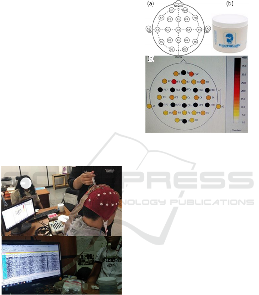
Experiments were carried out in a room that was
conditioned away from noise and provided comfort
for the subject (Figure 1). Before conducting an EEG
signal recording session, the subjects first directed
interviews with the medical team, filled out
information of concern, and follow the urine tests.
Then the subject is attached to an instrument in the
form of an electro-cap on the head and also tied a belt
to the chest of the subject so that the electro-cap does
not shift. Subjects were briefed regarding the
experimental scenario. The EEG signals are recorded
through 19 channel electrodes, including Fp1, Fp2,
F7, F3, Fz, F4, F8, T3, C3, Cz, C4, T4, T5, P3, Pz,
P4, T6, O1, and O2. The reference in this experiment
uses electrodes mounted on the ear, the A1 and A2
channel electrodes, which are A1 for the left ear and
A2 for the right ear. When installing electrodes, a gel
is applied to each EEG sensor to increase
conductivity while maintaining impedance between
ICAE 2020 - The International Conference on Applied Engineering
6
the scalp and electrodes below 5 k. The electrode
mounting position, the electrolyte liquid used in the
form of an electro gel, and the impedance shown in
Figure 2. The electrode impedance can be monitored
in WinEEG software before the signal recording
process is started. The dark color on the electrode
indicator indicates a higher impedance, while the
bright color indicates a low impedance level.
In addition to fix the electrode impedance, another
thing to consider is setting the recording process in
the WinEEG system. Settings include the sampling
frequency used which is 500 Hz, the list of channels
to be used and their references, and interconnection
with PCs / laptops to display the stimuli used when
recording EEG signals. During the recording process,
subject was asked to sit relaxed while closing his
eyes. Experimental time allocated for each trial is 1
hour and 10 minutes. The time is divided into 3
sessions, namely 5 minutes before, 10 minutes after,
and 60 minutes after consuming methadone. After the
recording session is finished, the recording of the
EEG signal is exported into a file with EEG format,
which the file can later be processed using a signal
processor.
Figure 1: The experiment design.
Figure 2: (a) Electrode position, (b) electrogel for
conductivity, (c) electrode conductivity with around 5 k
impedance.
3 SIGNAL PROCESSING
EEG raw data is processed with C2 references using
18 channels from 8 subjects. The 18 channels that are
used are grouped according to the brain lobes, namely
Central: C3, C4; Frontal: Fp1, Fp2, F3, F2, F4, F7,
F8; Parietal Occipital: P3, Pz, P4, O1, O2; Temporal:
T3, T4, T5, T6. The average amplitude (after
extraction) of each channel group is calculated.
Before the data is processed montage reference is
changed to the middle part of the brain with the Cz
channel. Recording of each subject is done for ± 5
minutes per session. Data is taken from 10 seconds to
130 seconds because data processing will be more
effective if taken 2 minutes of data that is clean and
free of artefacts. Data recording before the 10th
second is cut because the initial 10 seconds are
considered to be still corrupted by noise where the
subject is still adjusting to the experimental
conditions.
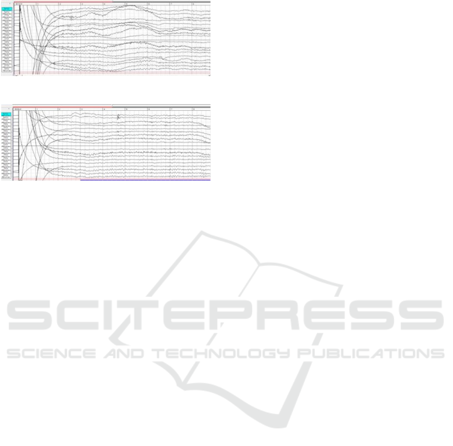
From Figure 3 raw data generated, clearly visible
on the EEG signal there are still many artifacts which
make it difficult in understanding the character of the
signal, therefore the next processes are needed.
Methadone Effects on Frontal Brain Lobe based EEG-P300 Waves in Drug Rehabilitation Patients
7
(A)
(B)
Figure 3: Raw data of EEG in relax and close aye condition:
before and (b) after Methadone intake.
To reduce background noise, the filtering process
for EEG raw data is carried out. Bandpass filter is a
circuit that is designed to pass the frequency within
certain limits and reject other frequencies outside the
desired frequency. And bandpass filter is a
combination of highpass and lowpass filter. In the
experiment, the cut off frequency used is 0.5 and 50
Hz. As for feature extraction, the wavelet method
with the symlet model and the 5 level decomposition
process is used.
Wavelet transform is a signal processing method
by resembling signal analysis using Fourier
transforms, namely by breaking the signal to be
analyzed into several parts. The difference, if the
Fourier transform signal is broken down into signal
sinusioda with different frequencies, then the wavelet
transformation of the analyzed signal is broken down
into a number of signals resulting from shifting and
scaling of a small signal called a wavelet. The wavelet
transform method in Equation (1) (Jawabri &
Sharma, 2019), mainly used to identify true
components and remove noise from the raw data
W
f
(
j
,
k
)
f
(
t
)
j
,
k
*(
t
)
dt
,
with
(1)
where,
j,k
(t) 2
j
/
2
(2
j
t k , where
(t) is the
mother wavelet, f (t) is the series analyzed, and t
indicates the time; integer j indicates the
decomposition level, and k indicates the time
translation factor, and
*(t) is the complex
conjugate.
The first step of the wavelet application starts
from the original signal then the coefficients set is
approximated on each level. In each step except the
first one, only the approximated coefficients are
analyzed. The wavelet used must meet the regularity
of order N condition in Eq. (2)
t
(
t
)
dt
0,
k
1,
,
N
1 (2)
Under the level of j, the original signals can be
reconstructed using Eq. (3).
f
j
(
t
)
W
f
(
j
,
k
)
*(2
j
t
k
)
(3)
By increasing the decomposition level j, the
detailed information of signals at larger temporal
scales would obtained. The more contribute
information we have, the better performance of the
model is achieved. However, more input could reduce
the computing efficiency and decrease the stability of
the model. Therefore, it is important to select an
appropriate decomposition level for wavelet
modeling.
4 RESULTS AND DISCUSSION
Drugs provide a dominant effect on the functioning
of the four brain lobe: frontal, parietal, temporal, and
occipital lobes. These effects can be observed if brain
activities is record and processed. In the experiment
of brain activity record, a group of subject is asked to
sit in relax while closing their eyes. The brain wave is
recorded about three times: before, 10 minutes, and
60 minutes after taking methadone. Assumption that
subjects who are follow the rules of experiment will
fill craving in the first record, starts to comfort after
10 minutes, and feel comforts after 60 minutes of
consuming methadone. The differences in the
amplitude of the extracted EEG before and 1 hour
after the subject consume the methadone in four
region of brain is observed. Subjects who have not
been given methadone have a higher level of interest
in methadone (craving), consequently the amplitude
after consuming methadone must be lower. The
decrease in amplitude value is also supported by the
influence of methadone which tends to make the
subject sleepy where theta waves increase and beta
waves decrease.
The cerebral cortex of the brain can be divided
into four lobes (see Figure 4): The frontal, parietal,
occipital, and temporal lobes. They are associated
with different functions into the body ranging from
reasoning to auditory perception. The frontal lobe is
associated with motivation, thinking, movements,
cognition, and expressive language. Damage to the
frontal lobe can lead to changes in sexual habits,
socialization, and attention as well as increased risk-
taking. The parietal lobe is associated with processing
ICAE 2020 - The International Conference on Applied Engineering
8
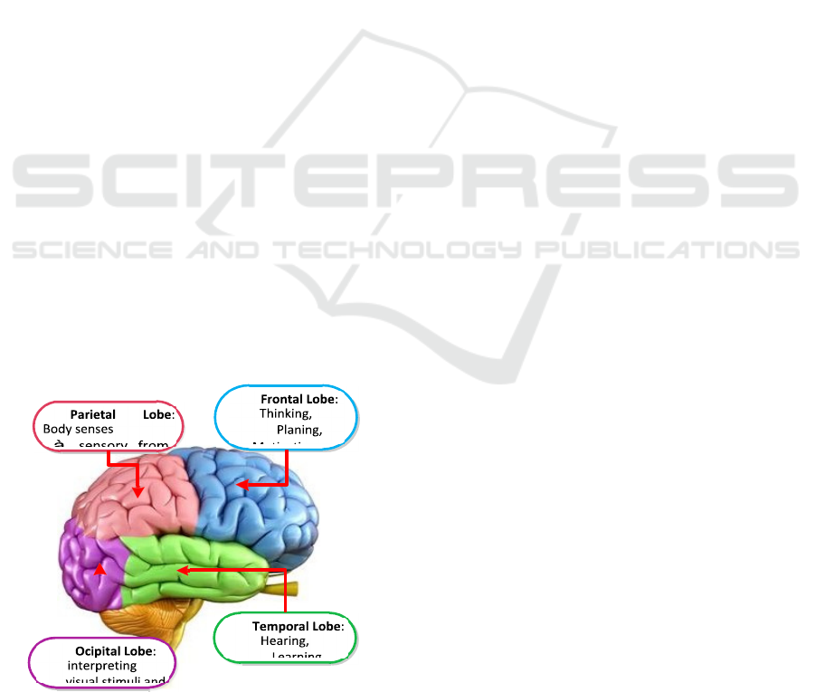
tactile sensory information such as pressure, touch,
and pain. The temporal lobe is important for
interpreting sounds and the language. Damage to the
temporal lobe can lead to problems with memory,
speech perception, and language skills. The occipital
lobe is associated with interpreting visual stimuli and
information. Damage to this lobe can cause visual
problems such as difficulty recognizing objects, an
inability to identify colors, and trouble recognizing
words (Jawabri & Sharma, 2019).
Because drugs affect the work of brain, drugs can
change the mood of feelings, ways of thinking,
awareness, and behavior of the wearer. That is why
narcotics are called psychoactive substances. There
are several kinds of effects of drugs on the brain, such
as inhibiting the work of brain, called depression.
This state could reduce awareness resulting in
drowsiness. Drugs can also stimulate the work of the
brain or what is often called a stimulant, so that arises
a sense of freshness and enthusiasm, increased
confidence, and relationships with others become
close. However, this can cause inability to sleep,
restlessness, faster heart palpitations, and increased
blood pressure. Some drugs could cause delusions, or
what are often called hallucinogens. Narcotics abuse
has an influence on the work of the nervous system,
including: Sensory nerve disorders (central and
occipital lobes) that cause numbness and blurred
vision that can cause blindness; Autonomic nerve
disorders (frontal lobe) that cause unwanted
movements through motor motion. Impaired motor
nerves (central and frontal lobes) that cause loss of
coordination with the motor system. Vegetative nerve
disorders (frontal, temporal, and central lobes) cause
language to come out of consciousness and cause fear
and lack of confidence.
Figure 4: Brain lobe effected by Methadone.
Table 1 shows the mean amplitude of each lobe
before and after consuming Methadone. The results
of previous studies show that drug use automatically
affect brain performance in each lobe such as
disturbing vision for the occipital, movement, and
language for central and frontal, emotions for
temporal. In theory, if someone who is craving is
given Methadone then the subject should feel more
comfortable after an hour. Based on the experimental
results in Table 1, except for subjects 5 and 6, the
average amplitude in each lobe has decreased.
Individually, subjects 2, 3, 4, 7, and 8 have their
respective amplitudes increasing at central, occipital,
frontal & temporal, occipital, central, and frontal
parts.
When compared with subjects 5 and 6, the
increase is not that significant. Subjects 5 and 6 had
lobes of increased amplitude after taking Methadone.
Based on the history of substance use, the two
subjects used almost the same drugs and the most
compared to other subjects. Both have almost the
same age with few mental disorders and a relatively
high value of impulsivity. They also took high doses
of Methadone even though they had been undergoing
rehabilitation for a long time (subject 5 had rehab for
11 years). Subjects 5 and 6 also consumed
benzodiazepines during the experiment.
Based on the demographic conditions of two
subjects, it can be understood that the increase in the
amplitude value of brain activity in each lobe is the
result of impaired brain function in the related area.
The highest increase was seen in subject, 6 which is
about 6.5 times the time of craving. Meanwhile, the
increase in subject 5 is only about 2.5 times from the
condition at craving. Based on demographic
conditions, subject 6 still consumed very high doses
of methadone or the maximum dose during the
experiment. It is suspected that subject 6 does not
follow routine and adequate rehabilitation.
Meanwhile, subject 5 had obtained a significant
reduction in dose to the maximum dose. However,
because the age of using the drug is quite long, which
is 11 years, it is likely that a lot of brain nerve tissue
has been damaged so that even though it has been
given Methadone, brain function cannot return to
normal.
Subjects 4 and 8 both had two lobes in which the
amplitude of brain activity did not decrease. Both
subjects had the second-highest history of substance
use than other subjects, and the dose was almost the
same as the maximum dose. When viewed as a whole,
changes in the brain activity amplitude after
consuming Methadone are closely related to history
of substance use and decreased Methadone dose.
Methadone Effects on Frontal Brain Lobe based EEG-P300 Waves in Drug Rehabilitation Patients
9

When compared with subjects 2 and 3, subject 7
experienced a significant increase in the amplitude of
the occipital region, which is almost 4 times
compared to the craving condition. Based on the
results of urine tests, subject 7 is suspected to
consume benzodiazepines and methamine during the
experiment. These conditions sufficiently state the
reasons for the increase in the associated amplitude.
Table 1: Amplitude of brain activity in the lobe of central,
frontal, occipital, and temporal: before, 10 minutes, one
hours of methadone intake.
Amplitude
S
Lobes Before 10 m 1 hours
1
Central
28.29 20.82 10.01
Frontal
119.80 63.73 8.34
Ocipital
48.23 37.78 15.40
Temporal
69.81 63.78 20.52
2
Central
7.80 5.82 18.12
Frontal
10.08 12.06 9.10
Ocipital
9.85 14.40 3.27
Temporal
10.55 17.04 2.71
3
Central
216.88 131.25 55.29
Frontal
84.02 61.15 33.26
Ocipital
89.55 105.08 124.45
Temporal
36.77 31.04 11.39
4
Central
42.69 28.47 32.42
Frontal
26.47 28.07 29.34
Ocipital
34.85 17.94 32.73
Temporal
8.70 8.46 9.34
5
Central
14.46 28.87 49.23
Frontal
15.17 21.83 50.79
Ocipital
20.55 59.43 25.65
Temporal
4.58 7.32 18.28
6
Central
24.49 20.47 120.79
Frontal
24.50 22.86 122.04
Ocipital
13.93 22.32 197.58
Temporal
16.74 5.6 92.36
7
Central
40.58 11.69 5.498
Frontal
38.51 11.85 8.81
Ocipital
1.25 6.76 5.46
Temporal
16.65 3.37 3.49
8
Central
27.05 28.41 33.38
Frontal
23.26 23.49 31.73
Ocipital
16.40 14.85 16.89
Temporal
7.93 9.83 7.53
5 CONCLUSIONS
Methadone intake by the drug rehabilitation patients
causes a decrease in the brain's impulsivity to given
stimuli, which indicates a decrease in the level of
desire for drugs after being given Methadone. The
main results of present analysis indicated that the
subjects have a longer P300 latency and a lower P300
amplitude after consuming Methadone. This study
revealed that drug patients have abnormalities in the
P300 component, which may reflect deficits in
cognitive function.
ACKNOWLEDGEMENTS
This research was supported by Technical
Implementation Unit for Instrumentation
Development, Indonesian Institute of Sciences and
funded by RISTEKDIKTI by INSINAS 2019,
Indonesia.
REFERENCES
Attou, A., Figiel, C., Timsit-Berthier, M., 2001. Opioid
addiction: P300 assessment in treatment by methadone
substitution. Neurophysiologie Clinique, 31,171.
BNN (Badan Nasional Narkotika), 2007. Survei nasional
penyalahgunaan dan peredaran gelap narkoba tahun
2003. Retrieved November 10
th
, 2018, from
http://www.bnn.go.id.
Hu, B., et al, 2017. Effective brain network analysis with
resting-state EEG data: a comparison between heroin
abstinent and non-addicted subjects. Journal of Neural
Engineering, 14 (4).
Iskandar, S., Kusumandari, D. E., Turnip, A., 2019. Artifact
removal with independent component analysis for 2D
brain mapping of drugs user before and after taking
Methadone. Internetworking Indonesia Journal, 1 (1),
29-33.
Jawabri, K. H., Sharma, S., 2019. Physiology, Cerebral
Cortex Functions, StatPearls Publishing. Treasure
Island (FL).
Li Q., Wang Y., Li W., Yang W. C., Zhu J., Chang H. F.,
Wu N., Zheng Y., Wang W., 2012. fMRI study on
craving and brain activity in response to heroin-related
cues in patients with methadone maintenance treatment.
Journal of Practical Radiology, 28, 1-5.
Lin C. Y., Chang K. C., Wang J. D., et al., 2016. Quality of
life and its determinants for heroin addicts receiving a
methadone maintenance program: Comparison with
matched referents from the general population. Journal
of the Formosan Medical Association, 115(9), 714-727.
Maeyer, J. D., Vanderplasschen, W., Camfield, L.,
Vanheule, S., Sabbe, B., Broekaert, E., 2011. A good
ICAE 2020 - The International Conference on Applied Engineering
10

quality of life under the influence of Methadone: s
qualitative study among opiate-dependent individuals.
International Journal of Nursing Studies, 48, 1244-
1257.
Malik, E., Adelson, M., Sason, A., Schreiber, S., Peles, E.,
2019. Outcome of patients with high depressive
symptoms on admission to Methadone maintenance
treatment. Journal of Dual Diagnosis, 1-10. doi:
10.1080/15504263.2019.1656353.
Motlagh, F., Ibrahim, F., Rashid, R., Shafiabady, N.,
Seghatoleslam, T., Habil, H., 2018. Acute effects of
methadone on EEG power spectrum and event-related
potentials among heroin dependents.
Psychopharmacology, 235 (11), 3273– 3288.
Pastor, A., Conn, J., O'Brien, C. L., Teng, J., Loh, M.,
Collins, L., MacIsaac, R.J., Bonomo, Y., 2019.
Clinicians feel comfortable discussing alcohol but not
illicit drug use with young adults with type 1 diabetes:
a survey of clinicians. Diabetic Medicine. doi:
10.1111/dme.14136.
Turnip, A. et al, 2018. Brain mapping of drug addiction in
withdrawal condition based P300 Signals. Journal of
Physics: Conference Series, 1007, 012060. IOPscience.
Turnip, A., et al., 2018. Detection of drug effects on brain
activity using EEG-P300 with similar stimuli. IOP
Conference Series: Materials Science and Engineering,
220. IOPScience.
Turnip, A., Kusumandari, D. E., Hidayat, T., Hidayat, T.,
2018. Brain mapping of low and high implusivity based
P300 Signals. Journal of Physics: Conference Series,
1007. IOPScience.
Turnip, A., Kusumandari, D. E., Pamungkas, D. S., 2018.
Drug abuse identification based EEG-P300 amplitude
and latency with Fuzzy Logic classifier. In
International Conference on Applied Engineering
(ICAE). IEEE.
Undang-Undang Republik Indonesia No. 5 tentang
Psikotropika, 1997. Retrieved from
http://hukum.unsrat.ac.id/uu/uu_5_97.htm.
Verdejo, A., Toribio, I., Orozco, C., Puente, K. L., Perez-
Garca, M., 2005. Neuropsychological functioning in
methadone maintenance patients versus abstinent
heroin abusers. Drug and Alcohol Dependence, 78,
283-288.
Wang, G. Y., Kydd, R., Wouldes, T. A., Jensen, M.,
Russell, B. R., 2015. Changes in resting EEG following
methadone treatment in opiate addicts. Clinical
Neurophysiology, 126 (5), 943-950.
Yang, L., et. al., 2015. The effects of methadone
maintenance treatment on heroin addicts with response
inhibition function impairments: Evidence from event-
related potentials. Journal of Food and Drug Analysis,
23, 260-266.
Zhang, X., Yao, L., Kanhere, S. S., Liu, Y., Gu, T., Chen,
K., 2017. MindID: Person identification from brain
waves through attention-based recurrent neural
network. ACM Journal on Computing and Cultural
Heritage, 9(4), 39.
Methadone Effects on Frontal Brain Lobe based EEG-P300 Waves in Drug Rehabilitation Patients
11
