
Potential Use of Smartphone as a Tool to Capture Embryo Digital
Images from Stereomicroscope and to Evaluate Them by an Artificial
Neural Network
Diego de Souza Ciniciato
1,*
, Maria Beatriz Takahashi
1,*
, Marcelo Fábio Gouveia Nogueira
2
and José Celso Rocha
1
1
Laboratório de Matemática Aplicada, School of Sciences and Languages, Universidade Estadual Paulista (Unesp),
Av. Dom Antonio 2100, Assis, Brazil
2
Laboratório de Micromanipulação Embrionária, School of Sciences and Languages, Unesp,
Av. Dom Antonio 2100, Assis, Brazil
*
Both authors contributed equally to the study
Keywords: Smartphone, Embryo Classification, Artificial Neural Network-based Software, Graphic User Interface.
Abstract: An online graphical user interface connected to a server was developed aiming to facilitate access to
professionals worldwide that face problems with bovine blastocysts classification. The blastocysts
assessment is carried on using images taken from an inverted microscope, which usually requires more
expensive devices such as digital camera and computer software. Smartphone camera quality and tasks
processing are getting better with technology advances. Therefore, a smartphone can be attached to the
eyepiece lens to provide Real-Time evaluation, and thus reducing costs when comparing to computers,
cameras, and software that are commonly used for this purpose.
1 INTRODUCTION
Brazilian cattle production has an important
contribution to the economy and social aspects of
this country. With approximately 215 million
livestock units and leader in meat exportations since
2004, Brazil is also the leader in in vitro production
of bovine embryos worldwide (Ministério do
Planejamento, Desenvolvimento e Gestão - Instituto
Brasileiro de Geografia e Estatística IBGE, 2016).
This production has utmost importance for
international and national improvement in cattle
genetics and productivity. The production of cattle
embryos for commercial purposes follows the steps:
they are produced in vitro and transferred to
synchronized receptors when they reach the
blastocyst stage (Hyttel et al., 2010). To help to
identify the quality embryos, which is associated
with the success of pregnancy, the International
Embryo Technology Society (IETS) recommends an
embryo classification system. This system is based
on morphological evaluation and establishes three
quality grades: excellent or good, “1”; fair, “2”; or
poor, “3” (Bó and Mapletoft, 2013).
However, this classification is directly affected
by the embryologist’s accuracy and experience to
evaluate the embryo variables related to the
development and pregnancy potential (Lindner and
Wright, 1983; Bó and Mapletoft, 2013). The reason
for this interference is that the morphological
analysis does not measure any objective variables to
determine the embryo classification. Moreover, the
analysis by the human vision is based on a
comparison between objects or images. In this
regard, human vision has difficulties at judging color
or brightness of shapes and features, which requires
measuring scales or relative size, angle and positions
of several objects to identify their characteristics
(Russ, 2016). Thus the analysis by an embryologist
is subjective and has low reproducibility (Bényei et
al., 2006). Indeed, the same embryo can be
classified with different degrees of quality by
different embryologists (inter-evaluator error) or
even by the same embryologist at different moments
(intra-evaluator error), especially in cases when the
quality grade is borderline (Farin et al., 1995).
Together with inexperience, the tiredness and the
mood of the evaluator could contribute to the major
Ciniciato D., Takahashi M., Gouveia Nogueira M. and Rocha J.
Potential Use of Smartphone as a Tool to Capture Embryo Digital Images from Stereomicroscope and to Evaluate Them by an Artificial Neural Network.
DOI: 10.5220/0006518501850189
In Proceedings of the International Conference on Computer-Human Interaction Research and Applications (CHIRA 2017), pages 185-189
ISBN: 978-989-758-267-7
Copyright
c
2017 by SCITEPRESS – Science and Technology Publications, Lda. All rights reserved

causes of the subjective and low reproducibility of
this standard system of embryo assessment.
Therefore, several methods have been or are
being developed to provide an optional evaluation
for embryo classification that does not have external
effects. Some of them includes a semi-automatized
image segmentation process with the use of artificial
intelligence (AI) for human embryos (Gonzalez,
2004), an automatic segmentation procedure of
bovine embryos without AI (Melo et al., 2014), a
semi-automatized grading method of human
blastocyst using a support vector machine (Santos
Filho et al., 2012), embryo metabolism analysis,
cellular respiration measurements, the use of zona
pellucida birefringence, microRNA profile
determination, analysis based on logistic regression
and evaluation by time-lapse video (reviewed by
Rocha et al., 2016). However, none of these
methods are totally effective, and, despite being
subjective and old, the visual morphological analysis
is still widely used (Lindner and Wright, 1983; Farin
et al., 1995; Richardson et al., 2015).
Recently, there have been attempts at creating a
method based on digital image processing to
determine the viability of human embryos by
detecting blastomeres (Singh et al., 2014; Tian et al.,
2014) or trophectoderm (Singh et al., 2015).
Additionally, using processing and digital image
analysis in the quality evaluation of mouse
blastocysts, a previous study used an artificial neural
network technique with significant success (Matos,
Rocha and Nogueira, 2014). However, as far as we
can determine from the studied literature, a
classification method using digital image processing
has not been applied to bovine blastocysts.
In this context, a method based on artificial
neural network (ANN) combined with genetic
algorithm (GA) was developed to train an ANN to
classify bovine blastocyst images based on the IETS
standards (Rocha et al., 2017). In this study, a 482
bovine blastocysts images dataset were used to train
some ANNs, from which the best obtained 76.4% of
accuracy. The input set was the variables extracted
from image processing and the output was the mode
from grading of three experienced embryologists.
The use of three evaluations avoids the bias of using
a single evaluation as the standard for the ANN
training. The Kappa index of the inter-evaluator
agreement was 0.571 (482 images, P<0.001), and the
three ANNs obtained 0.616 for the same dataset
(482 images, P<0.001). This represents that the
ANN technique was more consistent than the
embryologists’ evaluation. Moreover, the intra-
evaluator agreement was 0.28, 0.41 and 0.47 (48
images, P<0.001), and when compared to the ANNs,
there were 100% agreement (Kappa index of 1.0),
which supports the robustness and low subjectivity
of an ANN.
The present position paper is a continuation in a
deeper way of the previous work (Rocha et al.,
2017), aiming the development of a Graphical User
Interface due to users that could not be familiar with
the programming environment and do not use/have
an inverted microscope. In addition, embryologists
from around the world can access the technique
online, without downloading or install the software.
Furthermore, we describe the application of
smartphone adapters for stereomicroscope ocular
lens to classify embryos in Real-Time.
2 METHODOLOGY
A server for image processing and classification of
bovine blastocysts was developed aiming to
democratize the technology available in our research
group. The access to the server is by the link below:
http://blasto3q.com. The image processing and
evaluation are carried out by the algorithm
Blasto3Q, which is described in (Matos, Nogueira
and Rocha, 2012, 2014). The users can access this
computational tool by a multiplatform application
available on the same server. The application has a
friendly and intuitive interface for users, and it has
additional functionalities comparing to the desktop
version, such as the evaluation of multiple images in
parallel. Due to the high processing cost for each
image, we choose to centralize this operation on the
server. If this action were carried on in the
smartphone, the execution time should increase
considerably, which is not desired by users.
Therefore, the smartphone just captures the
blastocyst images and receive the results from
classification.
On the server-side, there is a MATLAB
®
application (version R2017a) that works in service
mode, which executes several scanning of databases
to search non-processed requisitions. Each service
runs one process at a time, however, it can process
several instances, and thus the processing of
different requests will be performed in parallel and
simultaneously.
For a greater user experience, an intuitive user
interface was developed to general users, which runs
on the client-side. This interface communicates by
requests to the server. Each new processing request
is initialized by the desired image uploaded into the
server. This request is added to the database in the
end of the requests queue and it will be processed
according to its rank. The process is finished with
the output that the users want.
Therefore, there is an extremely light and fast
application that can perform in devices compatible
with HTML5 (more modern navigators). Nowadays
a large part of devices provides this markup
language, and an advantage is that it is possible to
use the application in different operating systems
(for example, both Android and iOS can execute the
application). The user can access this software
wherever they are, if they have an internet
connection. The results are processed in few
seconds.
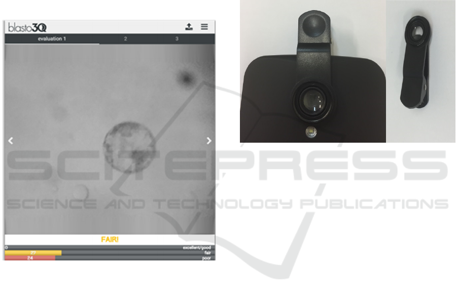
Figure 1: Graphical User Interface evaluating bovine
blastocyst as grade 2 (“fair”) since the highest vector was
the yellow that is related to the fair degree.
In Figure 1, there is an example of the
application interface describing an in vitro produced
bovine embryo image taken from a smartphone
juxtaposed to an eyepiece of the stereomicroscope.
For this purpose, we used a Samsung S6 coupled to
a macro lens (Figure 2) to allow the proximity of the
smartphone lens with the eyepiece. With this
apparatus, the image of the embryo on the eyepiece
could be captured by the smartphone lens and using
the zoom of the phone to fill the screen with the
image. Moreover, we used the maximum of
magnification of the stereomicroscope (i.e., 60 x,
Leica M80).
3 DISCUSSIONS
The smartphone development allowed the creation
of new technologies and applications. Nowadays the
daily tasks made by a computer and a smartphone
are very similar, as we can access websites and
software on both devices. The smartphones have
advantages due to easy portability, and they are
lighter and cheaper than computers. Moreover, the
recent improvement of new generation smartphones’
cameras allowed taking images with higher quality
than previous generations.
Figure 2: Illustrative image of the macro lens attached to
the lens of the smartphone (left) and the macro lens alone
with its clipper (right).
In this context, the application of cheaper and
robust technology in research laboratories is
required to reduce costs. The image records using
microscopy in any laboratory usually requires
desktop computers and expensive digital cameras
and software to analyze them. Also, expensive
inverted microscopes are often required to obtain
those images with a high quality when recording
mammalian oocytes and embryos. The development
of an application that is functional in any device
(smartphone or other devices, such as desktop and
tablet) to evaluate images of blastocysts from
microscopy allows Real-time assessment, reduce
costs, and solve a subjective issue in blastocysts
classification.
Several adapters were developed to attach the
smartphones to the eyepieces of a microscope,
which provide better ergonomic, simple and fast
ways to take pictures of the sample, as in anatomic
pathology analysis using mobile devices (Lehman
and Gibson, 2013; Roy et al., 2014) and diagnostic
of diseases using deep learning (Quinn et al., 2016)
both attached to conventional light microscope. In
this way, besides the macro lens attached to the
smartphone lens, an adapter to a better stabilization

and focal plane quality could useful. Also, the
standardization of the blastocysts images by image
processing steps keeps the features from the
blastocysts, which allows the software interprets it
properly.
The improvement of this technology processing
can be achieved by cloud computing, which is a
model related to applications called ‘Software as a
service’. The basis for this model is to run on distant
computers linked by a cloud that is owned and
operated by others and that connect to users’
computers via a web browser. The advantages of this
method are the access to applications and data from
different computers that are connected to the cloud,
less risk of missing data and dynamically scalable
(Armbrust et al., 2010).
4 CONCLUSIONS
The bovine blastocyst classification by Artificial
Neural Network available as a graphical user
interface provides a robust method to embryologists
that are not familiar with programming languages.
Also, the smartphone adapters for microscope
eyepiece should provide better ergonomic and a
Real-Time assessment of bovine embryos.
ACKNOWLEDGEMENTS
The authors’ research is supported by grants #
2012/50533-2, 2013/05083-1, 2006/06491-2,
2011/06179-7, 2012/20110-2 and 2016/19004-4
from São Paulo Research Foundation (FAPESP).
We also thank Agência UNESP de Inovação (AUIN)
for processing the national and international patents
of the invention.
REFERENCES
Armbrust, M. et al. (2010) ‘A view of cloud computing’,
Communications of the ACM, 53(4), p. 50. doi:
10.1145/1721654.1721672.
Bényei, B. et al. (2006) ‘The effect of internal and
external factors on bovine embryo transfer results in a
tropical environment’, Animal reproduction science,
93(3–4), pp. 268–279.
Bó, G. and Mapletoft, R. (2013) ‘Evaluation and
classification of bovine embryos’, Animal
Reproduction, 54(935), pp. 344–348.
Farin, P. W. et al. (1995) ‘Agreement among evaluators of
bovine embryos produced in vivo or in vitro’,
Theriogenology, (95), pp. 339–349.
Gonzalez, R. (2004) Digital Image processing using
Matlab. Upper Saddle River New Jersey: Pearson
Prentice Hall.
Hyttel, P. et al. (2010) Essentials of domestic animal
embryology. Saunders/Elsevier.
Lehman, J. S. and Gibson, L. E. (2013) ‘Smart
teledermatopathology: a feasibility study of novel,
high-value, portable, widely accessible and intuitive
telepathology methods using handheld electronic
devices’, pp. 513–518. doi: 10.1111/cup.12108.
Lindner, G. and Wright, R. W. J. (1983) ‘Bovine embryo
morphology and evaluation’, Theriogenology,
20(313), pp. 407–416.
Matos, F. D., Nogueira, M. F. G. and Rocha, J. C. (2012)
‘Método para determinação da viabilidade e qualidade
de embriões’. Brazil.
Matos, F. D., Nogueira, M. F. G. and Rocha, J. C. (2014)
‘Method for determining embryo viability and
quality’. Brazil.
Matos, F. D., Rocha, J. C. and Nogueira, M. F. G. (2014)
‘A method using artificial neural networks to
morphologically assess mouse blastocyst quality’,
Journal of Animal Science and Technology, 56.
Melo, D. H. et al. (2014) ‘Algorithms for automatic
segmentation of bovine embryos produced in vitro’,
Journal of Physics: Conference Series 490, pp. 1–4.
Ministério do Planejamento, Desenvolvimento e Gestão -
Instituto Brasileiro de Geografia e Estatística IBGE.
Produção da pecuária municipal 2015. IBGE 43, 1-47
(2016). e-Book (bulletin) <http://biblioteca.ibge.
gov.br/visualizacao/periodicos/84/ppm_2015_v43_br.
pdf>.
Quinn, J. A. et al. (2016) ‘Deep Convolutional Neural
Networks for Microscopy-Based Point of Care
Diagnostics’, in Proceedings of International
Conference on Machine Learning for Health Care.
Richardson, A. et al. (2015) ‘A clinically useful simplified
blastocyst grading system’, Reproductive BioMedicine
Online, 31, pp. 523–530.
Rocha, J. C. et al. (2016) ‘Methods for assessing the
quality of mammalian embryos: How far we are from
the gold standard?’, JBRA Assisted Reproduction,
20(3), pp. 150–158.
Rocha, J. C. et al. (2017) ‘A method based on artificial
intelligence to fully automatize the evaluation of
bovine blastocyst images’, Scientific Reports,
7(Article number: 7659).
Roy, S. et al. (2014) ‘Smartphone adapters for digital
photomicrography’, J Pathol Inform, (October). doi:
10.4103/2153-3539.137728.
Russ, J. C. (2016) The Image Processing Handbook.
Seventh. CRC press.
Santos Filho, E.
et al. (2012) ‘A method for semi-
automatic grading of human blastocyst microscope
images’, Human Reproduction, 27, pp. 2641–2648.
Singh, A. et al. (2014) ‘Automatic blastomere detection in
day 1 to day 2 human embryo images using partitioned
graphs and ellipsoids’, IEEE International Conference
on Image Processing (ICIP), pp. 917–921.

Singh, A. et al. (2015) ‘Automatic segmentation of
trophectoderm in microscopic images of human
blastocysts’, IEEE Transactions on Biomedical
Engineering, 62(1), pp. 382–393.
Tian, Y. et al. (2014) ‘Automatic Blastomere Recognition
from a Single Embryo Image’, Computational and
Mathematical Methods in Medicine, 2014.
