
Effects of Hunger on Sympathetic Activation and Attentional
Processes for Physiological Computing
Ferdinand Pittino
1
, Sandra Mai
2
, Anke Huckauf
1
and Olga Pollatos
2
1
General Psychology, Ulm University, Albert-Einstein-Allee 47, 89069 Ulm, Germany
2
Clinical and Health Psychology, Ulm University, Albert-Einstein-Allee 41, 89069 Ulm, Germany
Keywords: Hunger, Eye-tracking, Pupillometry, Visual Attention.
Abstract: Assessing users’ states becomes increasingly important also for technical systems. In the present study, we
assessed the influence of hunger on processing food versus household items by monitoring eye movements
in a picture categorization task. As indicator for sympathetic activation, pupil dilation was additionally
assessed in hungry and satiated participants. Food and household items were presented in the left and right
visual field and the task of the participants was to indicate whether the pictures in both visual fields
represented the same (household vs. food) or different categories (household and food). Although behavioural
data did not differ between hungry and satiated participants, more thorough investigations of gaze behaviour
showed that hungry participants were more impaired in processing household items than the satiated ones. In
addition, mean pupil dilation differed between hungry and satiated participants. Pupil size was shown to
correlate with hunger ratings suggesting that gaze-based measures can indeed serve as diagnostic tool for
sensing user states.
1 INTRODUCTION
Current technical systems are expected to react to the
intentions and dispositions of users. Hence,
knowledge about basic motivations of users and their
respective changes in behaviour are essential when
realizing affective computing systems. The technical
systems need to acquire their knowledge about the
user’s states by means of physiological measures.
Therefore, it is important to know how users react to
changes in bodily states as well as to respective
relevant stimuli. In the current study, we assessed
such physiological changes using the bodily state of
hunger and corresponding food versus household
images as relevant stimuli as an example.
Hunger is one of the most basic bodily
dispositions. One might assume that hunger affects
human attention and arousal. In addition, one might
suspect that it affects attending either towards all
stimuli or towards potentially eating-related stimuli
only. In the current study, this was investigated by
measuring eye movements and pupillary changes in
hungry versus satiated participants watching
comparable food and household images.
Hunger is known to influence many cognitive and
emotional processes in everyday life, which is
especially apparent in food-related behaviour:
Empirical data demonstrate that food deprivation
alters brain activation and subjective appeal to
pictures of high- and low-calorie food (Giel et al.,
2010; Goldstone et al., 2009; Piech et al., 2010) and
modulates activity in the food reward system (Siep et
al., 2009). It is also known that hunger is a potent
activator of the sympathetic nervous system
(Andersson et al., 1988; Chan et al., 2007; Pollatos et
al., 2012); various studies could demonstrate that
short-term food deprivation (up to 72 hours) leads to
an increased sympathetic and decreased
parasympathetic activation. For example, Chan et al.
(2007) demonstrated that short-term fasting increased
sympathetic activity as measured by heart rate
variability (HRV) and 24-hour urinary
catecholamines and decreased parasympathetic tone
(HRV) in humans.
In this context the model of neurovisceral
integration proposed by Thayer and Brosschot (2005)
is highly interesting. It states that autonomic
imbalance and reduced parasympathetic tone may be
the final common pathway linking negative affective
states to health problems, probably modulated by
interface regions like the prefrontal cortex, which is a
target region both for information from the central
152
Pittino, F., Mai, S., Huckauf, A. and Pollatos, O.
Effects of Hunger on Sympathetic Activation and Attentional Processes for Physiological Computing.
DOI: 10.5220/0006008301520160
In Proceedings of the 3rd International Conference on Physiological Computing Systems (PhyCS 2016), pages 152-160
ISBN: 978-989-758-197-7
Copyright
c
2016 by SCITEPRESS – Science and Technology Publications, Lda. All rights reserved

nervous system and attention, emotion and motivated
behaviour networks (Thayer and Brosschot, 2005). A
sympathetic activation and a parasympathetic
withdrawal has been demonstrated to be linked to
hypervigilance and inefficient allocation of
attentional and cognitive resources (Thayer and
Brosschot, 2005). A former study could show that
food deprivation provokes a parasympathetic
decrease and heightened sympathetic activity which
was associated with a hypervigilance to pain stimuli
both on a perceptual and emotional level (Pollatos et
al., 2012).
There are already reports indicating that eye
movements between hungry and satiated persons do
not differ (e.g. Nijs et al., 2010). However, there are
also observations that eye movements can be
diagnostic for eating behaviour (Werthmann et al.,
2011) in over-weight participants. Nevertheless,
whether hungry and satiated person process food
versus other images differently is still unclear. This
was to be examined in the present study.
Moreover, in video-based eye tracking, also pupil
dilation is given. As is evident, pupils of the eye are
activated by sympathetic nerves (e.g. Ehlers et al.,
2016; Partala and Surakka, 2003). Hence, pupillometry
can serve as an indicator of sympathetic activation. As
stated above, hunger is known to raise sympathetic
activation. Hence, one would expect observing larger
pupils in hungry relative to satiated participants.
Concerning food stimuli, numerous studies
demonstrated that nutrition state of the subjects
changed brain responses to food stimuli (Goldstone et
al., 2009; Siep et al., 2009; Stice et al., 2013). For
example, Goldstone and colleagues (2009) reported
that fasting increased activation to pictures of high-
calorie foods in various brain regions including the
ventral striatum, the amygdala, the anterior insula,
and medial and orbitofrontal cortex. Furthermore,
fasting enhanced the subjective appeal of high-calorie
foods, and the change in appeal bias towards high-
calorie foods was positively correlated with medial
and orbitofrontal cortex activation. The authors
concluded that fasting biased brain reward systems
towards high-calorie foods. Supporting these results,
also Stice and co-workers (2013) reported that the
duration of experimentally manipulated caloric
deprivation correlated positively with activation in
regions implicated in attention, reward, and
motivation in response to food images (including the
anterior cingulate cortex and the orbitofrontal cortex).
They suggested that self-imposed caloric deprivation
increases responsivity of attention, reward, and
motivation regions to food, which may explain why
caloric deprivation weight loss diets typically do not
produce lasting weight loss (Stice et al., 2013).
These results are in accordance to Siep et al.
(2009) who reported that hunger interacts with the
energy content of foods, modulating activity in
several regions (e.g. cingulate cortex, orbitofrontal
cortex, insula). They showed that food deprivation
increased activity following the presentation of high
calorie foods and also followed that this fact may
explain why treatments of obesity energy restricting
diets often are unsuccessful. Extending these results,
Frank and co-workers (2010) reported that high-
caloric pictures compared to low-caloric pictures led
to increased activity in food processing and reward
related areas, like the orbitofrontal and the insular
cortex, but furthermore they found activation
differences in visual areas (occipital lobe), despite the
fact that the stimuli were matched for their physical
features. Frank et al. (2010) concluded that hunger
and the calorie content of food pictures also modulate
the activation of early visual areas. Having this in
mind, other measures than imaging techniques that
are rather slow in their response profile might help to
disentangle early effects of hunger on visual
processing of food stimuli.
Using other measures than imaging techniques,
Hepworth and co-workers (2010) manipulated mood
in healthy participants before using food stimuli in a
visual-probe task assessing attentional bias. They
showed that negative mood increased both attentional
bias for food cues and subjective appetite. Attentional
bias and subjective appetite were positively inter-
correlated, suggesting a common activation of the
food-reward system. Giel and colleagues (2011) used
eye tracking in a free viewing paradigm: They
reported that anorectic patients allocated overall less
attention to food. Interestingly, attentional
engagement for food pictures was most pronounced
in fasted healthy control subjects.
Till now, the question of whether different levels
of visual processing of food stimuli can be
distinguished using eye tracking when manipulating
hunger state in participants remains unanswered. As
hunger essentially influences everyday behaviour and
is an important variable in sensing the state of a
person in interaction to his/her environment, the aim
of the present study was twofold: First, we wanted to
clarify whether hunger leads to an attentional bias for
food pictures using different measures of eye tracking
(direction of the initial fixation, first fixation duration,
fixation count) allowing to distinguish perceptual
from evaluating processes. Second, we aimed to
elucidate whether pupillometry was a valid measure
for a sympathetic increase associated with hunger.
Effects of Hunger on Sympathetic Activation and Attentional Processes for Physiological Computing
153
2 METHOD
2.1 Participants
In total 51 psychology students (40 females and 11
males, M
age
= 22.61, SD
age
= 3.92) of Ulm University
participated in this experiment for partial fulfilment
of course credit. All participants did not report a
history of eating disorder, had normal or corrected to
normal vision and provided informed consent based
on the guidelines of the German Research Foundation
(DFG). Due to technical difficulties (calibration and
data recording) the data of four participants were not
included in the analysis of the gaze data during the
food categorization task and six subjects were not
included in the analysis of the influence of hunger on
pupil size. Therefore, the sample for the food
categorization task (see Procedure) consisted of 47
participants (26 hungry, 38 females, M
age
= 22.77,
SD
age
= 4.05) and the final sample for the influence of
hunger on pupil size consisted of 45 participants (25
hungry, 37 females, M
age
= 22.93, SD
age
= 4.06).
2.2 Stimuli and Apparatus
As stimuli the pictures of food (e.g. bread) and
household items (e.g. handkerchief) used by Koch et
al. (2014) to study the attentional bias towards food
in overweight and obese children, were applied. The
advantage of this stimulus set consists in the similar
depiction of food and household items. Stimuli of
both categories were presented on a white plate.
The experiment was run on a Windows 7 PC and
was implemented using PsychoPy (Version 1.81.02;
Peirce, 2007). The stimuli were presented on a Dell
P2210 (resolution 1680 x 1050 px, refresh-rate 60 Hz)
which was stationed approximately 60 cm from the
participant. A remote eye-tracking system (RED250
with a sampling-rate of 120 Hz; SensoMotoric
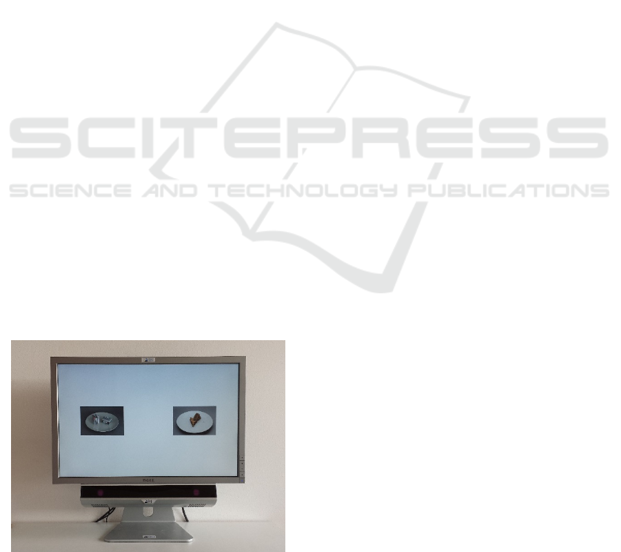
Figure 1: Overview of the experimental setup.
Instruments, Teltow Germany) was attached to the
monitor and recorded gaze as well as pupillometric
data. The experimental setup is illustrated in Figure 1.
2.3 Questionnaire Data
Besides the assessment of sociodemographic
variables (such as age and gender), the participants
were also asked whether they have suffered from any
kind of eating disorder in their lifetime. Height and
weight were assessed with a customary measuring
device and a customary digital scale. Furthermore, the
participants were asked to rate their subjective
feelings of hunger on the 8-item visual analogue scale
ranging from 0 to 10 of Flint, Raben, Blundell, and
Astrup (2000, e.g. “How hungry are you?”).
2.4 Procedure
In preparation for the study, the participants in the
food deprivation condition (n = 27) were asked not to
consume any food, alcohol or caffeine beginning at
8:00 pm on the day prior to their individual study
appointment, which was either until 8:00 am, 9:00 am
or 10:00 am. In contrast, the participants in the
satiated condition (n = 24) were asked not to change
their eating habits.
After arriving in the laboratory the weight and size
of the subjects were assessed and the participants
rated their subjective hunger feelings on the visual
analogue scale of Flint et al. (2000).
The experiment consisted of two parts. First, pupil
size was measured and afterwards, the experiment
aiming at exploring the processing of food and
household items in satiated and hungry participants
using eye-tracking was carried out.
The two parts of this experiment are now
described in more detail.
2.4.1 Pupillometric Measurement Phase
Participants’ pupil size was measured for five seconds
using the SMI RED250 eye-tracking system. To
avoid eye-movements, participants were instructed to
fixate on a black fixation cross, which was presented
centrally with a size of 0.6° on a homogenously white
screen (141.7 cd/m²). The luminance of the room was
kept constant at 75.3 lx (including the luminance of
the monitor).
2.4.2 Food Categorization Task
After the pupillary measurement phase, the food
categorization task was carried out. At its beginning
a 9 point calibration and 4 point validation was
PhyCS 2016 - 3rd International Conference on Physiological Computing Systems
154

performed.
In the food categorization task, a trial started with
the central presentation of a black fixation cross with
a size of 0.6° for 1 s. Subsequently, two pictures (each
9° x 6°) were peripherally presented in the left and
right visual field at an eccentricity of 9°. These
pictures either consisted of food (F) or household (H)
items (Koch et al., 2014; see Figure 1 for an
illustration). Therefore, there were four different
stimulus constellations: food items are presented on
both sides (FF), household items are presented on
both sides (HH), food items are presented on the left
and household items on the right side (FH) and
household items are presented on the left and food
items are presented on the right side (HF). The task
of the participants was to decide whether the objects
shown in both visual fields were representing the
same category (either both food or both household
items) or whether mixed categories were presented in
the left and right visual field. If the stimuli presented
on both sides represented the same category
participants were instructed to press the key ‘l’ on the
right hand-side of a standard QWERTZ-keyboard. If
the stimuli presented on both sides represented
different categories participants were instructed to
press the key ‘a’ on the left side of the keyboard.
The experimental block started with a short
training phase consisting of four trials, in which the
participants were familiarized with the task and the
stimuli’s appearances and categories. The test phase
consisted of 64 trials, 16 for each stimulus
combination (FF, HH, FH, HF). The order of the trials
was randomized. During this test phase, the gaze
position was continuously recorded.
2.5 Processing of the Oculomotor Data
2.5.1 Processing of Pupillometric Data
For processing of the pupillary signal, first, blinks and
saccades were removed from the data stream. Further,
values deviating more than 1.5 times the interquartile
range from the median were treated as artefacts and
were removed from the signal. The resulting missing
values were replaced using linear interpolation.
2.5.2 Processing of the Eye-movements Data
For processing the gaze data, we utilized the Be Gaze
(Version 3.5) software of SMI (SensoMotoric
Instruments, Teltow Germany).
As areas of interest (AOI), the left and right
stimuli were regarded. For these two AOIs, event
statistics were computed and exported. Afterwards,
the raw data were processed using Matlab R2015b
(Mathworks Inc.). Trials with very high or low
reaction times (median ± 1.5 interquartile range) were
dismissed. Additionally, for each variable, subject
and stimulus category an outlier analysis was
performed. Values deviating more than three times
the interquartile range of the median were not
considered in the adjunct analysis. Finally, all
dependent variables (initial fixation direction, first
fixation duration and fixation count) were extracted
separately for the different stimulus configuration
(FF, HH, FH, HF) and both visual fields (left, right).
In order to control for reliable computation of the
mean, only data of participants were considered,
which had at least eight remaining correct responses
per condition after the procedure, described above,
was carried out.
The data were descriptively and inferentially
analysed using SPSS (Version 21, IBM).
3 RESULTS
3.1 Hunger Ratings
In order to check whether the instruction to resign for
food during the last hours yielded in hungrier
participants, the reported feelings of hunger were
assessed. Since the eight items of the hunger scale of
Flint et al. (2000) showed high internal consistency (α
= .887), the items were averaged and combined into
one scale.
A t-test revealed that participants in the food-
deprivation condition indeed reported higher hunger
feelings than participants in the satiated condition
(t(30.47) = 4.92, p < .001, M
hungry
= 7.12 SD
hungry
=
1.30, M
satiated
= 4.41 SD
satiated
= 2.24). Therefore, we
conclude that the short-term food deprivation was
successful in manipulating the participants’ hunger
state.
3.2 Analysis of the Behavioral Data
One can assume that hunger speeds up motor
reactions and the processing of either stimuli
independent of their content (food and non-food) or
specific to the motivational relevant stimulus food.
To examine this question, we analyzed the
correctness and reaction times in the food
categorization task. The purpose of this analysis was
to investigate whether hungry and satiated
participants differed on a behavioral level in
processing the different stimuli (FF, HH, FH, HF).
The percentage of correct responses of the food
Effects of Hunger on Sympathetic Activation and Attentional Processes for Physiological Computing
155
categorization task was entered in a repeated
measures ANOVA with the within-subject factor
stimulus (FF: food pictures are presented in both
visual fields; HH: household items in both visual
fields; FH: food items in the left and household items
in the right visual field; HF: household items in the
left and food items in the right visual field) and the
between-subject-factor hunger (satiated vs. hungry).
This analysis revealed a main effect of stimulus
(F(1.39,62.74) = 25.55, p < .001, η² = .362).
Participants gave most correct responses, when food
items were presented in both visual fields (M =
95.3%, SE = 0.7%) and least correct response when
household items were presented in both visual fields
(M = 72.4%, SE = 3.6%). Mixed stimulus
combinations were rated equally good at 89.3% (SE
= 1.3%).
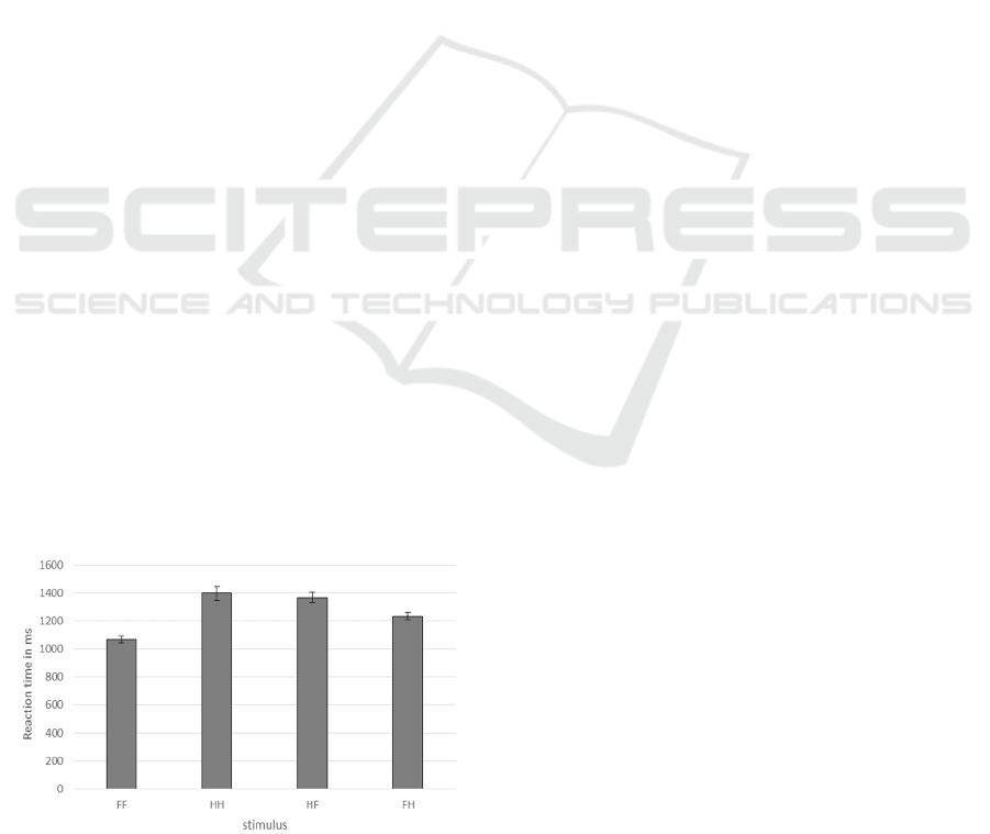
Reaction times for correct trials were entered in a
repeated measures ANOVA with the within-subject
factor stimulus (FF vs. HH vs. FH vs. HF) and the
between-subject-factor hunger (satiated vs. hungry)
for all 43 participants (24 hungry) who answered at
least eight trials per stimulus combination (FF, HH,
FH, HF) correctly. Again, the analysis revealed a
main effect for stimulus (F(1.74,71.35) = 36.98, p <
.001, η² = .474, see Figure 1). Participants reacted
especially fast when food was displayed in both
visual fields. They were slower when only household
items were presented and when a combination of food
and household items was shown. Furthermore, for
trials with mixed stimulus categories, reactions were
faster when food was displayed on the left side (FH)
compared to when it was presented on the right side
(HF). All other effects were not statistically
significant (F < 1).
Summarizing, there was no difference between
hungry and satiated participants, neither in percentage
of correct responses nor in reaction times. Both
dependent variables indicated that it was easiest to
react to stimuli, when food was presented in both
Figure 2: Reaction time in ms depending on the stimulus
configuration (FF, HH, HF, FH).
visual fields and that it was most difficult to react,
when household items were presented on both sides.
3.3 Analysis of the Eye Movements
Data
3.3.1 Initial Fixation Direction
One might assume that hunger produces a salience
signal for food items, thus boosting the amount of
initial fixations hitting the AOI containing food.
Hence, the direction of the initial fixation towards
either of the AOIs can be interpreted as the priority of
a certain visual field.
The present data clearly show that most of the first
fixations (M = 85.5%, SE = 0.02%) hit the left AOI.
The percentage of first fixations hitting the left AOI
was entered in a repeated-measures ANOVA with the
within-subject factor stimulus (FF vs. HH vs. HF vs.
FH) and the between-subject factor hunger (satiated
vs. hungry). This analysis revealed that the
percentage of initial fixations on the left AOI was
independent of the factors stimulus, hunger and the
interaction of both factors (all F < 1.5).
Summarizing, the data show that most of the
initial fixations hit the left AOI independently of the
stimulus which is displayed in the left or the subject’s
hunger state. This suggests that initial fixations
mainly indicate a certain processing strategy rather
than current user states.
3.3.2 First Fixation Duration
Considering the influence of hunger one might also
suppose that hunger results in a faster processing of
food stimuli. To examine this idea, the duration of the
first fixations on food versus household items was
analysed. The durations of the first fixation towards
the left AOI were entered in a repeated-measures
ANOVA with the within-subject factors stimulus-left
(household vs. food) and stimulus-right (household
vs. food) and the between-subject-factor hunger
(satiated vs. hungry).
The ANOVA revealed a significant main effect of
stimulus-left (F(1,36) = 5.11, p = .03, η² = .124): That
is, independently of the hunger state, participants
fixated longer on the left AOI, when household items
were presented compared to food items (M
household
=
234.56 ms, SE
household
= 6.45 ms; M
food
= 222.27 ms,
SE
food
= 6.29 ms, see Table 1 for an overview of the
pattern of results). The other effects were not
significant (all F < 1.5).
PhyCS 2016 - 3rd International Conference on Physiological Computing Systems
156
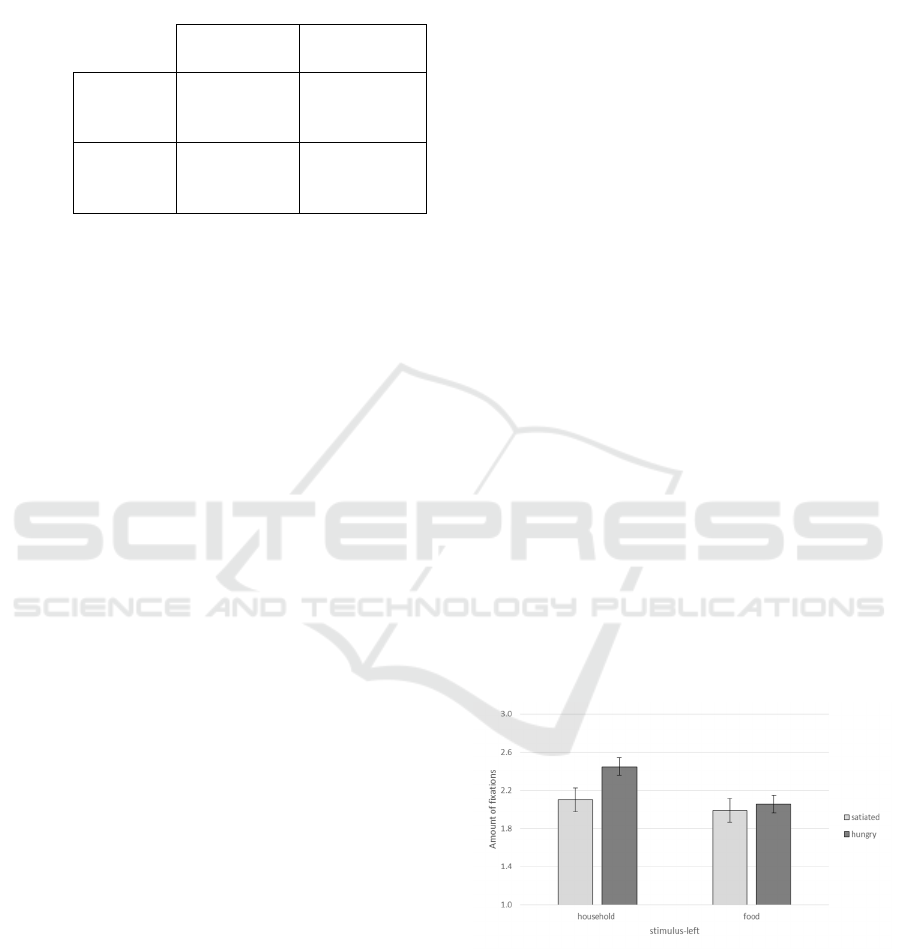
Table 1: Mean duration of first fixation in ms on the left
AOI depending which stimulus category is presented in the
left (household vs. food) and in the right visual field
(household vs. food).
right visual field
household food
left
visual
field
household
M = 234.74
SE = 8.37
M = 234.38
SE = 6.93
food
M = 224.04
SE = 6.98
M = 220.49
SE = 7.07
A similar picture emerged for the right AOI. The
analysis revealed a main effect of stimulus-right
(F(1,36) = 7.96, p = .008, η² = .181), also reflecting the
fact that participants fixated longer on the right AOI
when household items were presented compared to
food items (M
household
= 308.78 ms, SE
household
= 9.81 ms;
M
food
= 288.22 ms, SE
food
= 8.17 ms). Besides this
effect of stimulus category, the analysis showed a main
effect of hunger (F(1,36) = 6.62, p = .014, η² = .155):
The duration of the first fixation on the right AOI was
longer for hungry participants compared to satiated
ones (M
hungry
= 319.75 ms, SE
hungry
= 11.05 ms; M
satiated
= 277.26 ms, SE
satiated
= 12.28 ms). The other effects
were not statistically significant (all F < 2.5).
Summarizing, the data showed that participants’
first fixation on an AOI was longer when household
items were presented at the respective side compared
to food items. Furthermore, on the right AOI the first
fixation was longer for hungry participants.
3.3.3 Fixation Count
Since hunger can be thought to increase the interest
into food stimuli and the ease of processing of either
all or only motivational relevant stimuli, also, the
number of fixations on either AOI was investigated.
We again conducted repeated-measures ANOVAs
with the within-subject factors stimulus-left
(household vs. food) and stimulus-right (household
vs. food) and the between-subject factor hunger
(satiated vs. hungry).
First, the results regarding the amount of fixations
on the left AOI are considered. The analysis revealed
that fixations on the left AOI were more frequent
when household items were presented compared to
when food was presented in the left visual field (main
effect for stimulus-left: F(1,31) = 18.62, p < .001, , η²
= .375). The interaction of stimulus-left and hunger
was significant (F(1,31) = 6.03, p = .020, η² = .163,
see Figure 2), indicating that hungry participants
fixated more often on household compared to food
items than satiated ones did. Besides, the interaction
of stimulus-left and stimulus-right reached
significance (F(1,31) = 12.03, p = .002, η² = .280):
When household items were presented in the right
visual field, the amount of fixations on the left AOI
was independent of stimulus category in the left
(M
household
= 2.17, SE
household
= .079; M
food
= 2.15, SE
food
= .10). However, for food items in the right visual
field the amount of fixations was higher when
household items (M
household
= 2.38, SE
household
= .091)
compared to food items were presented in the left
visual field (M
food
= 1.90, SE
food
= .078). The other
effects were not statistically relevant (all F < 2.5)
Second, the analysis for the amount of fixations on
the right AOI is reported. The repeated-measures
ANOVA revealed a main effect for the factor stimulus-
right (F(1,31) = 6.40, p = .017, η² = .171). As for the
left AOI, there were more fixations on the right AOI
when household-items were presented than when food
items were presented (M
household
= 1.85, SE
household
=
0.05; M
food
= 1.73, SE
food
= 0.06). Furthermore, there
were more fixations on the right AOI, when household
items relative to food items were presented in the left
visual field (stimulus-left: (F(1,31) = 12.60, p = .001,
η² = .289; M
household
= 1.87, SE
household
= 0.06; M
food
=
1.70, SE
food
= 0.05). The other effects were not
statistically significant (all F < 1.5).
Summarizing the important results concerning our
question at issue, the amount of fixations was higher
on household items than on food items. For the left
AOI the amount of fixations on household items was
even higher for hungry compared to satiated
participants.
Figure 3: Amount of fixations on the left AOI depending on
the stimulus presented in the left visual field and hunger.
Error bars reflect the standard error of the mean.
3.4 Analysis of the Pupillometric Data
Due to the algorithm outlined in the method section
in average 9.7% (SD = 8.2%) of data points were
Effects of Hunger on Sympathetic Activation and Attentional Processes for Physiological Computing
157
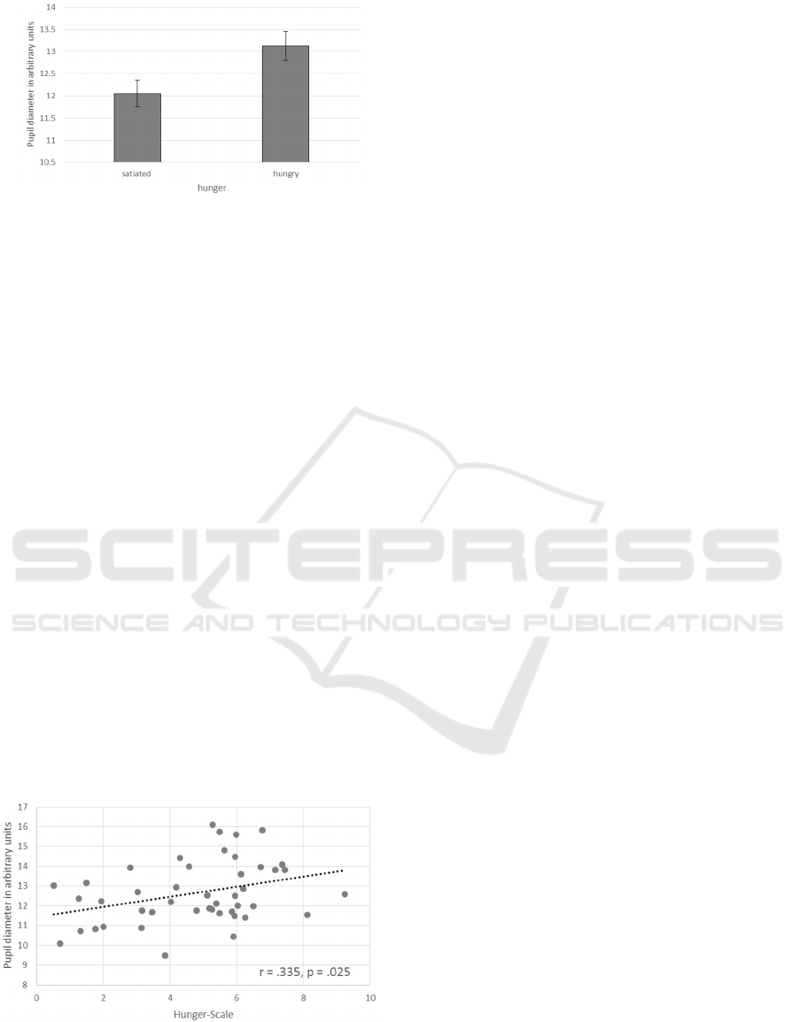
Figure 4: Mean pupil diameter in dependence of hunger.
Error bars reflect the standard error of the mean.
treated as artefacts and were replaced using linear
interpolation. The pupillary data were then averaged
over the 5 s recording time. An independent samples
t-test revealed larger pupils for hungry compared to
satiated participants (t(43) = 2.40, p = .021, ΔM =
1.08, SE = 0.45, see Figure 3). Importantly, pupil size
and the individual ratings on the hunger scale
correlated positively and highly (r = .375, p = .011;
see Figure 4). That is, the higher the ratings on the
hunger scale, meaning that participants felt hungrier,
the larger the pupils.
Therefore, the data do not only indicate that
hungry participants showed larger pupil sizes than
satiated ones, but that their individual feelings of
hunger are correlated with this physiological
measure.
4 DISCUSSION
In the present study we investigated whether a bodily
drive like hunger leads to an attentional bias towards
relevant (i.e., food) pictures. This was examined using
measures of eye movements allowing an examination
of early perceptual processes. Second, we aimed to
Figure 5: Correlation between pupil size and the ratings on
the hunger-scale of Flint et al. (2000).
elucidate whether pupillometry is a valid measure for a
sympathetic increase associated with hunger.
First of all, we derived at investigating differences
between more and less hungry participants, as
instructions and subjective reports confirmed. In
addition, referring to overt performances in a
classification task using food and household items as
stimuli, there was no difference between hungry and
satiated participants observable. Also the direction of
the first fixation was unaffected by hunger as well as
by the stimulus category.
Nevertheless, effects of hunger could be observed
in more implicit gaze signals: The first fixation
duration was longer on the right AOI for hungry
compared to satiated ones. While all participants had
fixated more often on household items, this effect was
more pronounced for hungry participants. Hence, we
observed an interaction of hunger and stimulus
category. The results are now discussed in more detail.
Using the direction of the initial fixation we aimed
at examining an early attentional bias towards food
items for hungry participants and investigated the
priority of a visual field. We found that most of the
initial fixations hit the left AOI independent of the
displayed stimulus configuration and participants’
state of hunger. Thus, in completing the task
participants followed normal reading direction and
started at the left and later changed to the right AOI.
This effect suggests that in the current set-up, the
initial fixation direction indicates more a routine
behaviour being less influenced by the bodily
disposition of hunger and the stimulus category. Our
results are in contrast to the study of Giel et al. (2011)
who found that hungry participants initially fixated
more often on food items. However, there are some
important differences in the study design which might
account for the different results. Giel et al. (2011)
employed a free viewing paradigm without a specific
task whereas we instructed the participants to classify
whether the same category or different categories
were presented in the visual fields. Furthermore, we
instructed our participants to react as fast and as
accurately as possible. This could have resulted in a
more rigid deployment of practiced search
techniques. Additionally, the distance between the
two AOI was larger in our study. This difference
might be important, as a greater distance between the
centered fixation cross and the AOI might have
impaired peripheral preprocessing of the stimuli.
Additionally, peripheral preprocessing may have
been impaired by the similar depiction of food and
non-food items on a white plate: This similar
depiction and the fact that both stimuli were presented
on a white plate, which may be a cue for food items,
PhyCS 2016 - 3rd International Conference on Physiological Computing Systems
158

could have diminished the effects of peripheral
perception on the direction of the initial fixation.
More research in this field is needed to clarify the
boundaries of peripheral preprocessing in hungry
participants, when food and non-food items are
presented while also controlling for stimulus
characteristics of these two categories.
The results for first fixation durations showed that
first fixations were longer when household items were
shown compared to food items. This also becomes
clear when considering that household items were
presented on plates. It is obviously odd to see
household items (like keys) presented on a white plate.
We found longer first fixation durations on the right
AOI for hungry participants. When assuming that
fixations towards the left in the current set-up reflect
rather strategic processes, fixations towards the right
might be regarded as more prone to user states.
A similar picture emerged for the amount of
fixations on the AOIs: Overall, there were more
fixations towards household items than towards food
items supporting the assumption that household items
presented on a plate are unfamiliar and therefore more
difficult to process. But again, this effect was more
pronounced for hungry subjects. This indicates that
processing of non-food stimuli – the less relevant or
more distracting category when being hungry - is
impaired in hungry participants.
Our results are in accordance to former studies
showing that hunger and the calorie content of food
pictures also modulates the activation of early visual
areas (Frank et al., 2010). The present study
substantially extends these findings by showing that
hunger might also affect effectiveness of visual
search as indicated by longer fixation duration in
hungry participants when food is presented in the
paradigm. As we did not use a separate task with non-
food stimuli only, potential expectation effects
concerning food might have influenced the HH-
categorization too. In addition to that, presenting
household objects like keys on a plate might have
increased the association with food for these objects
as only food is usually presented or served on plates.
Therefore, future research should consider using a
more naturalistic display of household items.
Previous studies already confirmed the increase in
sympathetic activation and decrease in
parasympathetic activation of hunger (Chan et al.,
2007). Given that pupil size is influenced by
sympathetic activation (e.g. emotional arousal, Ehlers
et al., 2016; Partala and Surakka, 2003), it was
hypothesized that pupil dilation can be linked to
hunger. The results of the present study indeed
demonstrate the first time that the pupils of hungry
participants are more dilated than the pupils of
satiated ones. Moreover, the data show that pupil size
and subjective hunger are positively correlated
suggesting that this measure can also serve for
diagnostic purposes. For user sensing, this means that
pupil dilations have to be carefully interpreted with
regard to potentially activating sources. That is,
whether or not this method allows to discriminate
between different sources of bodily arousal such as
mental stress has to be further elucidated in future
research. Besides, taking additional sources of bodily
arousal into account, further studies should also
examine whether user characteristics such as weight,
height or psychological disorders such as eating
disorders influence the results of hunger on
sympathetic activation and attentional processing.
Hence, our study indicates that pupillometry is a
feasible way to quantify bodily arousal as associated
with hunger feelings. This method is therefore an
innovative way to assess physiological processes in
the context of bodily states.
ACKNOWLEDGEMENTS
We want to thank Andreas Stegmaier for his help in
data collection.
REFERENCES
Andersson, B., Wallin, G., Hedner, T., Ahlberg, A. C., &
Andersson, O. K. (1988). Acute effects of short-term
fasting on blood pressure, circulating noradrenaline and
efferent sympathetic nerve activity. Acta Medica
Scandinavica, 223(6), 485–490.
Chan, J. L., Mietus, J. E., Raciti, P. M., Goldberger, A. L.,
& Mantzoros, C. S. (2007). Short-term fasting-induced
autonomic activation and changes in catecholamine
levels are not mediated by changes in leptin levels in
healthy humans. Clinical Endocrinology, 66(1), 49–57.
Ehlers, J., Strauch, C., Georgi, J., & Huckauf, A. (2016).
Pupil Size Changes as an Active Information Channel
for Biofeedback Applications. Applied
Psychophysiology and Biofeedback.
Flint, A., Raben, A., Blundell, J. E., & Astrup, A. (2000).
Reproducibility, power and validity of visual analogue
scales in assessment of appetite sensations in single test
meal studies. International Journal of Obesity, 24(1),
38–48.
Frank, S., Laharnar, N., Kullmann, S., Veit, R., Canova, C.,
Hegner, Y. L., et al. (2010). Processing of food pictures:
Influence of hunger, gender and calorie content. Brain
Research, 1350, 159–166.
Giel, K. E., Friederich, H.-C., Teufel, M., Hautzinger, M.,
Enck, P., & Zipfel, S. (2011). Attentional processing of
Effects of Hunger on Sympathetic Activation and Attentional Processes for Physiological Computing
159

food pictures in individuals with anorexia nervosa--an
eye-tracking study. Biological Psychiatry, 69(7), 661–
667.
Giel, K. E., Teufel, M., Friederich, H.-C., Hautzinger, M.,
Enck, P., & Zipfel, S. (2010). Processing of pictorial
food stimuli in patients with eating disorders-A
systematic review. International Journal of Eating
Disorders, 105–117.
Goldstone, A. P., Prechtl de Hernandez, Christina G.,
Beaver, J. D., Muhammed, K., Croese, C., Bell, G., et
al. (2009). Fasting biases brain reward systems towards
high-calorie foods. European Journal of Neuroscience,
30(8), 1625–1635.
Hepworth, R., Mogg, K., Brignell, C., & Bradley, B. P.
(2010). Negative mood increases selective attention to
food cues and subjective appetite. Appetite, 54(1), 134–
142.
Koch, A., Matthias, E., & Pollatos, O. (2014). Increased
Attentional Bias towards Food Pictures in overweight
and Obese Children. Journal of Child and Adolescent
Behavior, 2(2), 1000130.
Nijs, I. M., Muris, P., Euser, A. S., & Franken, I. H. (2010).
Differences in attention to food and food intake
between overweight/obese and normal-weight females
under conditions of hunger and satiety. Appetite, 54(2),
243–254.
Partala, T., & Surakka, V. (2003). Pupil size variation as an
indication of affective processing. International Journal
of Human-Computer Studies, 59(1-2), 185–198.
Peirce, J. W. (2007). PsychoPy - Psychophysics software in
Python. Journal of Neuroscience Methods, 162(1-2), 8–
13.
Piech, R. M., Pastorino, M. T., & Zald, D. H. (2010). All I
saw was the cake. Hunger effects on attentional capture
by visual food cues. Appetite, 54(3), 579–582.
Pollatos, O., Herbert, B. M., Füstös, J., Weimer, K., Enck,
P., & Zipfel, S. (2012). Food deprivation sensitizes pain
perception. Journal of Psychophysiology, 26(1), 1–9.
Siep, N., Roefs, A., Roebroeck, A., Havermans, R., Bonte,
M. L., & Jansen, A. (2009). Hunger is the best spice: an
fMRI study of the effects of attention, hunger and
calorie content on food reward processing in the
amygdala and orbitofrontal cortex. Behavioural Brain
Research, 198(1), 149–158.
Stice, E., Burger, K., & Yokum, S. (2013). Caloric
deprivation increases responsivity of attention and
reward brain regions to intake, anticipated intake, and
images of palatable foods. NeuroImage
, 67, 322–330.
Thayer, J. F., & Brosschot, J. F. (2005). Psychosomatics
and psychopathology: looking up and down from the
brain. Psychoneuroendocrinology, 30(10), 1050–1058.
Werthmann, J., Roefs, A., Nederkoorn, C., Mogg, K.,
Bradley, B. P., & Jansen, A. (2011). Can(not) take my
eyes off it: Attention bias for food in overweight
participants. Health Psychology, 30(5), 561–569.
PhyCS 2016 - 3rd International Conference on Physiological Computing Systems
160
