
BIOLOGICALLY INSPIRED EDGE DETECTION USING
SPIKING NEURAL NETWORKS AND HEXAGONAL IMAGES
Marine Clogenson
1
, Dermot Kerr
2
,
Martin McGinnity
2
,
Sonya Coleman
2
and Qingxiang Wu
2
1
CPE Lyon, Domaine Scientifique de la Doua, BP 82077, 69616, Villeurbanne, France
2
Intelligent Systems Research Centre, University of Ulster, Magee, Derry, BT48 7JL, U.K.
Keywords: Spiking neural network, Edge detection, Multi-scale hexagonal receptive fields.
Abstract: Inspired by the structure and behaviour of the human visual system, we extend existing work using spiking
neural networks for edge detection with a biologically plausible hexagonal pixel arrangement. Standard
digital images are converted into a hexagonal pixel representation before being processed with a spiking
neural network with scalable hexagonally shaped receptive fields. The performance is compared with
different sized receptive fields implemented on standard rectangular images. Results illustrate that using
hexagonal-shaped receptive fields provides improved performance over a range of scales compared with
standard rectangular shaped receptive fields and images.
1 INTRODUCTION
The human vision system (HVS) processes a visual
scene starting in the retina where light intensity is
converted into nerve signals within the
photoreceptors. The signals are pre-processed and
propagated through the various layers within the
retina. The resulting spike train output from the
retinal ganglion cells travels along the optic nerve
for further processing in the lateral geniculate
nucleus and visual cortex. The powerful
performance of the HVS is achieved through
massive parallel processing using neurons and their
complex interconnections formed by synapses.
Taking inspiration from the HVS research has tried
to improve image processing techniques, via the use
of neural networks (NNs) (Egmont-Petersen et al.,
2002). Spiking neural networks (SNNs) are a class
of NNs that mimic more accurately the biological
information processing in the brain. Using a
temporal coding scheme SNNs improve upon NNs
increasing computational power, speed and therefore
enabling real-time processing (Kunkle and
Merrigan, 2002). SNNs use simple neuronal models
and communicate using spikes in a manner similar
to action potentials found in biological neurons. In
(Wu et al., 2007); (Meftah et al., 2008); (Buhmann
et al., 2005) SNN approaches have been developed
for image segmentation. In (Escobar et al., 2009) a
SNN is used to model two areas of the brain
concerned with motion with the aim of action
recognition. A SNN model is proposed in (Meftah et
al., 2010) that performs segmentation and edge
detection, however images must first be segmented
before the edge detection stage can be performed. A
SNN is proposed in (Hugues et al., 2002) to detect
contours in images through the synchronisation of
integrate and fire neurons using simple synthetic
images. In (Wu et al. 2007) a SNN is proposed for
real-time edge detection. In (Chevallier et al., 2006)
a distributed SNN is proposed for extracting
saliencies in an image and in (Chevallier and
Dahdouh, 2009) a SNN is used to perform
Difference of Gaussian filtering. In (Delorme and
Thorpe, 2003) a SNN is proposed that uses a rank
order coding scheme.
In computer vision the apparent strength of a
feature in an image may depend on the scale at
which the appropriate feature detection operator is
applied and many standard approaches to multi-scale
feature detection have been developed (Lindeberg,
1994). However, scalable feature extraction
algorithms using bio-inspired approaches have been
researched and developed to a much lesser extent
with the exception of the approach by (Gao et al.,
2006) where a NN that simulates the multi-scale
receptive field of the biological vision is proposed.
In this paper we present a novel approach to feature
extraction using scalable receptive fields.
381
Clogenson M., Kerr D., McGinnity M., Coleman S. and Wu Q..
BIOLOGICALLY INSPIRED EDGE DETECTION USING SPIKING NEURAL NETWORKS AND HEXAGONAL IMAGES.
DOI: 10.5220/0003682103810384
In Proceedings of the International Conference on Neural Computation Theory and Applications (NCTA-2011), pages 381-384
ISBN: 978-989-8425-84-3
Copyright
c
2011 SCITEPRESS (Science and Technology Publications, Lda.)

Within the fovea the cone photoreceptors are
tightly packed into a hexagonal lattice, resulting in
photoreceptors that are generally surrounded by 6-
neighbours (small irregularities occasionally exist).
Most existing methods for feature extraction are
based on rectangular lattices. Allen (2003) has
shown that curved structures are not well
represented on a rectangular lattice leading us to
question why we use them when nature has chosen a
hexagonal lattice for photoreceptors? Using an
artificial hexagonal sampling lattice, both spatial and
spectral advantages may be derived: namely,
equidistance of all pixel neighbours and improved
spatial isotropy of spectral response. Pixel spatial
equidistance facilitates the implementation of
circular symmetric kernels that are associated with
an increase in accuracy when detecting straight and
curved edges (Allen, 2003). Better spatial sampling
efficiency is achieved by the hexagonal structure
compared with a rectangular grid of similar pixel
separation, leading to improved computational
performance. A hexagonal grid with unit separation
of pixel centres has approximately 13% fewer pixels
than the same image resolution on a rectangular grid
with unit horizontal and vertical separation of pixel
centres (Vitulli, 2002).
In this paper we present a biologically inspired
approach to feature extraction using spiking neural
networks, hexagonal pixel-based images that mimic
the hexagonal arrangement found in the retina, and
scalable hexagonally arranged receptive fields.
2 MODEL IMPLEMENTATION
We use a method proposed in (Middleton and
Sivaswamy, 2001) to create hexagonal pixels (and
images) from clusters of sub-pixels which limits the
loss of image resolution whilst complying with the
main hexagonal properties (Middleton and
Sivaswamy, 2001). The hexagonal pixel is
comprised of 56 sub-pixels closely representing the
shape of a single hexagonal pixel, thus enabling us
to mimic the hexagonal structure used by the HVS
for image capture.
We define our spiking neural network structure
as illustrated in Figure 1. Suppose that the first layer
represents photoreceptors where each pixel in the
hexagonal image corresponds to a photoreceptor. A
receptive field is where a spiking neuron integrates
the spikes from a group of afferent neurons, and in
our model this intermediate layer is composed of
four types of neurons corresponding to four different
receptive fields respectively.
Each of the four parallel arrays of neurons in the
intermediate layer are the same dimension as the
receptor layer with only one neuron in each array
illustrated in Figure 1 for simplicity. Each of these
layers performs the processing for different edge
directions and is connected to the receptor layer by
differing weight matrices. These weight matrices can
be of varying sizes to represent the width of the
receptive field under consideration. We use the
conductance-based integrate-and-fire spiking neuron
model as this offers biological realism whilst
providing a reduction in computational complexity
(Izhikevich, 2004). ‘X’ in the synapse connections
represents an excitatory synapse and ‘Δ’ represents
an inhibitory synapse. Each neuron in the output
layer integrates four corresponding outputs from
intermediate neurons. The firing rate map of the
output layer forms an edge graphic corresponding to
the input image. The receptive fields illustrated in
the intermediate layer in Figure 1 can be receptive
fields of any size and we will use 7, 19, and 37-point
hexagonal receptive fields.
The network model was implemented in Matlab
using the network parameters found in (Wu, et al.,
2007) that are consistent with biological neurons
(Masland, 2001). Synapse strengths can be adjusted
to ensure that the neuron does not fire in response to
a uniform image within its receptive field.
3 RESULTS AND EVALUATION
We present edge detection results at three different
scales using 7- 19- and 37-point sized hexagonal
receptive fields (denoted as HSNN-7, HSNN-19 and
HSNN-37). For comparison we also present results
using the SNN approach in (Wu et al., 2007) which
uses a standard rectangular structure (denoted as
SNN). In Figure 2 we present the edge maps
generated by using the well known Lena image as
input to the SNN models. The edge brightness
increases as the firing rate of the neuron becomes
stronger, thus the firing rate may be set as a
threshold to determine the presence or absence of an
edge. The output from the HSSN is much clearer
and less noisy than the corresponding output from
the SNN.
We evaluate the performance of both the HSNN
and SNN approaches using the Figure of Merit
(FOM) technique (Abdou and Pratt, 1979) in Figure
3. The FOM is compared over a range of noise
levels. Figure 3 illustrates that the HSNN shows
improved performance over the SNN for all edge
types, in particular in areas of high noise and this is
NCTA 2011 - International Conference on Neural Computation Theory and Applications
382
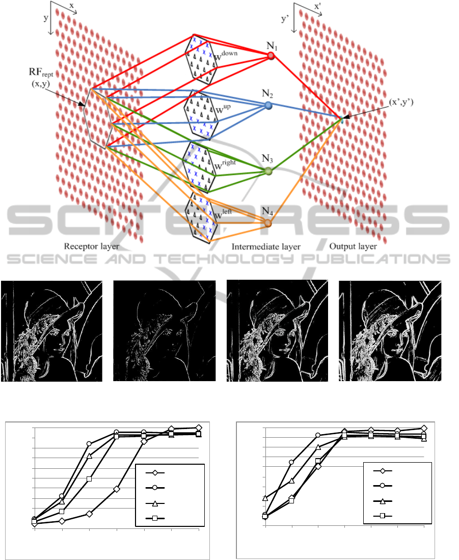
Figure 1: Spiking Neural Network Structure.
(a) SNN (Wu et al., 2007). (b) 7-Point HSNN (c) 19-Point HSNN (d) 37-Point HSNN
Figure 2: Example Edge Detection Outputs.
0
0.1
0.2
0.3
0.4
0.5
0.6
0.7
0.8
0.9
1
1 5 10 20 50 100 No
noiseSNR
FoM
HSNN‐7
HSNN‐19
HSNN‐37
SNN
0
0.1
0.2
0.3
0.4
0.5
0.6
0.7
0.8
0.9
1
1 5 10 20 50 100 No
noise
SNR
FoM
HSNN‐7
HSNN‐19
HSNN‐37
SNN
(a) Vertical edge (b) Diagonal edge
Figure 3: Figure of Merit result.
becomes more evident as the receptive field size
increases. The simulation is run and spikes are
computed over a time interval of 100ms. Table 1
compares the time to run this simulation and
illustrates an improvement in computation time with
the hexagonal arrangement.
BIOLOGICALLY INSPIRED EDGE DETECTION USING SPIKING NEURAL NETWORKS AND HEXAGONAL
IMAGES
383

Table 1: Algorithm run times (seconds).
RF Size Processing time
SNN 3.92
HSNN 7-Point 3.16
HSNN 19-Point 3.47
HSNN 37-Point 3.78
4 DISCUSSION AND FUTURE
WORK
We present a biologically inspired approach to
feature detection that is mimics the human visual
system. The presented SNN is constructed by a
hierarchical structure that is composed of spiking
neurons with various receptive fields. The input
image has a hexagonal pixel arrangement and
correspondingly the receptive fields used are also
arranged in a hexagonal structure. The spiking
neuron models provide powerful functionality for
integration of inputs and generation of spikes.
Synapses are able to perform different complicated
computations. This paper demonstrates how a
spiking neural network can detect edges in an image
using a hexagonal structure over a wide range of
scales and demonstrates performance and
computational improvements over rectangular pixel-
based SNN approaches.
ACKNOWLEDGEMENTS
This work was supported by the Centre of
Excellence in Intelligent Systems project, funded by
InvestNI and the Integrated Development Fund.
REFERENCES
Abdou, I. E. and Pratt, W. K., (1979). Quantitative design
and evaluation of enhancement/thresholding edge
detectors. Proc. of the IEEE, 67(5) pp. 753-763.
Allen J. D., (2003). Filter Banks for Images on Hexagonal
Grid. Signal Solutions.
Buhmann, J. M., Lange, T. M and Ramacher, U., (2005).
Image segmentation by networks of spiking neurons.
Neural computation, 17, 5, 1010-1031.
Chevallier, S., Tarroux, P. and Paugam-Moisy, H., (2006).
Saliency extraction with a distributed spiking neural
network. Proc. of ESANN, 209-214.
Chevallier, S. and Dahdouh, S., (2009). Difference of
Gaussians Type Neural Image Filtering with Spiking
Neurons”. Proc. of Int. Conf. on Neural Compuation,
Maderia, Portugal.
Delorme, A. and Thorpe, S. J., (2003). SpikeNET: an
event-driven simulation package for modelling large
networks of spiking neurons. Computation in Neural
Systems,14(4), 613-627.
Egmont-Petersen, M., De Ridder, D. and Handels, H.,
(2002). Image processing with neural networks-a
review. Pattern Recognition,35(10), 2279-2301.
Escobar, M. J., Masson, G. S., Vieville, T. and
Kornprobst, P., (2009). Action Recognition Using a
Bio-Inspired Feedforward Spiking Network. IJCV,
82(3), 284-301.
Gao Y., Zhang L., Wang Y. and Jiang J., (2006). Multi-
scale receptive field neural networks for object
tracking. International Congress Series, 1291, 229–
232.
Hugues, E., Guilleux, F. and Rochel, O., (2002). Contour
Detection by synchronization of Integrate and Fire
Neurons. LNCS. 2525, 60-69.
Izhikevich, E. M., (2004). Which model to use for cortical
spiking neurons?. IEEE Trans. on NN, Vol. 15, No. 5.
Kunkle, D. R. and Merrigan, C., (2002). Pulsed neural
networks and their application. Computer Science
Dept., College of Computing and Information
Sciences, Rochester Institute of Technology.
Lindeberg, T., (1994). Scale-Space Theory in Computer
Vision. Kluwer, Academic Publisher/Springer,
Dordrecht, Netherlands.
Masland, R. H., (2001). The fundamental plan of the
retina. Nature Neuroscience, vol. 4, pp. 877-886.
Meftah, B., Lezoray, O. and Benyettou, A., (2010).
Segmentation and Edge Detection based on Spiking
Neural Network Model. Neural Processing Letters.
32(2), 131-146.
Middleton L. and Sivaswamy, J., (2001). Edge Detection
in a Hexagonal-Image Processing Framework. Image
and Vision Computing.19, pp. 1071-1081.
Paugam-Moisy, H. and Bohte, S. M., (2009). Computing
with spiking neuron networks. Handbook of Natural
Computing. 40. Springer, Heidelberg.
Vitulli R., (2002). Aliasing Effects Mitigation by
Optimized Sampling Grids and Impact on Image
Acquisition Chains. Geoscience and Remote Sensing
Symposium, pp. 979-981.
Wu, Q. X., McGinnity, M., Maguire, L., Belatreche, A.
and Glackin, B., (2007). Edge Detection Based on
Spiking Neural Network Model.
Advanced Intelligent
Computing Theories and Applications. With Aspects
of Artificial Intelligence, 26-34.
NCTA 2011 - International Conference on Neural Computation Theory and Applications
384
