
UNDERSTANDING CEREBRAL ACTIVATIONS IN
NEUROMARKETING
A Neuroelectrical Perspective
Giovanni Vecchiato
1,2
, Laura Astolfi
2,3
, Fabrizio De Vico Fallani
1,2
, Jlenia Toppi
2,3
, Fabio Aloise
2
Febo Cincotti
2
, Donatella Mattia
2
and Fabio Babiloni
1,2
1
Department of Physiology and Pharmacology, University “Sapienza”, Rome, Italy
2
IRCCS "Fondazione Santa Lucia", Rome, Italy
3
Department of Computer Science and Systems, University “Sapienza”, Rome, Italy
Keywords: Neuromarketing, MEG, High Resolution EEG, SSVEP, Functional connectivity.
Abstract: This paper aims to be a survey of recent experiments performed in the Neuromarketing field. Our purpose is
to illustrate results obtained by employing the popular tools of investigation well known in the international
neuroelectrical community such as the MEG, High Resolution EEG techniques and steady-state visually
evoked potentials. By means of temporal and frequency patterns of cortical activations we intend to show
how the neuroscientific community is nowadays sensible to the needs of companies and, at the same time,
how the same tools are able to retrieve hidden information about the demands of consumers. These
instruments could be of help both in pre- and post-design stage of a product, or a service, that a marketer is
going to promote.
1 INTRODUCTION
For a long time in the neuroscience the experimental
psychology has been the main method to investigate
the human being. In that field, they measure the
execution time of the experimental subject as well as
his/her reaction time to particular stimuli. In
particular, the subject can answer, move the finger,
do free mental associations and the experimenter
will measure some variables correlated with the
internal processes of the subject. Of course, the
access to the internal state of the subject is restricted,
since it is observed by means of his/her behavioural
answers. The experimental context plays an
important role in these kind of experiments, since
the subjects are required to answer in a short time in
order to cause errors. The measure of the amount
and frequency of such errors is important as an
indirect measure of the internal processes of the
subject. These behavioural techniques can be applied
with a low cost to a large amount of people. The
brain imaging techniques have the capability to
show images of the cerebral activity during the
execution of a particular experimental task. The
most popular method is the functional Magnetic
Resonance Image (fMRI), which returns a sequence
of images of the cerebral activity by means of the
measure of the cerebral blood flow. Although such
as images are “static”, i.e. they are related to around
ten seconds activity, they have a high special
resolution that no other neuroimaging method can
offer. Nowadays, fMRI scanners are employed in the
neuromarketing field and in literature there exists
some scientific studies showing the activation of
particular cerebral areas during the tasting of a
couple of popular drinks such as Coca-Cola and
Pepsi (Mc Clure et al., 2004). It must be noted that
the design of the fMRI studies rely on highlighting
cortical areas which differ between the experimental
task and the control one. The issue is connected with
the data interpretation which can be given by the
activation of particular cerebral areas during the
experimental task proposed. In order to solve this
problem there is the need to generate a proper
experimental design in order to remove these kind of
confounding factors. As stated above, the brain
imaging allows us to observe cerebral areas
activating during a particular task but it does not
explain why, neither in which way the information is
91
Vecchiato G., Astolfi L., De Vico Fallani F., Toppi J., Aloise F., Cincotti F., Mattia D. and Babiloni F..
UNDERSTANDING CEREBRAL ACTIVATIONS IN NEUROMARKETING - A Neuroelectrical Perspective.
DOI: 10.5220/0003354900910097
In Proceedings of the International Conference on Bio-inspired Systems and Signal Processing (BIOSIGNALS-2011), pages 91-97
ISBN: 978-989-8425-35-5
Copyright
c
2011 SCITEPRESS (Science and Technology Publications, Lda.)
processed within the experimental subject. In this
way we are not accessing the cerebral answer of the
subject by his/her behave but directly by means of
the activity of his/her neurons. The role of the brain
imaging in the neuromarketing field is to refine, on
the basis of a selected sample of people, some
theories and hypotheses the neuropsychological
research draw from the same experimentation on a
larger number of subjects.
In the following paragraphs we intend to give a
survey about the neuromarketing research performed
in the neuroelectrical community. Our purpose is to
show the results obtained by different group of
research, their congruence and similarity leading
towards a shared model which is able to extract
information about the memorization and the
pleasantness perceive by subjects while watching
TV advertisements. In particular, this paper will
focus on the results achieved by employing the
magnetoencephalography (MEG) and
electroencephalography (EEG) technique along with
a discussion about patterns of cortical functional
connectivity and indexes derived from the graph
theory.
Finally, we describe the experimental findings
obtained by a steady-state visually evoked potential
analysis.
2 TEMPORAL PATTERNS
OF CORTICAL ACTIVITY
In this context, the research team of Sven
Braeutigam (Braeutigam et al., 2005, 2004, 2001)
has employed the MEG in order to study the
temporal relationship of cerebral areas involved in
consumers’ choices when they have to make
decisions among different items within a laboratory.
In this study they wanted to analyse the cerebral
behaviour by distinguishing male subjects from
females during a simulated shopping.
Cerebral activations induced by choices to make
reflect the level of familiarity or the preference that a
particular experimental subject has with the
presented products. These factors can be considered
by taking into account the relationship between the
current choice of a product on the shelf and the
relative frequency of choice and usage of that
product in the past.
In particular, the main observation came to light
from these studies presents the consumer’s choice
like a complex sequence of cerebral activations that
greatly differ according to the consumer’s sex and to
the probability of choice. From a behavioural point
of view, choices with a high probability were faster
than those less predictable. This can be interpreted
by supposing that in the case of more difficult
choices the cortical activities are more complex than
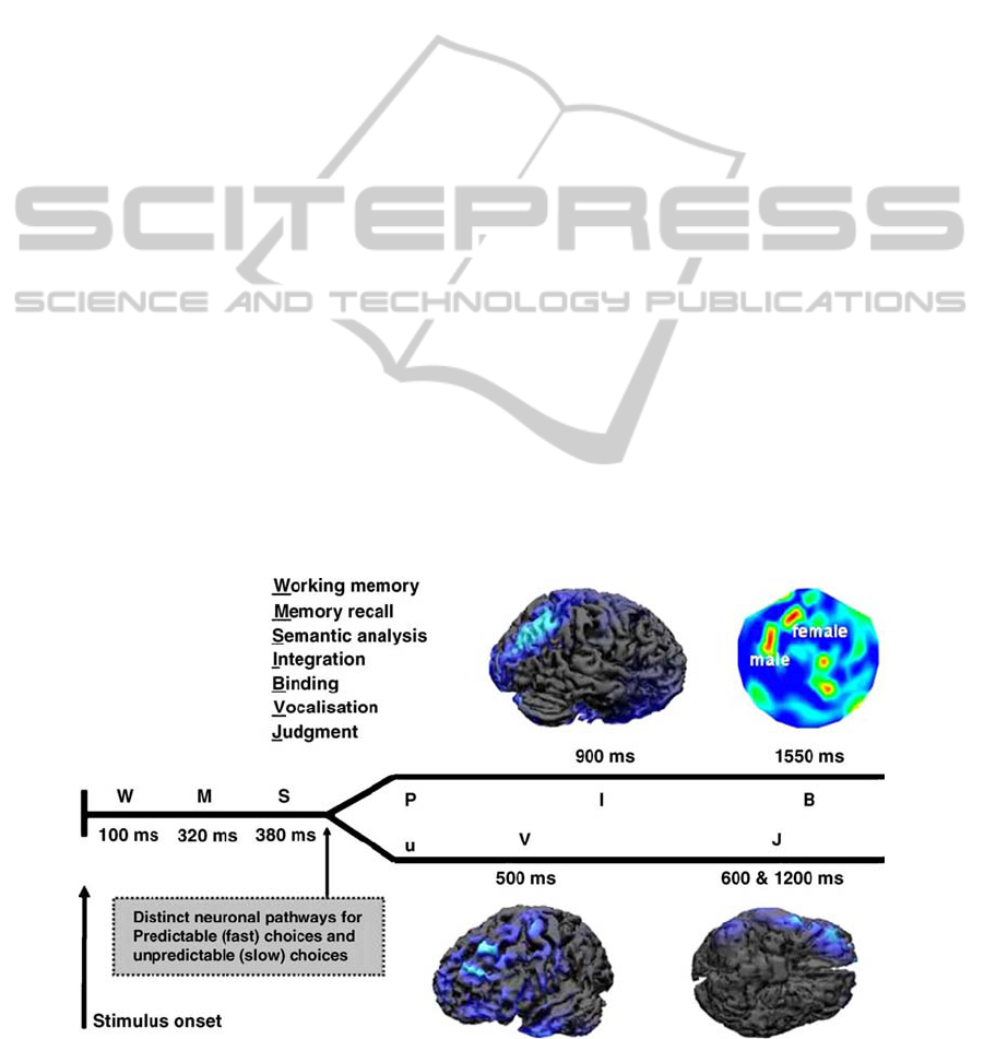
those simple to make. As illustrated in figure 1, they
distinguished two distinct cerebral paths. The first
Figure 1: Cortical activations associated to the decisions of an experimental subject. Predictable choices are the ones related
to familiar items which has been often bought or used in the past. Cortical maps present the brain areas activated during the
different decision stages in the frequency range of [30, 40] Hz. Modified with permission from Braeutigam et al., 2003.
BIOSIGNALS 2011 - International Conference on Bio-inspired Systems and Signal Processing
92

one is referred to predictable choices, i.e. associated
to products that the experimental subject already
used in the past or said to prefer; the second one is
related to unpredictable choices, i.e. associated to
unfamiliar products to the subject. In the experiment,
a first stage in the decisional process has been
individuated around 100 ms after the stimulus onset
with an activity located in the occipital cortex. At
this stage of decision (W) the subject compare the
product to choose with the list of products seen
before, by involving the working memory.
The sequence of cortical activations observed in
the experimental subjects continues with two
neuronal stages partially correlated (M, S) which can
be observed between 280 and 400 ms after the
beginning of the decisional process. In this period
the selective attention of the subject is oriented
towards images of products to identify, classify and
compare with those stored in the memory related to
the preferred products and brands. This memory can
involve the past experience to have bought the
particular item or to have watched the commercial of
the specific brand. The cerebral activation differs in
this time interval between men and women. In
particular, female subjects showed a stronger
activation with respect to the males in the left
parieto-occipital lobule of the brain while males
presented a stronger cortical activity in the right
temporal lobe. These differences connected with the
sex of the subjects characterize both the stage of
choice of the product and its discrimination. This
observation suggest that, at this temporal stage,
women tend to employ a strategy based on the
knowledge of the product to buy, while men tend to
act according to a spatial memory strategy (Kimura,
1996).
After 500 ms from the beginning of the
decisional process, two patterns of cortical
activations can be identified according to the
predictability of the choices adopted by the subjects.
In particular, as to the predictable choices, we can
observe a strong activation in the right parietal areas
around 900 ms after the beginning of the experiment
(I). In later time latencies, predictable choices of
products recall strong MEG oscillations in the
frequency band between [30, 40] Hz in the left
prefrontal cortex (B). Parietal cortex receive inputs
from many cortical areas since it is involved in the
spatial integration of sensorial information.
Differences in the cortical activity between men and
women can strengthen the hypothesis of two
different groups of strategies. On the contrary,
unpredictable choices generate a strong activation in
the right inferior frontal cortex (V), at a latency of
around 500 ms, and in the left orbitofrontal cortex
(J) between 600 and 1200 ms after the stimulus
presentation. In the case V, the cortical patterns are
consistent with the activity in the Broca’s area,
which is involved in the spoken language, which is
also active during the observation of videoclips.
Hence, the cortical activity at this latency may
indicate a tendency to vocalize brands, as a part of
strategy which helps in the decision when it is
difficult. The activity in the orbitofrontal cortex (J)
can be explained by stating that during an
unpredictable choice we have to evaluate the
outcome in terms of convenience. Overall, these
results explain a complex neuronal network which is
active during a simple decisional process connected
to the purchase of a product. The generation of a
choice is considered as an information processing
which can be highly influenced, sensible to the
complexity of the decision to make and to the rush
in which the decision is made and many other
factors.
A strong involvement of parietal areas during the
observation of the TV commercials with an affective
and cognitive content was also noted in a previous
study, performed by using sophisticated MEG
recordings (Ioannides et al., 2000). In this study,
cognitive frames elicited a stronger activity in the
parietal areas and superior prefrontal cortex while
the observation of the affective ones is correlated
with the activation of the orbitofrontal and
retrosplenial cortex, amygdala and brainstem. The
magneto field tomography (MFT) results showed an
increasing activity during the observation of
cognitive stimuli rather than affective commercials
in parietal and superior prefrontal areas known to be
associated with executive control of working
memory and maintenance of highly processed
representation of complex stimuli (Summerfield et
al., 2005). Although the affect related activations are
more variable across subjects, these findings are
consistent with previous PET and fMRI studies
(Cahill et al., 1996; Maddock 1999; Grabenhorst et
al., 2008) showing that stimuli with affective content
modulates activity in the orbitofrontal and
retrosplenial cortex, amygdala and brainstem.
3 FREQUENCY PATTERNS OF
CORTICAL ACTIVITY AND
FUNCTIONAL CONNECTIVITY
The research in the neuromarketing field performed
in the recent years showed result suggesting that the
cortical activity elicited during the observation of the
UNDERSTANDING CEREBRAL ACTIVATIONS IN NEUROMARKETING - A Neuroelectrical Perspective
93
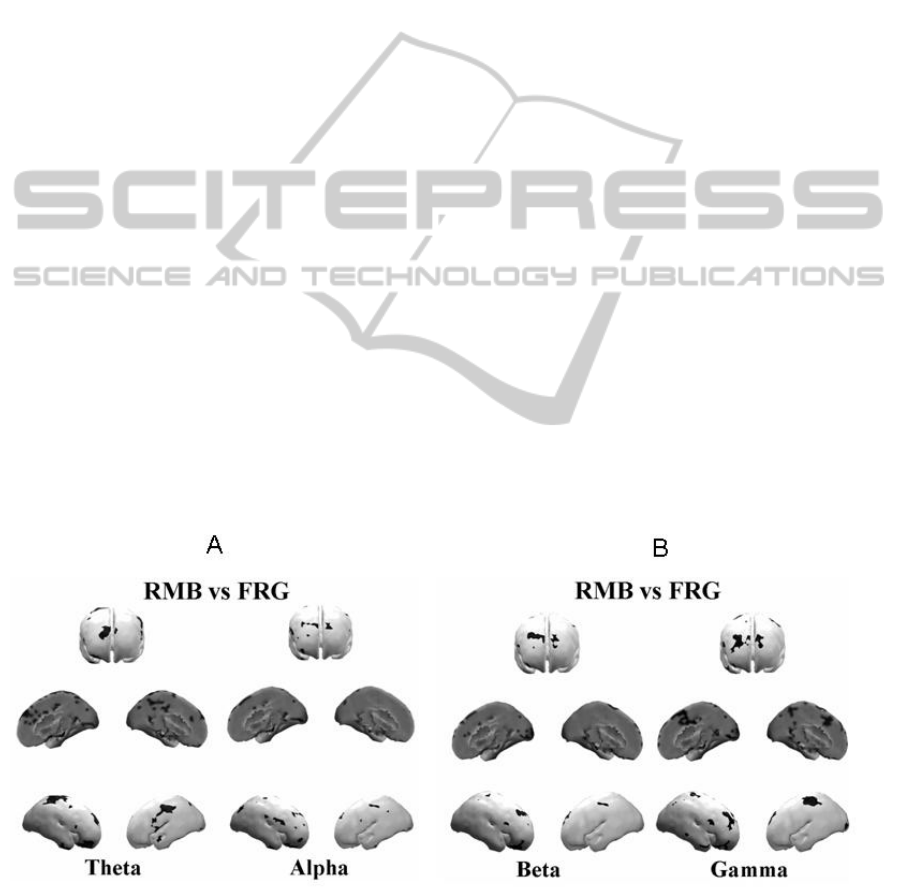
TV commercials that were forgotten (FRG) is
different from the cortical activity observed in
subjects that remembered the same TV commercials
(RMB). In fact, the principal areas of statistical
differences in power spectra between such
conditions are located almost bilaterally in the
prefrontal Broadmann Areas (BAs) 8, 9 as well as in
the parietal BAs 7, as showns in figure 2. The
spectral amplitude in the RMB condition was always
higher than the power spectra in the FRG conditions
over the BAs 8, 9 and 7 (Astolfi et al., 2008). A
statistical increase of EEG spectral power in the
prefrontal and parietal areas for the RMB dataset
compared with the FRG one is in agreement with the
suggested role of these regions during the transfer of
sensory percepts from short-term memory to long-
term memory storage. Although in this study the
differences in the cortical power spectra between the
RMB and FRG conditions are relatively insensitive
to the particular frequency bands considered, there
are experimental evidences showing that there is a
stronger engagement of the left frontal areas in all
the subjects analyzed during the observation of the
TV commercials that were remembered (Vecchiato
et al., 2010).
In particular, the analysis of the statistical
cortical maps in the condition RMB vs FRG
suggested that the left frontal hemisphere was highly
active during the RMB condition, especially in the
theta and gamma band. These results are in
agreement with different observations on the RMB
condition performed in literature by studying
different experimental paradigm (Summerfield et al.,
2005; Werkle-Bergner et al., 2006). Taken together,
the results indicated the cortical activity in the theta
band on the left frontal areas was increased during
the memorization of commercials, and it is also
increased during the observation of commercials that
were judged pleasant by subjects. These results are
in agreement with the role that has been advocated
for the left pre and frontal regions during the transfer
of sensory percepts from the short-term memory
toward the long-term memory storage by the HERA
model (Tulving et al., 1994; Habib et al., 2003). In
fact, in such model the left hemisphere plays a key
role during the encoding phase of information from
the short term memory to the long term memory,
whereas the right hemisphere plays a role in the
retrieval of such information.
It must be noted, however, that the role of the
right cortices in storing images has been also
recognized for many years in neuroscience
(Braeutigam et al., 2005, 2004; Babiloni et al., 2000;
Astolfi et al., 2009, 2008). It is worthy of note that
the subjects were unaware of the kind of questions
that the researcher asked them after the viewing of
the documentary. Hence, the cortical areas elicited
by this study are likely to be involved just in the
process of the memorization of the pictorial
material, owing to an increase of attention during the
observation of the TV commercial. In addition, there
was no particular set of commercials remembered
that was common to all the subjects.
Figure 2: Figure presents the results of statistical comparisons of RMB and FRG groups in the theta and alpha frequency
ranges (panel A), and in the beta and gamma bands (panel B). In particular, the picture is composed by three rows: the first
one shows the brain from a frontal perspective, the second one is related to a medial- sagittal perspective while the third one
presents statistically significant images associated to the left and right lateral vision. Modified with permission from Astolfi
et al., 2008.
BIOSIGNALS 2011 - International Conference on Bio-inspired Systems and Signal Processing
94
The results also suggest an active involvement of
the Anterior Cingulate Cortex (ACC) and the
Cingulate Motor Area (CMA) as sources of links to
all the other cortical areas during the observation of
the TV commercials remembered after ten days. In
this case the increased activity related to an increase
of the outflow of PDC links from ACC and CMA
towards other cortical regions could be taken as a
sign of increased ‘emotive’ attention to the stories
proposed by different TV commercials that
significantly aid successive memorization.
The EEG spectral and cortical network analyses
performed in these study also suggest a key role of
the parietal areas as targets of the incoming
information flow from all the other cortical areas.
Functional networks in the frequency domain were
also estimated by evaluating the global- and local-
efficiency indexes derived from the graph theory,
employed as a measure of the level of
communication in the networks (De Vico Fallani et
al., 2008). The changes of these indexes could be
related both to memory coding activity as well as to
increase/decrease of attentive state of the subjects.
As to the RMB condition, the functional network in
the beta and gamma band state a significant non-
homogeneous allocation of the involved information
flows and a consequent reduction of the efficiency in
the overall communication between the network
nodes. In the beta and gamma frequency bands, the
respective reduction of global-efficiency, as well as
the reduction of local-efficiency for the alpha band
of the cortical network communication could
represent a predictive measure for the accurate recall
of the commercials that will be remembered.
A contrast of the activity elicited by observing
pleasant (LIKE dataset) and unpleasant (DISLIKE
dataset) audiovisual content has been performed in a
previous study (Vecchiato et al., 2010). The result of
this experiment shows that the activity of the brain is
greater in the LIKE condition than in the DISLIKE
except that in beta band, being the activity in the
LIKE condition for the gamma band rather
symmetrical. The results here obtained for the LIKE
condition are also congruent with other observations
performed with EEG in a group of 20 subjects
during the observation of pictures from the
International Affective Picture System (IAPS,
Aftnas et al., 2004). Such observations indicated an
increase of the EEG activity in the theta band for the
anterior areas of the left hemisphere.
4 STEADY-STATE VISUALLY
EVOKED POTENTIALS
These results are also supported by findings obtained
from the group of Richard Silberstein which
measured the steady-state visually evoked potential
(SSVEP) by means of the steady-state probe
topography (SSPT), which is a particular version of
Figure 3: Figure presents four cortical z-score maps, in the four frequency bands employed. Colour bar represents cortical
areas in which increased statistically significant activity occurs in the RMB group when compared to the FRG group in red,
while blue is used otherwise (p < 0,05 Bonferroni corrected). Grey colour is used to map cortical areas where there are no
significant differences between the cortical activity in the RMB and FRG groups (panel A). Panel B refers to the statistical
comparison LIKE vs DISLIKE with the same conventions of panel A. Modified with permission from Vecchiato et al.,
2010.
UNDERSTANDING CEREBRAL ACTIVATIONS IN NEUROMARKETING - A Neuroelectrical Perspective
95

the EEG technology (Silberstein et al., 1990, 2000;
Silberstein, 1995). In this study (Silberstein et al.,
2000), they collected the cerebral activity from thirty
five women that were subjected to the exposition of
eighteen minutes documentary in which 12 US TV
commercials were inserted within. Seven days after
the recording, the participants were asked to recall
the viewed advertisements from a series of frames
taken from the same commercials. They found out
that images corresponding to a minima of the
posterior frontal latency were more likely to be
recognized than images associated to a SSVEP
latency maxima. Moreover, they showed a
significant correlation between the recognition
performance and SSVEP latency measured at
electrode sites located in the left posterior frontal
site suggesting that this kind of result can be
employed in order to asses the strength of long-term
memory encoding for the audiovisual stimuli they
proposed.
5 CONCLUSIONS
In recent years, Neuromarketing has gained always
more interest and attention in both the scientific
community and mass media. Findings obtained so
far show that results from these studies can be of
help for many areas of marketing. For instance,
marketers could exploit neuroimaging tools in order
to achieve hidden information about products and
services to advertise that is impossible to acquire.
This information could be employed both during the
design process of an item and during its commercial
campaign. In fact, one could think to adopt these
neuroelectrical tools to test different versions of the
same object, evaluate the cerebral results and use
them according to the requirements of the company
and at the same time facing with the consumers’
need. In addition, marketers could use an ad pre-test
in order to create a TV commercial which is as
closer as possible to the demand of the same. In this
case we can distinguish two different point of view
of the powerful and innovative tool: from one hand,
product manufacturers could use cerebral
information in order to force people to buy and
consume products that they don’t want and neither
need; from the other hand, we hope that
neuromarketing will be of help in design objects and
the environment following the pleasures of each of
us and for identifying new and exciting products that
people want and find useful.
REFERENCES
Aftanas, L.I. et al., 2004. Analysis of evoked EEG
synchronization and desynchronization in conditions
of emotional activation in humans: temporal and
topographic characteristics. Neuroscience and
Behavioral Physiology, 34(8), 859-867.
Astolfi, L. et al., 2005. Assessing cortical functional
connectivity by linear inverse estimation and directed
transfer function: simulations and application to real
data. Clinical Neurophysiology: Official Journal of the
International Federation of Clinical Neurophysiology,
116(4), 920-932.
Astolfi, L. et al., 2007. Imaging functional brain
connectivity patterns from high-resolution EEG and
fMRI via graph theory. Psychophysiology, 44(6), 880-
893.
Astolfi, L. et al., 2004. Estimation of the effective and
functional human cortical connectivity with structural
equation modeling and directed transfer function
applied to high-resolution EEG. Magnetic Resonance
Imaging, 22(10), 1457-1470.
Astolfi, L. et al., 2008. Neural basis for brain responses to
TV commercials: a high-resolution EEG study. IEEE
Transactions on Neural Systems and Rehabilitation
Engineering: A Publication of the IEEE Engineering
in Medicine and Biology Society, 16(6), 522-531.
Astolfi, L. et al., 2009. The track of brain activity during
the observation of TV commercials with the high-
resolution EEG technology. Computational
Intelligence and Neuroscience, 652078.
Babiloni, F. et al., 2000. High-resolution electro-
encephalogram: source estimates of Laplacian-
transformed somatosensory-evoked potentials using a
realistic subject head model constructed from
magnetic resonance images. Medical & Biological
Engineering & Computing, 38(5), 512-519.
Braeutigam, S. et al., 2001. Magnetoencephalographic
signals identify stages in real-life decision processes.
Neural Plasticity, 8(4), 241-254.
Braeutigam, S., 2005. Neuroeconomics--from neural
systems to economic behaviour. Brain Research
Bulletin, 67(5), 355-360.
Braeutigam, S. et al., 2004. The distributed neuronal
systems supporting choice-making in real-life
situations: differences between men and women when
choosing groceries detected using
magnetoencephalography. The European Journal of
Neuroscience, 20(1), 293-302.
Cahill, L. et al., 1996. Amygdala activity at encoding
correlated with long-term, free recall of emotional
information. Proceedings of the National Academy of
Sciences of the United States of America, 93(15),
8016-8021.
De Vico Fallani, F. et al., 2008. Structure of the cortical
networks during successful memory encoding in TV
commercials. Clinical Neurophysiology: Official
Journal of the International Federation of Clinical
Neurophysiology, 119(10), 2231-2237.
BIOSIGNALS 2011 - International Conference on Bio-inspired Systems and Signal Processing
96

Grabenhorst, F., Rolls, E.T. & Parris, B.A., 2008. From
affective value to decision-making in the prefrontal
cortex. The European Journal of Neuroscience, 28(9),
1930-1939.
Habib, R., Nyberg, L. & Tulving, E., 2003. Hemispheric
asymmetries of memory: the HERA model revisited.
Trends in Cognitive Sciences, 7(6), 241-245.
Ioannides, A.A. et al., 2000. Real time processing of
affective and cognitive stimuli in the human brain
extracted from MEG signals. Brain Topography,
13(1), 11-19.
Kimura, D., 1996. Sex, sexual orientation and sex
hormones influence human cognitive function.
Current Opinion in Neurobiology, 6(2), 259-263.
Maddock, R.J., 1999. The retrosplenial cortex and
emotion: new insights from functional neuroimaging
of the human brain. Trends in Neurosciences, 22(7),
310-316.
McClure, S.M. et al., 2004. Neural correlates of behavioral
preference for culturally familiar drinks. Neuron,
44(2), 379-387.
Silberstein, 1995. Steady State Visually Evoked
Potentials, Brain Resonances and Cognitive Processes.
In Neocortical Dynamics and Human EEG Rhythms.
Oxford University Press.
Silberstein, R.B. et al., 2000. Frontal steady-state potential
changes predict long-term recognition memory
performance. International Journal of
Psychophysiology: Official Journal of the
International Organization of Psychophysiology,
39(1), 79-85.
Silberstein, R.B. et al., 1990. Steady-state visually evoked
potential topography associated with a visual vigilance
task. Brain Topography, 3(2), 337-347.
Summerfield, C. & Mangels, J.A., 2005. Coherent theta-
band EEG activity predicts item-context binding
during encoding. NeuroImage, 24(3), 692-703.
Tulving, E. et al., 1994. Hemispheric encoding/retrieval
asymmetry in episodic memory: positron emission
tomography findings. Proceedings of the National
Academy of Sciences of the United States of America,
91(6), 2016-2020.
Vecchiato, G. et al., 2010. Changes in brain activity during
the observation of TV commercials by using EEG,
GSR and HR measurements. Brain Topography,
23(2), 165-179.
Werkle-Bergner, M. et al., 2006. Cortical EEG correlates
of successful memory encoding: implications for
lifespan comparisons. Neuroscience and
Biobehavioral Reviews, 30(6), 839-854.
UNDERSTANDING CEREBRAL ACTIVATIONS IN NEUROMARKETING - A Neuroelectrical Perspective
97
