
3D HUMAN ANATOMY LEARNING
Demonstration of 3D Tools used in Teaching: 3D Videos, Podcasts, PDF
Patrice Thiriet, Christophe Batier, Olivier Rastello, Emmanuel Sylvestre, Nora Van Reeth
iCAP (Innovation,Conception, Accompagnement pour la Pédagogie), University Lyon 1
43 Bd du 11 novembre, 69622 Villeurbanne, France
Aymeric Guillot, Christian Collet, Nady El Hoyek
CRIS (Centre de Recherche et d’Innovation sur le Sport), University Lyon 1
43 Bd du 11 novembre, 69622 Villeurbanne, France
Keywords: 3D technology, PDF, Instructional design, Human anatomy.
Abstract: Human anatomy courses are based on 3D technology since 2006 in Lyon 1 University. The major
instructional tools will be presented: 3D Video animation –some of them integrated into podcast – and PDF.
Every PDF file contains one 3D image of an anatomical structure that can be moved and turned in space or
zoomed. We can even hide or show parts of an anatomical structure and name them, make cut sections,
move apart a joint or bones and then put it together. All these 3D images of anatomical structures can be
assembled in a portfolio or integrated and animated in a PDF. 3D instructional tools help the students in
creating mental images of the anatomical structures, make them rotate in space and better understand their
spatial organization which is essential in learning anatomy. An instructional design using this 3D
technology is implemented in an online server called Spiral. Our educational evaluations show that students
give very positive feedbacks our teaching method using 3D technology.
1 INTRODUCTION
Lyon 1 university encourages lecturers to produce
and use multimedia instructional tools. A big
funding, probably one of the biggest of all French
universities, is dedicated to this purpose. The ICAP
department has the mission to create instructional
tools on lecturers’ demand. Higher education
ministry and French universities are financially
supporting this project through the French-speaking
numerical university of sports and health sciences
(UNF3S). Producing 3D tools has been one of the
major priorities of Lyon 1 university (since 2006)
and UNF3S (since 2010). One of the main projects
is enhancing human anatomy teaching. Students who
have to learn human anatomy encounter difficulties
in regards to the anatomical vocabulary as well as
understanding the spatial orientation of an
anatomical structure. Using 3D instructional tools
seems to bring an adequate solution to these
difficulties. An instructional design is implemented
using technological innovation. The 3D tools that we
created are used at the first undergraduate level in
kinesiology and other paramedical courses. Lyon 1
university is the only one in France offering this type
of teaching.
2 THE INSTRUCTIONAL TOOLS
Producing 3D images of anatomical structures
enabled setting up a publishing chain for teaching
human anatomy. These document model images
were re-adapted in order to be used in various
contexts:
2.1 3D Animated Videos
Videos are played during lectures. Using Quicktime
player enables to show a slow motion forward or
backward playing or to play different videos
simultaneously.
Free online access to our videos: http://
anatomie3d.univ-lyon1.fr/.
408
Thiriet P., Batier C., Rastello O., Sylvestre E., Van Reeth N., Guillot A., Collet C. and El Hoyek N..
3D HUMAN ANATOMY LEARNING - Demonstration of 3D Tools used in Teaching: 3D Videos, Podcasts, PDF.
DOI: 10.5220/0003339804080411
In Proceedings of the 3rd International Conference on Computer Supported Education (CSEDU-2011), pages 408-411
ISBN: 978-989-8425-49-2
Copyright
c
2011 SCITEPRESS (Science and Technology Publications, Lda.)
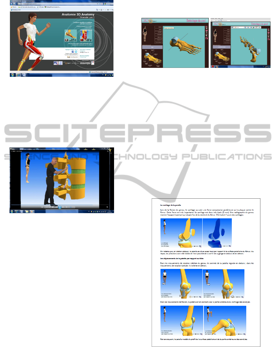
Figure 1: Screen of the online access.
2.2 Podcasts
With the aim to enhance this project, we used our
3D videos commented by a teacher to create
podcasts.
Free online access to our podcasts (two last lines
in the bottom): http://anatomie3d.univ-lyon1.fr/
Figure 2: Screen of a podcast.
2.3 3D Adobe PDF
We also embedded 3D model images into PDF files.
Thus the latest version of Adobe Acrobat Reader
enabled us to move, turn in space, zoom, hide and
make cut sections of anatomical structures.
The portability of these PDF files associated with
their small size make them easy to send and load
through emails. We can even turn around an
anatomical structure while it is moving as well as
integrating text information, thus assembling
illustrations comments and captions into the same
PDF. Furthermore different PDFs can be assembled
in one electronic portfolio.
An application enables us to create different
animations of 3D objects, a teacher can thus create
his own screenplay instead of using the predefined
fixed screenplay of our 3D videos.
Free online access to our PDF: http://
anatomie3d.univ-lyon1.fr/
These instructional tools are also available in the
French-speaking university of health science and
sports (UV2S) web site: http://www.uv2s.fr/
index2.php? page=nouveautes
Figures 3 and 4: Example of PDF 3D.
NB: At that time, these PDF cannot show a change
of shape of an anatomical 3D object, e.g. showing
how a muscle can lengthen or shorten during
contraction. A new software called Unity will have
this feature from June 2011.
2.4 Integrating 3D Images into other
Educational Purposes
• Multiple choice questions for students’
assessment via the E-learning server Spiral,
• The Quicktime format enables the integration
of 3 images into a Word file document or
Multiple choice questions by simply using the
copy/paste function:
Figure 5: Integration 3D images into a Word file
document.
The use of these teaching tools justifies the fact
that our courses of practical skill are held in a
3D HUMAN ANATOMY LEARNING - Demonstration of 3D Tools used in Teaching: 3D Videos, Podcasts, PDF
409
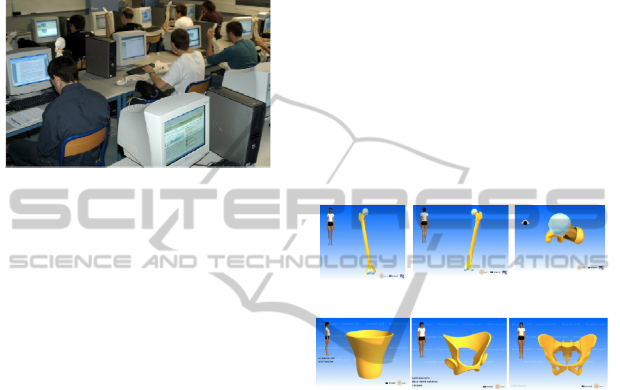
computer classroom. In addition to our electronic
teaching tools we provide a skeleton for every
student, in order to make possible the relationships
between the image and the actual anatomical
structure:
Figure 6: Practical skill lessons in a computer classroom.
2 PEDAGOGICAL
BACKGROUND AND
HYPOTHESES
Our teaching design aims at providing courses in
human anatomy to undergraduate students,
kinesiology, physical and psychomotor education,
occupational therapy and kinesiotherapy.
Our main aim is to help students at better
understanding the verbal and graphical knowledge
of human anatomy. As all scientific fields, human
anatomy requires learning a specific vocabulary and
competence. Although, teaching this vocabulary and
enhancing these specific competences in novice
students is not systematically included in official
teaching programs nor in the instruction methods
used by lecturers.
Furthermore, the new students’ generation is
being more and more reluctant to human anatomy.
On the other hand, they are accustomed to using
digital technologies in daily life activities. The
French Ministry of National Education noticed that
if marketing, advertising, gaming and cinema are at
the edge of social manners, education will stay
aside.
Using a specific vocabulary, human anatomy
describes geometrical shapes in a three-dimensional
space. These are often described statically or
dynamically. Thus, ownership of such knowledge
requires creating mental images based on a well
structured space perception. In France, learning
anatomy is not however preceded by specific spatial
ability training, thus making anatomy a highly
theoretical discipline, requiring a lot of memory.
Even though, the difficulties encountered by
students are well known, the number of lectures,
teaching tools and equipment are still reduced.
We assume that our 3D instructional tools may in
different ways help the students facing their
difficulties by providing:
- A better understanding of spatial organization
and mental rotation (the ability to imagine an
anatomical structure turning in space): Vivid mental
images of anatomical structures. This process may
start by a simple mental image that is progressively
complicated:
- A video screenplay adapted to students’
difficulties and constructed upon the student’s,
expectations and level.
Thus, for novices in anatomy learning, the 3D
images can replace the complex verbal vocabulary
and make the information smoother to understand.
Figures 7, 8 and 9: Mental rotation (a bone, the femur).
Figures 10, 11 and 12: Simple mental image progressively
complicated (coxal bone).
The first arguments in favor of these hypotheses
were mainly mentioned in the doctoral thesis of the
first author and the supervisor of this project
(Thiriet, 1982). He stated that students having bad
scores in human anatomy examination had low
abilities in spatial representation. He thus concluded
that 3D images should take place of verbal
explanation. Therefore a research group of the
University focused on studying the relationships
between spatial orientation and learning anatomy.
The studies were conducted in collaboration with the
“ICAP” department specialized in 3D technologies,
web-based and computer supported education. All
our 3D based teaching tools were developed by
ICAP team.
Our first experimental results confirmed the main
conclusions underlined in the thesis by P. Thiriet.
Several publications showed that scores in human
anatomy is correlated with scores on spatial tasks on
the one hand (mental rotation, field dependence…)
and that training in mental rotation may enhance
CSEDU 2011 - 3rd International Conference on Computer Supported Education
410

anatomy understanding (Guillot et al. 2007; Hoyek
et al. 2009).
3 THE PROJECT CHRONOLOGY
The first 3D video was designed in April 2005 for
students in kinesiology. In September 2006, the first
3D videos were displayed during lectures. Then, the
videos were immediately uploaded on the online
server (Spiral). Alternatively, our practical lessons
were conducted for the first time in a computer
room. In February 2007, the first PDFs were created,
used during lectures and uploaded directly from the
Spiral wbsite. In September 2008, our teaching
design was implemented in psychomotor education
and occupational therapy programs. In November
2008, we published a book with a DVD assembling
all our 3D videos. (Thiriet & Rastello, 2008, De
Boeck Editions: http://universite.deboeck.com/livre
/?GCOI=28011100121510&fa=description).
Figure 13: Book with a DVD edited by De Boeck.
4 EVALUATIONS
Since 2008, our students completed two evaluation
questionnaires. The first one was completed before
starting the lectures during which the students did
not know that they will attend a 3D-based learning.
The second questionnaire was an evaluation
conducted at the end of the courses. A total number
of 346 students completed the questionnaires. This is
a brief overlook at our main results:
- 94% of the students confirmed the interest of using
3D in human anatomy
- 95% of the students confirmed the interest of using
colored images.
- 91% of the students confirmed the interest of using
a skeleton simultaneously with 3D-images.
- 92% of the students confirmed that 3D-videos
helped them at better understanding what the
lecturer was aimed to teach, especially when the
delivered information required spatial ability.
REFERENCES
Guillot A., Champely S., Batier S., Thiriet P., Collet C.
Relationship Between Spatial Abilities, Mental
Rotation and Functional Anatomy Learning. Advances
in Health Sciences Education 2007; 12, 491-507.
Hoyek N., Collet C., Rastello O., Fargier P., Thiriet P.,
Guillot, A. (2009). Enhancement of Mental Rotation
Abilities and its Effect on Anatomy Learning.
Teaching and Learning in Medicine, 21 (3), 201-206.
Thiriet P. 3D basis of functional anatomy (Bases
d’anatomie fonctionnelle en 3D). Editions De Boeck.
2008.
Thiriet P. (1982). La formation scientifique des
professeurs africains d’éducation physique.
Contribution à une didactique de l’anatomie et de la
physiologie. Université Lyon II. Thesis on line.
3D HUMAN ANATOMY LEARNING - Demonstration of 3D Tools used in Teaching: 3D Videos, Podcasts, PDF
411
