
ACCURATE EYE CENTRE LOCALISATION BY MEANS OF
GRADIENTS
Fabian Timm and Erhardt Barth
Institute for Neuro- and Bioinformatics, University of L
¨
ubeck, Ratzeburger Allee 160, D-23538 L
¨
ubeck, Germany
Pattern Recognition Company GmbH, Innovations Campus L
¨
ubeck, Maria-Goeppert-Strasse 1, D-23562 L
¨
ubeck, Germany
Keywords:
Eye centre localisation, Pupil and iris localisation, Image gradients, Feature extraction, Shape analysis.
Abstract:
The estimation of the eye centres is used in several computer vision applications such as face recognition
or eye tracking. Especially for the latter, systems that are remote and rely on available light have become
very popular and several methods for accurate eye centre localisation have been proposed. Nevertheless, these
methods often fail to accurately estimate the eye centres in difficult scenarios, e.g. low resolution, low contrast,
or occlusions. We therefore propose an approach for accurate and robust eye centre localisation by using
image gradients. We derive a simple objective function, which only consists of dot products. The maximum
of this function corresponds to the location where most gradient vectors intersect and thus to the eye’s centre.
Although simple, our method is invariant to changes in scale, pose, contrast and variations in illumination. We
extensively evaluate our method on the very challenging BioID database for eye centre and iris localisation.
Moreover, we compare our method with a wide range of state of the art methods and demonstrate that our
method yields a significant improvement regarding both accuracy and robustness.
1 INTRODUCTION
The localisation of eye centres has significant impor-
tance in many computer vision applications such as
human-computer interaction, face recognition, face
matching, user attention or gaze estimation (B
¨
ohme
et al., 2006). There are several techniques for eye
centre localisation, some of them make use of a head-
mounted device, others utilise a chin rest to limit head
movements. Moreover, active infrared illumination is
used to estimate the eye centres accurately through
corneal reflections. Although these techniques allow
for very accurate predictions of the eye centres and are
often employed in commercial eye-gaze trackers, they
are uncomfortable and less robust in daylight appli-
cations and outdoor scenarios. Therefore, available-
light methods for eye centre detection have been pro-
posed. These methods can roughly be divided into
three groups: (i) feature-based methods, (ii) model-
based methods, and (iii) hybrid methods. A survey on
video-based eye detection and tracking can be found,
for example, in (Hansen and Ji, 2010).
In this paper we describe a feature-based approach
for eye centre localisation that can efficiently and ac-
curately locate and track eye centres in low resolution
images and videos, e.g. in videos taken with a web-
cam. We follow a multi-stage scheme that is usually
performed for feature-based eye centre localisation
(see Figure 1), and we make the following contribu-
tions: (i) a novel approach for eye centre localisation,
which defines the centre of a (semi-)circular pattern
as the location where most of the image gradients in-
tersect. Therefore, we derive a mathematical function
that reaches its maximum at the centre of the circu-
lar pattern. By using this mathematical formulation
a fast iterative scheme can be derived. (ii) We incor-
porate prior knowledge about the eye appearance and
increase the robustness. (iii) We apply simple post-
processing techniques to reduce problems that arise
in the presence of glasses, reflections inside glasses,
or prominent eyebrows. Furthermore, we evaluate the
accuracy and the robustness to changes in lighting,
contrast, and background by using the very challeng-
ing BioID database. The obtained results are exten-
sively compared with state of the art methods for eye
centre localisation.
2 EYE CENTRE LOCALISATION
Geometrically, the centre of a circular object can be
detected by analysing the vector field of image gradi-
ents, which has been used for eye centre localisation
125
Timm F. and Barth E..
ACCURATE EYE CENTRE LOCALISATION BY MEANS OF GRADIENTS.
DOI: 10.5220/0003326101250130
In Proceedings of the International Conference on Computer Vision Theory and Applications (VISAPP-2011), pages 125-130
ISBN: 978-989-8425-47-8
Copyright
c
2011 SCITEPRESS (Science and Technology Publications, Lda.)
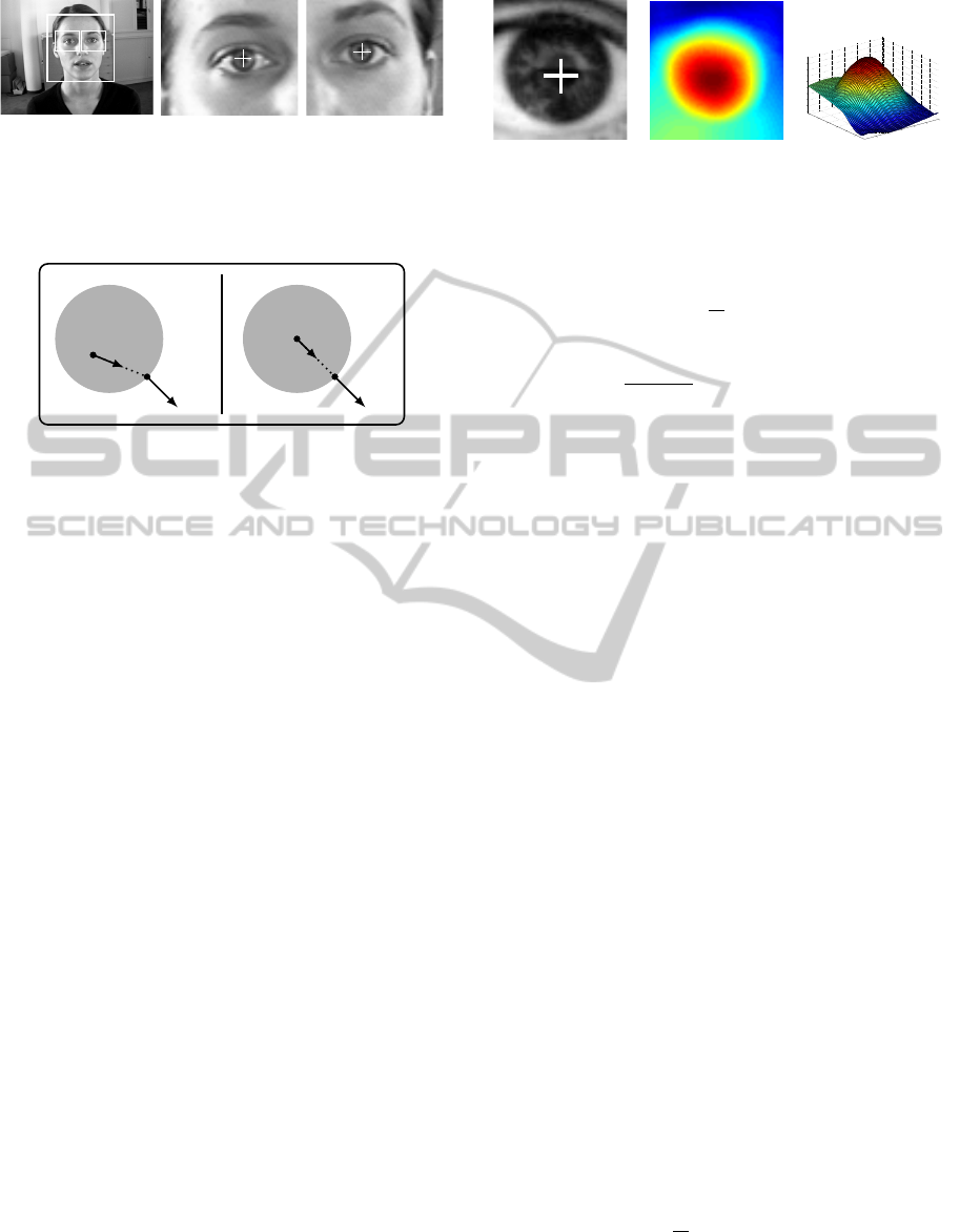
Figure 1: Multi-stage approach for eye centre localisation.
A face detector is applied first; based on the face loca-
tion rough eye regions are extracted (left), which are then
used for a precise estimation of each eye centre (middle and
right).
g
i
d
i
c
x
i
g
i
d
i
c
x
i
Figure 2: Artificial example with a dark circle on a light
background, similar to the iris and the sclera. On the left
the displacement vector d
i
and the gradient vector g
i
do not
have the same orientation, whereas on the right both orien-
tations are equal.
previously. Kothari and Mitchell, for example, pro-
posed a method that exploits the flow field character
that arises due to the strong contrast between iris and
sclera (Kothari and Mitchell, 1996). They use the ori-
entation of each gradient vector to draw a line through
the whole image and they increase an accumulator bin
each time one such line passes through it. The ac-
cumulator bin where most of the lines intersect thus
represents the estimated eye centre. However, their
approach is only defined in the discrete image space
and a mathematical formulation is missing. More-
over, they don’t consider problems that arise due to
eyebrows, eyelids, or glasses.
In this work, we also analyse the vector field of
image gradients but derive a novel mathematical for-
mulation of the vector field characteristics. Therefore,
we mathematically describe the relationship between
a possible centre and the orientations of all image gra-
dients. Let c be a possible centre and g
i
the gradient
vector at position x
i
. Then, the normalised displace-
ment vector d
i
should have the same orientation (ex-
cept for the sign) as the gradient g
i
(see Fig. 2). If we
use the vector field of (image) gradients, we can ex-
ploit this vector field by computing the dot products
between the normalised displacement vectors (related
to a fixed centre) and the gradient vectors g
i
. The op-
timal centre c
∗
of a circular object in an image with
pixel positions x
i
, i ∈ {1,...,N}, is then given by
Figure 3: Evaluation of (1) for an exemplary pupil with the
detected centre marked in white (left). The objective func-
tion achieves a strong maximum at the centre of the pupil;
2-dimensional plot (centre) and 3-dimensional plot (right).
c
∗
= argmax
c
(
1
N
N
∑
i=1
d
T
i
g
i
2
)
, (1)
d
i
=
x
i
− c
kx
i
− ck
2
, ∀i : kg
i
k
2
= 1 . (2)
The displacement vectors d
i
are scaled to unit length
in order to obtain an equal weight for all pixel posi-
tions. In order to improve robustness to linear changes
in lighting and contrast the gradient vectors should
also be scaled to unit length. An example evalua-
tion of the sum of dot products for different centres is
shown in Fig. 3, where the objective function yields a
strong maximum at the centre of the pupil.
Computational complexity can be decreased by
considering only gradient vectors with a signifi-
cant magnitude, i.e. ignoring gradients in homo-
geneous regions. In order to obtain the image
gradients, we compute the partial derivatives g
i
=
(
∂I(x
i
,y
i
)
/∂x
i
,
∂I(x
i
,y
i
)
/∂y
i
)
T
, but other methods for com-
puting image gradients will not change the behaviour
of the objective function significantly.
2.1 Prior Knowledge and
Postprocessing
Under some conditions, the maximum is not well de-
fined, or there are local maxima that lead to wrong
centre estimates. For example, dominant eyelids and
eyelashes or wrinkles in combination with a low con-
trast between iris and sclera can lead to wrong es-
timates. Therefore, we propose to incorporate prior
knowledge about the eye in order to increase robust-
ness. Since the pupil is usually dark compared to
sclera and skin, we apply a weight w
c
for each pos-
sible centre c such that dark centres are more likely
than bright centres. Integrating this into the objective
function leads to:
argmax
c
1
N
N
∑
i=1
w
c
d
T
i
g
i
2
, (3)
where w
c
= I
∗
(c
x
,c
y
) is the grey value at (c
x
,c
y
) of
the smoothed and inverted input image I
∗
. The image
VISAPP 2011 - International Conference on Computer Vision Theory and Applications
126

needs to be smoothed, e.g. by a Gaussian filter, in or-
der to avoid problems that arise due to bright outliers
such as reflections of glasses. The values of the new
objective function is rather insensitive to changes in
the parameters of the low-pass filter.
The proposed summation of weighted squared dot
products yields accurate results if the image contains
the eye. However, when applying the multi-stage
scheme described in Figure 1, the rough eye regions
sometimes also contain other structures such as hair,
eyebrows, or glasses. Especially, hair and strong re-
flections in glasses show significant image gradients
that do not have the same orientation as the image gra-
dients of the pupil and the iris; hence the estimation
of the eye centres might be wrong. We therefore pro-
pose a postprocessing step in order to overcome these
problems. We apply a threshold on the objective func-
tion, based on the maximum value, and remove all re-
maining values that are connected to one of the image
borders. Then, we determine the maximum of the re-
maining values and use its position as centre estimate.
Based on our experiments the value of this threshold
doesn’t have a significant influence on the centre esti-
mates, we suggest to set this threshold to 90% of the
overall maximum.
3 EVALUATION
For our evaluation we have chosen the BioID
database, since it is the most challenging set of im-
ages for eye centre localisation and many recent re-
sults are available. The database consists of 1521
grey level images of 23 different subjects and has
been taken in different locations and at different day-
times, which result in variable illumination condi-
tions comparable to outdoor scenes. In addition to
the changes in illumination, the position of the sub-
jects change as well as their pose. Moreover, several
subjects wear glasses and some subjects have curled
hair near to the eye centres. In some images the eyes
are closed and the head is turned away from the cam-
era or strongly affected by shadows. In few images
the eyes are even completely hidden by strong reflec-
tions on the glasses. Because of these conditions, the
BioID database is considered as one the most chal-
lenging database that reflects realistic conditions. The
image quality and the image size (286 × 384) is ap-
proximately equal to the quality of a low-resolution
webcam. The left and right eye centres are annotated
and provided together with the images.
We perform the multi-stage scheme described in
Figure 1, where the position of the face is detected
first. Therefore, we apply a boosted cascade face de-
tector that proved to be effective and accurate on sev-
eral benchmarks (Viola and Jones, 2004). Based on
the position of the detected face and anthropometric
relations, we extract rough eye regions relative to the
size of the detected face. The rough eye regions are
then used to estimate the eye centres accurately by
applying the proposed approach.
As accuracy measure for the estimated eye cen-
tres, we evaluate the normalised error, which indi-
cates the error obtained by the worst of both eye esti-
mations. This measure was introduced by Jesorsky et
al. and is defined as:
e ≤
1
d
max(e
l
, e
r
) , (4)
where e
l
, e
r
are the Euclidean distances between the
estimated and the correct left and right eye centres,
and d is the distance between the correct eye centres.
When analysing the performance of an approach for
eye localisation this measure has the following char-
acteristics: (i) e ≤ 0.25 ≈ distance between the eye
centre and the eye corners, (ii) e ≤ 0.10 ≈ diame-
ter of the iris, and (iii) e ≤ 0.05 ≈ diameter of the
pupil. Thus, an approach that should be used for eye
tracking must not only provide a high performance for
e ≤ 0.25, but must yield good results for e ≤ 0.05. An
error of slightly less than or equal to 0.25 will only
indicate that the estimated centre might be located
within the eye, but this estimation cannot be used to
perform accurate eye tracking. When comparing with
state of the art methods we therefore focus on the per-
formance that is obtained for e 0.25.
Since in some other published articles the nor-
malised error is used in a non-standard way, we
also provide the measures e
better
≤
1
d
min(e
l
, e
r
) and
e
avg
≤
1
2d
(e
l
+ e
r
) in order to give an upper bound as
well as an averaged error.
3.1 Results
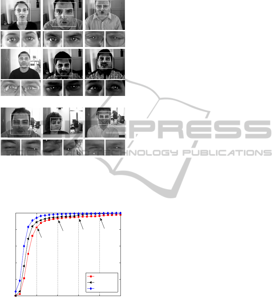
The qualitative results of the proposed approach are
shown in Figure 4. It can be observed that our ap-
proach yields accurate centre estimations not only for
images containing dominant pupils, but also in the
presence of glasses, shadows, low contrast, or strands
of hair. This demonstrates the robustness and proves
that our approach can successfully deal with several
severe problems that arise in realistic scenarios. Our
approach yields inaccurate estimations if the eyes are
(almost) closed or strong reflections on the glasses oc-
cur (last row). Then, the gradient orientations of the
pupil and the iris are affected by “noise” and hence
their contribution to the sum of squared dot products
is less than the contribution of the gradients around
the eyebrow or eyelid.
ACCURATE EYE CENTRE LOCALISATION BY MEANS OF GRADIENTS
127
(a) accurate eye centre estimations
(b) inaccurate eye centre estimations
Figure 4: Sample images of accurate and inaccurate results
for eye centre localisation on the BioID database. The esti-
mated centres are depicted by white crosses. Note that, the
estimated centres might be difficult to identify due to low
printer resolution.
0 0.05 0.1 0.15 0.2 0.25
0
20
40
60
80
100
normalized error
accuracy [%]
worse eye
avg. eye
better eye
82.5
95.2
93.4
96.4
Figure 5: Quantitative analysis of the proposed approach
for the BioID database. In order to give upper and lower
bounds, the accuracy versus the minimum (better eye,
e
better
), the maximum (worse eye, e) and the average (avg.
eye, e
avg
) normalised error are shown. Some characteristic
values are given explicitly.
The quantitative results of the proposed method
are shown in Figure 5, where the accuracy measures
e, e
better
, and e
avg
are illustrated. By using the stan-
dard definition of the normalised error, Eq. (4), our
approach yields an accuracy of 82.5% for pupil lo-
calisation (e ≤ 0.05), which indicates that the cen-
tres detected by our approach are located within the
pupil with high probability and can therefore be used
for eye tracking applications. For iris localisation
(e ≤ 0.10), the estimated centres lie within the iris
with a probability of 93.4%, which will further in-
crease if images with closed eyes are left out.
3.2 Comparison with State of the Art
We extensively compare our method with state of the
art methods that have been applied to the BioID im-
ages as well. For comparison we evaluate the per-
formance for different values of the normalised er-
ror e in order to obtain a characteristic curve (see
Fig. 5 “worse eye”), which we will call worse eye
characteristic (WEC). The WEC is roughly simi-
lar to the well-known receiver operator character-
istic (ROC) and can be analysed in several ways.
As mentioned previously, it depends on the appli-
cation which e should be applied in order to com-
pare different methods, e.g. for eye tracking appli-
cations a high performance for e ≤ 0.05 is required,
whereas for applications that use the overall eye po-
sition such as face matching comparing the perfor-
mance for e ≤ 0.25 will be more appropriate. In or-
der to compare the overall performance, i.e. for dif-
ferent e, the area under the WEC can be used. Un-
fortunately, the WEC of other methods is often not
available, and we therefore compare the methods for
a discretised e ∈ {0.05,0.10,0.15,0.20,0.25}. Fur-
thermore, we also evaluate the rank of each method,
which is roughly inversely proportional to the area un-
der the WEC.
The comparison between our method and state of
the art methods is shown in Tab. 1. If the performance
for e ∈ {0.05, 0.10, 0.15, 0.20, 0.25} was not provided
by the authors explicitly, but a WEC is shown, we
measured the values accurately from the WEC. Note
that, for some methods, the authors evaluated the per-
formance only for few values of e, see for example
(Chen et al., 2006) or (Zhou and Geng, 2004). It can
be seen that our method performs only 2% worse in
average compared to the best method for each e. For
example, the method proposed by Valenti and Gevers
yields a performance of 84.1% for e ≤ 0.05, whereas
our method yields a performance of 82.5%. How-
ever, Valenti and Gevers reported that their method,
which uses mean-shift clustering, SIFT features, and
a k nearest neighbour classifier, will produce unstable
centre estimations when applying it to eye tracking
with several images per second. Hence, our method
can be considered as one of the best methods for
VISAPP 2011 - International Conference on Computer Vision Theory and Applications
128

Table 1: Comparison of the performance for eye detection on the BioID database. Brackets indicate values that have been
accurately measured from author’s graphs. (∗) Images with closed eyes and glasses were omitted. (•) Methods that don’t
involve any kind of learning or model scheme. Since some authors didn’t provide any graphical evaluation of the performance,
e.g. by using a WEC curve, intermediate values couldn’t be estimated – these missing values are denoted by “–”.
Method e ≤ 0.05 e ≤ 0.10 e ≤ 0.15 e ≤ 0.20 e ≤ 0.25 Remarks
(Asadifard and Shanbezadeh, 2010) 47.0% 86.0% 89.0% 93.0% 96.0% (∗), (•)
(Kroon et al., 2008) 65.0% 87.0% – – 98.8%
(Valenti and Gevers, 2008) 77.2% 82.1% (86.2%) (93.8%) 96.4% MIC, (•)
(Valenti and Gevers, 2008) 84.1% 90.9% (93.8%) (97.0%) 98.5% MIC+SIFT+kNN
(T
¨
urkan et al., 2007)
(18.6%) 73.7% (94.2%) (98.7%) 99.6%
(Campadelli et al., 2006) 62.0% 85.2% 87.6% 91.6% 96.1%
(Niu et al., 2006) (75.0%) 93.0% (95.8%) (96.4%) (97.0%)
(Chen et al., 2006) – 89.7% – – 95.7%
(Asteriadis et al., 2006) (44.0%) 81.7% (92.6%) (96.0%) 97.4% (•)
(Hamouz et al., 2005) (58.6%) (75.0%) (80.8%) (87.6%) (91.0%)
(Zhou and Geng, 2004) – – – – 94.8% (•)
(Cristinacce et al., 2004) (57.0%) 96.0% (96.5%) (97.0%) (97.1%)
(Behnke, 2002) (37.0%) (86.0%) (95.0%) (97.5%) (98.0%)
(Jesorsky et al., 2001) (38.0%) (78.8%) (84.7%) (87.2%) 91.8%
our method 82.5% 93.4% 95.2% 96.4% 98.0% (•)
accurate eye centre localisation. Furthermore, our
method has significantly less computational complex-
ity compared to that of Valenti and Gevers, since it
requires neither clustering nor a classifier. Compar-
ing those methods that do not involve any kind of
learning scheme, our method achieves the best per-
formance by far (82.5% for e ≤ 0.05). For iris loca-
tion (e ≤ 0.10), our method achieves the second best
performance (93.4%); only the method by Cristinacce
et al. yields a significant improvement (96.0%) –
however, this improvement implies, again, a higher
computational complexity compared to our method,
which is solely based on dot products. For higher nor-
malised errors, e.g. e ≤ 0.15, e ≤ 0.20, or e ≤ 0.25,
our method performs comparable to other methods.
A comparison based on the ranks of the perfor-
mances is shown in Tab. 2. It can be seen clearly,
that there isn’t one single method that performs supe-
rior for all values of e. Exemplarily, the method pro-
posed by T
¨
urkan et al. achieves accurate estimations
for detecting the overall eye centres, i.e. e ≤ 0.20 and
e ≤ 0.25, but it fails for iris localisation (e ≤ 0.10)
and pupil localisation (e ≤ 0.05) with rank 13 in both
cases. In contrast, our method ranks 2nd for both
pupil and iris localisation and ranks 3rd and 4th for
larger e. Hence, our method doesn’t yield the best re-
sult for one single e, but if we evaluate the average
rank our method yields the best result (3.0). Com-
pared to the method with the second best average
rank (3.4, Valenti and Gevers, MIC+SIFT+kNN) our
method is not only superior according to the average
rank, but also the variance of the individual ranks is
significantly less, and the complexity is much lower.
In total, our method performs comparable to other
state of the art methods when looking for a particular
e, but it yields the best average performance over all
values of e. Hence, our method proves to be powerful
for several problems such as eye centre localisation
(e ≤ 0.05), iris localisation (e ≤ 0.10), and eye lo-
calisation (e ≤ 0.25). Comparing only those methods
that do not apply any learning scheme, our method
achieves significant improvements for the more diffi-
cult tasks, i.e. 5% improvement for e ≤ 0.05, 7% for
e ≤ 0.10, and 2.6% for e ≤ 0.15.
4 CONCLUSIONS
We propose a novel algorithm for accurate eye cen-
tre localisation based on image gradients. For ev-
ery pixel, we compute the squared dot product be-
tween the displacement vector of a centre candidate
and the image gradient. The position of the maxi-
mum then corresponds to the position where most im-
age gradients intersect. Our method yields low com-
putational complexity and is invariant to rotation and
linear changes in illumination. Compared to several
state of the art methods, our method yields a very high
accuracy for special scenarios such as pupil localisa-
tion (2nd place) and ranks in 1st place if the average
performance over several scenarios, e.g. pupil local-
isation, iris localisation, and overall eye localisation,
is evaluated. Our method can be applied to several
(real-time) applications that require a high accuracy
such as eye tracking or medical imaging analysis (cell
tracking).
ACCURATE EYE CENTRE LOCALISATION BY MEANS OF GRADIENTS
129

Table 2: Comparison of ranks of each method according to its performance shown in Tab. 1.
Method e ≤ 0.05 e ≤ 0.10 e ≤ 0.15 e ≤ 0.20 e ≤ 0.25 avg. rank
(Asadifard and Shanbezadeh, 2010) 9 7 8 7 10 8.2
(Kroon et al., 2008) 5 6 – – 2 4.3
(Valenti and Gevers, 2008) 3 9 10 6 8 7.2
(Valenti and Gevers, 2008) 1 4 6 3 3 3.4
(T
¨
urkan et al., 2007) 13 13 5 1 1 6.6
(Campadelli et al., 2006) 6 8 9 8 9 8.0
(Niu et al., 2006) 4 3 2 4 7 4.0
(Chen et al., 2006) – 5 – – 11 8.0
(Asteriadis et al., 2006) 10 10 7 5 5 7.4
(Hamouz et al., 2005) 7 12 12 9 14 10.8
(Zhou and Geng, 2004) – – – – 12 12.0
(Cristinacce et al., 2004) 8 1 1 3 6 3.8
(Behnke, 2002) 12 7 4 2 4 5.8
(Jesorsky et al., 2001) 11 11 11 10 13 11.2
our method 2 2 3 4 4 3.0
REFERENCES
Asadifard, M. and Shanbezadeh, J. (2010). Automatic adap-
tive center of pupil detection using face detection and
cdf analysis. In Proceedings of the IMECS, volume I,
pages 130–133, Hong Kong. Newswood Limited.
Asteriadis, S., Asteriadis, S., Nikolaidis, N., Hajdu, A., and
Pitas, I. (2006). An eye detection algorithm using
pixel to edge information. In Proceedings of the 2nd
ISCCSP, Marrakech, Morocco. EURASIP.
Behnke, S. (2002). Learning face localization using hi-
erarchical recurrent networks. In Proceedings of the
ICANN, LNCS, pages 135–135. Springer.
B
¨
ohme, M., Meyer, A., Martinetz, T., and Barth, E. (2006).
Remote eye tracking: State of the art and directions
for future development. In Proceedings of the 2nd
COGAIN, pages 10–15, Turin, Italy.
Campadelli, P., Lanzarotti, R., and Lipori, G. (2006).
Precise eye localization through a general-to-specific
model definition. In Proceedings of the 17th BMVC,
volume I, pages 187–196, Edingburgh, England.
Chen, D., Tang, X., Ou, Z., and Xi, N. (2006). A hierar-
chical floatboost and mlp classifier for mobile phone
embedded eye location system. In Proceedings of the
3rd ISNN, LNCS, pages 20–25, China. Springer.
Cristinacce, D., Cootes, T., and Scott, I. (2004). A multi-
stage approach to facial feature detection. In Proceed-
ings of the 15th BMVC, pages 277–286, England.
Hamouz, M., Kittler, J., Kamarainen, J., Paalanen, P.,
K
¨
alvi
¨
ainen, H., and Matas, J. (2005). Feature-based
affine-invariant localization of faces. IEEE Transac-
tions on PAMI, 27(9):1490.
Hansen, D. and Ji, Q. (2010). In the eye of the beholder: A
survey of models for eyes and gaze. IEEE Trans. on
PAMI, 32(3):478–500.
Jesorsky, O., Kirchberg, K., and Frischholz, R. (2001). Ro-
bust face detection using the Hausdorff distance. In
Proceedings of the 3rd AVBPA, LNCS, pages 90–95,
Halmstad, Sweden. Springer.
Kothari, R. and Mitchell, J. (1996). Detection of eye loca-
tions in unconstrained visual images. In Proceedings
of the IEEE ICIP, volume 3, pages 519–522. IEEE.
Kroon, B., Hanjalic, A., and Maas, S. (2008). Eye localiza-
tion for face matching: is it always useful and under
what conditions? In Proceedings of the 2008 CIVR,
pages 379–388, Ontario, Canada. ACM.
Niu, Z., Shan, S., Yan, S., Chen, X., and Gao, W. (2006).
2d cascaded adaboost for eye localization. In Proceed-
ings of the 18th IEEE ICPR, volume 2, pages 1216–
1219, Hong Kong. IEEE.
T
¨
urkan, M., Pard
`
as, M., and C¸ etin, A. E. (2007). Human
eye localization using edge projections. In Proceed-
ings of the 2nd VISAPP, pages 410–415. INSTICC.
Valenti, R. and Gevers, T. (2008). Accurate eye center lo-
cation and tracking using isophote curvature. In Pro-
ceedings of the CVPR, pages 1–8, Alaska. IEEE.
Viola, P. and Jones, M. (2004). Robust real-time face detec-
tion. IJCV, 57(2):137–154.
Zhou, Z. and Geng, X. (2004). Projection functions for eye
detection. Pattern Recognition, 37(5):1049–1056.
VISAPP 2011 - International Conference on Computer Vision Theory and Applications
130
