
OPTICAL FIBER CHARACTERIZED WITH A LOW
REFRACTIVE INDEX CAN DETECT BLOOD
Blood Increased Light Loss through an Air–cladding Optical Fiber
Akihiro Takeuchi, Tomohiro Miwa
Department of Medical Informatics, School of Allied Health Sciences, Kitasato University, Sagamihara, Japan
Graduate School of Medical Sciences, Kitasato University, Sagamihara, Japan
Minoru Sawada, Haruo Imaizumi, Hiroyuki Sugibuchi, Minoru Hirose, Noriaki Ikeda
Junkosha Co., Ltd., Ishibashi, Sakaigawa, Fuefuki, Yamanashi, Japan
Department of Clinical Engineering, School of Allied Health Sciences, Kitasato University, Sagamihara, Japan
Department of Medical Informatics, School of Allied Health Sciences, Kitasato University, Sagamihara, Japan
Keywords: Air-cladding plastic optical fiber, Light loss, Venous needle dislodgement.
Abstract: Large amounts of undetected blood loss during hemodialysis are caused by venous needle dislodgement. A
special air-cladding plastic optical fiber with a low refractive index, fluoropolymer, PFA fiber, JUNFLON
®
,
was developed to monitor oil and lipid leakage in industrial fields, and to monitor the dust in the air in clean
rooms. To apply the air-cladding plastic optical fiber as a bleed sensor, we studied the optical effects of
soaking the fiber with various liquids and porcine blood on light-loss experimental settings. Light intensity
through the fiber was studied with a light emitting diode and a photodiode under various conditions of
soaked fiber with reverse osmosis water, physiological saline, glucose, and porcine blood. The more the
soaked length increased with all mediums, the more the light intensity decreased. Although the slopes of the
decreased curves varied according to the mediums, the light scattering phenomena caused by the mediums
can be applied to a bleed sensor for clinical use.
1 INTRODUCTION
Although hemodialysis has evolved into a safe and
less stressful procedure for both patients and
caregivers (Sarkar, Kaitwatcharachai and Levin,
2005; Hawley, Jefferies, Nearhos and Van Eps,
2008), intradialytic complications still cause
considerable patient morbidity and rarely, mortality
(Sarkar et al., 2005). Venous needle dislodgment
(VND) is one of the most serious accidents that can
occur during hemodialysis (Hawley et al., 2008; Van
Waeleghem, Chamney, Lindley and Pancírová,
2008). The European Dialysis and Transplant Nurses
Association/ European Renal Care Association has
produced 12 practice recommendations to help
reduce the risk of VND and detect blood leakage as
soon as possible (Van Waeleghem et al., 2008). A
safety device from Redsense Medical, Halmstad,
Sweden for use during hemodialysis that uses fiber
optic technology to detect blood has been approved
as a Class I medical device with the intended
purpose of detecting VND (Van Waeleghem et al.,
2008; Ahlmén, Gydell, Hadimeri, Hernandez,
Rogland and Strömbom, 2008).
On the other hand, optical fibers are widely and
directly used in fiber optic communications, medical
endoscopes, and sensors (Goodyer, Fothergill, Jones
and Hanning, 1996; Zubia and Arrue, 2001; Sugita,
2001; Lee, 2003). An optical fiber generally consists
of a core and a surrounding layer called “cladding”
with a low refractive index. Based on the difference
in refractive indices, light is reflected at the
core-cladding interface. An air-cladding plastic
optical fiber characterized with a low refractive
index of 1.328 nD was developed by the Junkosha
Co., Ltd. (Yamanashi, Japan), that can monitor
contaminates or impurities in a clean room or
environs (Suzuki, 2004). When the air-cladding
optical fiber is contaminated with a liquid, the light
307
Takeuchi A., Miwa T., Sawada M., Imaizumi H., Sugibuchi H., Hirose M. and Ikeda N..
OPTICAL FIBER CHARACTERIZED WITH A LOW REFRACTIVE INDEX CAN DETECT BLOOD - Blood Increased Light Loss through an Air–cladding
Optical Fiber.
DOI: 10.5220/0003122003070310
In Proceedings of the International Conference on Biomedical Electronics and Devices (BIODEVICES-2011), pages 307-310
ISBN: 978-989-8425-37-9
Copyright
c
2011 SCITEPRESS (Science and Technology Publications, Lda.)

signal may partially be lost from the contaminated
site and may decrease when the area contaminated
with the liquid increases. Hence, it would be
possible to detect bleeding by monitoring the light
loss from the air-cladding plastic optical fiber
attached to the skin around the needle site of a
patient’s arm or leg. To our knowledge, any
relationship between lengths of the segment soaked
with a liquid and light loss has not yet been reported
in either PubMed or Optics InfoBase literature, nor
has utilizing this particular relationship to detect
VND been reported. The present study presents the
optical characteristics of air-cladding optical fiber to
detect blood or fluid leakages.
2 MATERIALS AND METHODS
2.1 Experiment Overview
Figure 1 shows an experimental work desk on which
an optical fiber was placed with a straight segment
to test the effect the mediums have on the optical
fiber. The working area was strictly cleaned to
prevent any contamination from other mediums, e.g.,
dust, hand oils, or any liquids.
The examined air-cladding optical fiber is a
fluoropolymer, PFA core fiber, JUNFLON
®
(Junkosha), with a 1-mm diameter, and a refractive
index of 1.328 nD. The fiber used in this experiment
was 2 m in length and weighed 2 g. It was resistive
to acids and alkalines, and to ethanol, ethylene oxide
gas, and heating for sterilization.
The sensor module, LEAKLEARN OPT
®
(Junkosha), consisted of a light emitting diode
(LED), a detector photodiode (PD), and electrical
circuits for monitoring and alarming. The sensor
module monitors voltage as light intensity. Thus, the
voltage decreases as the light transmitted to the PD
becomes darker.
2.2 Mediums used to Soak the Optical
Fiber
The applied liquids were: reverse osmosis (RO)
water, physiological saline, and glucose in water at
5%, 10% and 20%. All of which except the RO
water were pharmaceutical products. Porcine plasma
and blood (Hct 40% and 20%) were also applied and
tested in the same manner. The hematocrits were
prepared by adding porcine plasma but not saline.
To control a length of fiber soaked with a medium
without a longitudinal leak under the fiber, narrow
gauze strips were crossed over the fiber or wrapped
around the fiber. Then each medium was manually
dripped on each gauze strip. The mediums, thus,
soaked the gauze strips and circumferentially
surrounded the fiber for the whole width of the strips.
There was no expansion observed of the mediums
along the fibers.
For physiological saline, light intensities were
compared with two settings, the continuous and the
separate modes, of the gauze strips on the fiber. The
soaked lengths were the sum of the widths of the
gauze strips in the separate mode. For other
mediums, the light intensities were measured in the
continuous mode on the fibers.
Figure 1: Experimental setting.
Figure 2: Light intensity to soaked length for each test
medium. The horizontal axis indicates the soaked length in
centimeters.
3 RESULTS
Figure 2 shows all of the raw data obtained for each
medium tested. Data points for all experiments
including duplicates are shown with 5 symbols for
the 5 experiments. The light intensities clearly and
exponentially decreased with the longer soaked
length for all the mediums. For physiological saline,
BIODEVICES 2011 - International Conference on Biomedical Electronics and Devices
308
the raw data of the continuous mode were visually
the same as those of the separate mode. Porcine
plasma showed a slightly steeper curve than did the
porcine blood. The raw data of RO water showed a
milder decreasing curve than did those of the other
mediums.
4 DISCUSSION
The light intensity decreased as the length soaked
with the medium increased. Although Golnabi and
Azimi (2007) proposed a plastic optical fiber
leakage sensor by immersing liquids with the higher
index of refraction, the quantitative relationship
between soaked lengths and light loss was not
reported in detail in their study.
The phenomenon of scattering back to the front
end of a fiber is also utilized as a sensor, optical time
domain reflectometry (OTDR) (Sugita, 2001).
Although the OTDR is also widely used in medical
and chemical analyses and molecular biotechnology
(Lee, 2003; Barnoski and Jensen, 1976; Sensfelder,
Bürck and Ache, 1998), it is difficult for
commercially available OTDR to detect any clinical
events in a region less than 1 m from the sensor
module. The mechanism of our sensor is simpler
than that of OTDR, and only based on light intensity
without any chemical modifiers to sense bleeding or
liquids. The phenomena that the decrease of light
intensity depends on the length soaked can be
applied to monitor VND.
Although it is well known that bending a fiber
modifies its guiding properties and increases light
loss, the optical fiber used in the present study
showed no light loss even when bending it in a
1-cm-diameter loop. This flexibility would be useful
for attaching the optical fiber to the skin around a
needle site of a patient’s arm or leg.
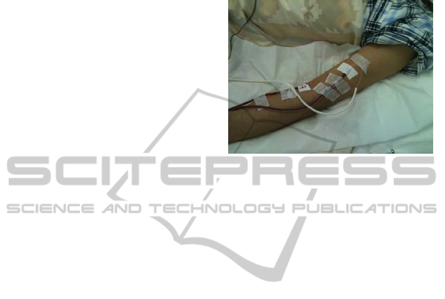
The optical fiber can be used as a non-invasive
disposable sensor to detect bleeding or leakage
during hemodialysis. The optical fiber was looped
on the surface of the skin around the puncture site
(Figure 3). Although the sensor can detect bleeding
from the needle site, it can not sense a subcutaneous
bleeding.
Although this sensor module may be made
smaller while maintaining its sophisticated functions
for clinical use, the air-cladding optical fiber offers
certain advantages, such as the fiber’s light weight,
flexibility, and the ability to adjust the fiber length,
loop size, and route, simple fixation with surgical
tape, and continuous real-time sensing. These
advantages allow the air-cladding optical fiber to be
used as a disposable sensor to quickly detect
bleeding and leakage during hemodialysis and
continuous venous infusion.
Figure 3: Clinical setting during hemodialysis. The optical
fiber was attached with 3M tape to the skin and partially
passed through a white tube.
5 CONCLUSIONS
We confirmed the phenomenon that light intensity
clearly decreased as the soaked length of a fiber
increased. This phenomenon can be used to quickly
detect bleeding and leakage and set off alarms for
patients undergoing hemodialysis and for those
receiving infusion therapy.
ACKNOWLEDGEMENTS
The authors thank Robert E. Brandt (CEO, MedEd
Japan) for helpful advice on the English language in
the preparation and editing of this manuscript.
REFERENCES
Ahlmén, J., Gydell, K., H., Hadimeri, H., Hernandez, I.,
Rogland, B. and Strömbom, U. (2008). A New Safety
Device for Hemodialysis. Hemodial Int, 12, 264-267.
Barnoski, M. K. and Jensen, S. M. (1976). Fiber
Waveguides: A Novel Technique for Investigating
Attenuation Characteristics. Appl Opt, 15, 2112-2115.
Golnabi, H. and Azimi, P. (2007). Design and Performance
of a Plastic Optical Fiber Leakage Sensor. Opt Laser
Technol, 39, 1346-1350
Goodyer, P. D., Fothergill, J. C., Jones, N. B. and Hanning,
C. D. (1996). The Design of an Optical Fiber Pressure
Transducer for Use in the Upper Airways. IEEE Trans
Biomed Eng, 43, 600-606.
OPTICAL FIBER CHARACTERIZED WITH A LOW REFRACTIVE INDEX CAN DETECT BLOOD - Blood Increased
Light Loss through an Air-cladding Optical Fiber
309

Hawley, C. M., Jefferies, J, Nearhos, J. and Van Eps, C.
(2008). Complications of Home Hemodialysis.
Hemodial Int, 12 (Suppl 1), S21-S25.
Lee, B. (2003). Review of the Present Status of Optical
Fiber Sensors. Opt Fiber Technol, 9, 57-79.
Sarkar, S. R., Kaitwatcharachai C. and Levin N.W. (2005).
Complications During Hemodialysis. In: Nissenson, A.
R., Fine, R. N., (Eds.), Clinical Dialysis. (4th ed., pp.
237-272). New York, NY: Mcgraw-Hill.
Sensfelder, E., Bürck, J. and Ache, H. J. (1998).
Characterization of a Fiber-Optic System for the
Distributes Measurement of Leakages in Tanks and
Pipelines. Appl Spectrosc, 52, 1283-1298.
Sugita, T. (2001). Optical Time-Domain Reflectometry of
Bent Plastic Optical Fibers. Appl Opt, 40, 897-905.
Suzuki, M., inventor (2004). Junkosha Co., Ltd., Japan
assignee. Optical Fiber and Liquid Sensor. Patent of
Japan 2004-101279. 02, Apr.
Van Waeleghem, J. P., Chamney, M., Lindley, E. J. and
Pancírová, J. (2008). Venous Needle Dislodgement:
How to Minimise the Risks. J Ren Care, 34, 163-168.
Zubia, J. and Arrue, J. (2001). Plastic Optical Fibers: An
Introduction to Their Technological Processes and
Applications. Opt Fiber Technol, 7, 101-140.
BIODEVICES 2011 - International Conference on Biomedical Electronics and Devices
310
