
SOLO-MEDICINE IN OPTICAL BIOPSIES
A Way to Practice Telemedicine
Olga Ferrer, Francesco Ettorre, Xiomara Santos
University of La Laguna, La Laguna, Canary Islands, Spain
Thomas Zinkl, Ruben Tous, Jaime Delgado
Department of Computer Architecture, Universitat Politecnica de Catalunya (UPC-BARCELONATECH), Barcelona, Spain
Keywords: Solo-medicine, Optical biopsy, Confocal laser endomicroscopy, CLE, CBIR, Medical image retrieval,
Query by image, ISO-15938-12, MPEG query format, MPQF, ISO 24800-3, JPSearch, JPEG query format,
JPQF, Artificial intelligence, Multimedia standard.
Abstract: A way to practice Telemedicine is to access a data-base capable to assist you in medical procedures
(diagnosis, treatment and prognosis), similarly to consult a book or to ask a college. In many countries the
lack of specialists and training capabilities demand to practice solo-medicine, that in the case of surgery
require robots capable to induce anesthesia or help in vision or handling instruments. A relevant case is the
diagnostic self-training requirements for optical biopsies (OBs) obtained with confocal laser
endomicroscopy (CLE) or the assistance in the diagnosis of pathology slides. In both cases it is required a
training set of digital images against which to compare the question case by means of image-queryformat.
The present paper present a content-based image retrieval system (CBIR) based on the MPEG Query
Format Standard in order to provide a set of similar pictures and the corresponding diagnosis to help on
diagnosis or just to train the doctor. The paper defined the Image Solo-Medicine Paradigm (ISMP)
architecture merging medical image standards and MPEG and JPEG standards. It tested the solution with
normal, and benign colon OBs with 90% congruency. The ISMP is of particular interest viewed the
proliferation of iPhone medical applications aiming to train doctors and support medical decisions.
1 INTRODUCTION
Nowadays the number of medical applications for
iPhone proliferate (CATAI, 2010), attracting the
interest of relevant medical Journals such as the
British Medical Journal (BMJ Group, n.d.) to build
applications that help doctor to make their decisions
and auto-train themselves. The time of training
books, with periodical updates for new diagnosis or
treatments, is arriving to an end; doctors will have
on-line and on mobile phones that information, and
will use mobile phones for a variety of medical
applications (Ferrer-Roca and Marcano, 2009;
Ferrer-Roca and Marcano, 2010; Ferrer-Roca,
2010).
We have been working in a diagnostic medical
application based on images and on which “gold-
standards” are still on the way (Hersch et al., 2005;
Kiesslich et al., 2008; Ferrer-Roca et al., 2010). This
is the so-called optical biopsy (OB) (Wang and
VanDam 2004). A non-intrusive optic diagnostic
method, capable to analyze the tissue in surface and
in deepness with one of the following techniques:
laser, OCT, infrared, fluorescence, spectroscopy etc.
This means, that it is not necessary to extract the
tissue from the body. Tissue is accessed through the
surface of the body through the skin or by
endoscopy.
In OBs images are obtained in real time together
with complementary information that allows
evaluating the disease in vivo, but “gold-standards”
are still lacking while in surgical pathology
standards lay on the histology of the normal fixed
tissue (Ferrer-Roca, 2009). To provide training and
self-confidence on OB diagnosis, two possibilities
are open: (a) Tele-consult to a pathologist or (b)
Train themselves with a non-supervised search for a
“similar image” on the Net using multimedia query
and image mining techniques (Chen et al., 2006).
441
Ferrer O., Ettorre F., Santos X., Zinkl T., Tous R. and Delgado J..
SOLO-MEDICINE IN OPTICAL BIOPSIES - A Way to Practice Telemedicine.
DOI: 10.5220/0003090804410445
In Proceedings of the International Conference on Health Informatics (HEALTHINF-2011), pages 441-445
ISBN: 978-989-8425-34-8
Copyright
c
2011 SCITEPRESS (Science and Technology Publications, Lda.)

The present proposes a standardized ISMP
(Image Solo-Medicine Paradigm) architecture based
in the usage of two novel standards, the MPEG
Query Format (MPQF) and the JPEG’s JPSearch
project (Tous, 2006). While MPQF provides a
uniform language for querying multimedia
databases, JPSearch provides an interoperable
architecture for images’ metadata management.
Preliminary results on Internet image search and
discovery system for diagnostic medical purpose are
showed. Results were based on a training-set of
CLE-OB images annotated with specific CLE
semantics.
2 MATERIAL AND METHODS
The proposed system to allow users to navigate
searching similar images considered to be golden
standards (due to the pathology confirmation and
availability of pathology image) in a pair image
database integrated by OB-CLE images together
with the histological counterpart.
Images used in the present paper were provided
by one of the authors (OFR) or taken from data
published in Internet. All were JPEG images.
2.1 ISMP (Image Solo Medicine
Paradigm) Architecture
The ISMP system provided tools to annotate an
unknown OB-CLE image with key-words and image
structural information for content based image
retrieval (CBIR). Figure 1 summarizes the overall
architecture.
CBIR CBIR
image
GUI ClientGUI Client
HTT P
GUI
Server
GUI
Server
Image
Analysis
Image
Analysis
MPEG
Query
Format
Interpreter
MPEG
Query
Format
Interpreter
Metadata
DB
Image
Processing
Steps
Image
Processing
Steps
Feature
Extraction
Algorithms
Feature
Extraction
Algorithms
Similarity
Functions
Similarity
Functions
image
image
imageimage
Image indexing
Search
Engi ne
Framework
Search
Engi ne
Framework
metadata
indexing
metadata
index
cbir index
MPQFMPQF
Figure 1: Overall architecture of Image Solo-medicine
Paradigm(ISMP).
Image Solo-Medicine Paradigm (ISMP) architecture
was integrated by four main modules:
1) Image processing: Offline extraction of medium-
level and high-level metadata from the images in the
database, and also to the on-the-fly extraction of the
same metadata from an example image submitted by
a user as a query. We used the ImageJ (ImageJ, n.d.)
Java library to implement an adhoc algorithm.
2) CBIR index construction: We generated an index
for query-by-example search by means of selection
of a feature vector and a similarity function.
3) Search Engine Framework: We built a query
processor capable of solving text-based queries,
CBIR queries and combinations of both.
4) MPEG Query Format Interpreter: In order to
effectively ensure interoperability with potential
third-party applications we built a standard interface
based on ISO/IEC 15938-12:2008 (MPEG Query
Format, MPQF).
3 RESULTS
3.1 ISMP Training Set
It is composed by 25 OB-CLE images obtained with
a PENTAX CLE with their re-sulting histological
images (50 images in total).
3.1.1 ISMP Preprocessing
ISMP preprocessing was done in two steps: 1-
Normalization (to minimize light in homogeneities
caused by laser light source) that included several
image processing steps (enhanced contrast,
equalization, etc.). 2- Grey level reduction using
pixel value range reduction and region merging
algorithms as seen in Figure 2.
Figure 2: ISMP pre-processing of original images (top).
Results in the bottom line.
3.1.2 ISMP Feature Extraction
ISMP feature extraction was done in two steps:
1) The Local Binary Pattern (LBP) (Pietikainen et
HEALTHINF 2011 - International Conference on Health Informatics
442
al., 2000) operator (A gray-scale invariant texture
measure derived from a general definition of texture
in a local neighbor-hood). The process included (a)
Integration: On each pixel, we calculated an array of
bits of 0 and 1 comparing the original pixel value
and its neighbors in a certain radius. (b) Decision
maker: The array values are summed up. The higher
lbpSum for a pixel indicated more likely to be the
center of one of the big black areas (Figure 2).
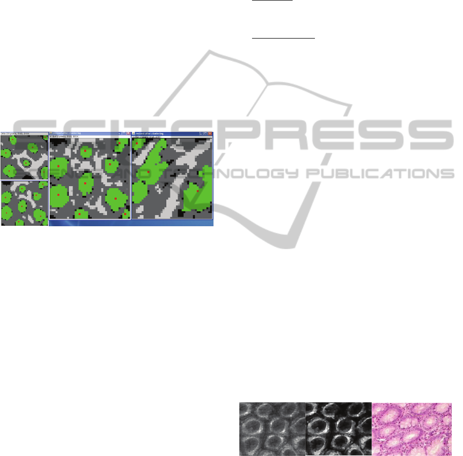
2) The modified density-based DBSCAN
algorithm, highlighted the various crypts and their
boundaries.
With the lbpSum value for every pixel, we apply a
clustering algorithm to cluster to a certain crypt. In
the clustering process we used a modified density-
based DBSCAN algorithm originally proposed in
(Sander et al., 2004). See Figure 3.
Figure 3: Clustering for gland identification. Normal (left)
and hiperplastic glands (right).
3.1.3 ISMP Feature Measurement
The results of this process allow us to extract and
measure certain features such as the silhouette
coefficient, the crypt compactness, the crypt
roundness or the inter-crypt distance.
3.1.4 ISMP-indexing and ISMP-retrieval in
Two Steps
1) We defined a feature vector and normalized it
applying linear scaling unit range normalization.
2) We retrieved similar images to a given one,
using the similarity function. The selected function
operated over the vector of selected features, whose
composition determines which is the nature of the
similarity being considered (similarity is relative in a
multidimensional space).
The test set demonstrated that the manhattan and the
euclidian distance, in combination with the linear
scaling unit range normalization, provide better
performance.
3.1.5 ISMP Data-base Query
MPQF queries were evaluated against one or more
multimedia databases which were unordered set of
Multimedia Contents-MC (combination of
multimedia data and its associated metadata).
1) Data-base: It was a dual database model (Figure
4) by content and by metadata. The MPQF operated
over sequences of evaluation items.
2) Condition Tree
: It was dual condition tree since
it (a) Combined filtering ele-ments (conditions) from
the BooleanExpressionType and (b) Interconnected
them with Boolean operators (AND, OR, NOT and
XOR).
The (image retrieve) IR-like condition used
QueryByExample query type and in-cluded the
Base64 encoding of the binary contents of a JPEG
image. The (data retrieve) DR-like condition
specified, in the present case, that the metadata field
FileSize must be less than 1000 bytes. Each
condition acted over a sequence of evaluation items
and, for each one, re-turned a value. For IR-like
conditions, returned any value in the range of [0.1].
For DR-like conditions returned 1 or 0 (true /false).
A threshold value within a condition was used to
indicate the minimum value the score to be
processed in the training set.
3.1.6 ISMP Image Retrieval
The ISMP retrieval system over the web interface
present de problem image for query and retrieve a
list of similar images (as many as possible) from the
data base.
3.2 Test Set
The web user interface used both 1) the query-by-
image in combination with 2) classic XML
metadata-based criteria.
Figure 4: ISMP retrieval in the Test set. Original OB
image (left), preprocessing (middle) and retrieved image
(right), in this case a histological image.
SOLO-MEDICINE IN OPTICAL BIOPSIES - A Way to Practice Telemedicine
443

Figure 5: ISMP retrieval in the Test set . Original OB
image (left) and retrieved image (right) only histological
images were retrieved.
The rate of adequate image retrieval from
normal, benign and hyperplasic images using the
threshold values indicated in the Section 3.1.5 was
90%.
4 DISCUSSION
The use of solo-medicine (Ferrer-Roca and
Marcano, 2009) will be a common practice in a near
future, and there-fore professional will require
support by any media, including mobile phone. Two
are the characteristics of this support: 1) is going to
be on line (books will be soon obsolete) and 2) it
will be required during patient interventions,
therefore the possibility to get access to mobile
phones is of paramount importance. The latter is
even more relevant considering that the majority of
this solo-medicine will be carried out in remote,
isolated or developing countries where satellite
mobile phones will be, probably, the only available
technology.
The solution brought in this paper merge the
standardization process of mass-used multimedia
standards with the medical image standardization
process, and built a ISMP (Image solo medicine
paradigm) architecture, that in the present paper was
applied to colon OBs (Optical Biopsies).
One of the main functionalities of the ISMP
system architecture is the ability to combine
conventional search criteria (keywords, metadata
ranges) with the direct usage of an example image
(query-by-example paradigm) to retrieve similar
precedent cases. In the data retrieve DR-like
conditions, the MPQF standard acts as a
conventional Boolean-based filtering language,
while with respect to (Information retrieve) IR-like
conditions MPQF acts preserving scores as a fuzzy-
logic system. The standard specifies the behaviour
of the provided Boolean operators in presence of
non-Boolean values.
The result showed that automatic feature-
extraction by image analysis on Black & White
images coming from the CLE, as well as colour
images from surgical specimens, reached the 90%
congruency. Thus indicating that the image-query
solution proposed in the ISMP architecture is an
adequate one to give professional support in
medicine at least in the normal and benign cases.
Nevertheless we have to test the borderline and
malignant ones to detect the sensitivity and
specificity of the proposed solution.
ACKNOWLEDGEMENTS
This work has been partly supported by the Spanish
government (TEC2008-06692-C02-01) and the
CATAI association.
REFERENCES
BMJ Group. Differential Diagnosis iPhone application.
<http://bestpractice.bmj.com/best-practice/marketing/
differentials.html>
CATAI 2010, En India: El hospital mas eficiente del
mundo. < http://catai.net/blog//2010/06/en-india-el-
hospital-mas-eficiente-del-mundo/>.
CATAI 2010, Anatomia Patologica en iPhone.
<http://catai.net/blog//2010/06/anatomia-patologica-
en-iphone/>.
Chen, N., Shatka, H., Blostein, D., 2006. Use of Figures in
Literature Mining for Biomedical Digital Libraries.
Dial, pp.180-197, Second International Conference on
Document Image Analysis for Libraries (DIAL'06).
Ferrer-Roca, O., 2009. Telepathology and optical biopsy.
Int J Telemed Appl Vol: 2009 Pages: 740712
doi:10.1155/2009/740712.
Ferrer-Roca, O., Marcano F., 2009. Anatomia Patologica
Digital. Control de calidad y patoinformatica.,
Rev.Esp.Patol 42(2): 85-85.
Ferrer-Roca, O., Marcano F., 2010. Computed assisted
microscopy. The era of the Small Size Virtual Slides
and the 4M microscopes. HEALTHINF 2010 Vol:
2010 Pp: 517-522. Portugal, ISBN: 978-989-674-018-
4.
Ferrer Roca, O., Duval V., Delgado J., Rolim C., Tous R,
2010 Query by image medical training. Optical Biopsy
with confocal endoscopy (OB-CEM). HEALTHINF
2010 Vol: 2010 Pp: 166-172. Portugal, ISBN: 978-
989-674-016-0.
Ferrer-Roca, O., 2010. Mobile phones in pathology.
J.Telemed & Telecare 16(3): 165.
Hersch W. R., Bhuptiraju R. T., Ross L., Johnson P.,
Cohen A. M., Kraemer D. F., 2005. TREC 2004
Genomics Track overview. Proc of TREC 2004 NIST
Special Publication <http://ir.ohsu.edu/genomics>.
HEALTHINF 2011 - International Conference on Health Informatics
444

ImageJ. <http://rsbweb.nih.gov/ij/>.
Ishii, N., Koike, A., Yamamoto, Y., Takagi T., 2008.
Figure Classification in Biomedical Literature
towards Figure Mining. bibm, pp.263-269, IEEE
InternationalConference on Bioinformatics and
Biomedicine, 2008.
Kiesslich R., Galle P. R., Neurath M. F., 2008. Atlas of
endomicroscopy. Springer-Verlag Heidelberg. ISBN
978-3-540-34757-6.
Pietikainen M., Ojala T. and Maenpaa T., 2000. Gray
Scale and Rotation Invariant Texture Classification
with Local Binary Patterns. In Proceedings of the
Sixth European Conference on Computer Vision
(ECCV2000, pages 404–420.
Sander, et al., 2000. Density-based Clustering In Spatial
Databases: The Algorithm Gdbscan and its
Applications. Data Min. Knowl. Discov., 2(2):169–
194.
Tous, R., 2008. Query formats for multimedia applications
ISO/IEC 15938-12 (MPEG Query Format) & ISO/IEC
24800 (JPSearch). In CATAI 2009: Super-resolution
and optical Biopsy. CATAI editions. Tenerife. Pp25-
32.
Wang, T. D., VanDam, J., 2004. Optical Biopsy: A New
Frontier in Endoscopic Detection and Diagnosis.. Clin
Gastroenterol Hepatol 2(9): 744–753.
SOLO-MEDICINE IN OPTICAL BIOPSIES - A Way to Practice Telemedicine
445
