
DEVELOPMENT OF A MECHANICAL INSTRUMENT
TO EVALUATE BIOMECHANICALLY THE SPINAL COLUMN
IN PREGNANT WOMEN
Cláudia Quaresma, Mário Forjaz Secca
CEFITEC, Dep. of Physics, Faculdade de Ciências e Tecnologia, UNL
Quinta da Torre, P-2829-516, Caparica, Portugal
João O’Neill
Faculdade de Ciências Médicas, UNL , Quinta da Torre, P-2829-516, Caparica, Portugal
Jorge Branco
Maternidade Dr. Alfredo da Costa, Lisboa, Portugal
Keywords: Biomechanics, Vertebral column, Non-invasive instrument, Evaluation, Biomechanical, Standing position.
Abstract: The incidence of problems related to rachialgiae is so frequent and usual that it must be studied as if it were
an epidemic and social disease (Knoplich, 2003). Instruments that evaluate the spinal column in a standing
position, in a global way, are needed to attain a better insight into this problem. This work presents a
completely safe instrument to assess the evolution of all the vertebra locations throughout time, in order to
study the biomechanical changes in women during pregnancy. A mechanical system, registered as Vertebral
Metrics, with the objective of evaluating the curvatures and lateral deviations of the spinal column in the
standing position was built. A measuring part, which is the body of the instrument, and a supporting part,
where the previous part is mounted, constitute the new non-invasive instrument. The measuring part
consists of 17 identical adjustable mechanical blocks that allow us to reproduce the position of each vertebra
of the spinal column, from the first cervical vertebra to the first sacral vertebra. Vertebral Metrics was
originally planned and built to be applied to pregnant women. However, after redefining dimensions of the
different parts it can be applied to any type of population, in the future.
1 INTRODUCTION
Rachialgiae constitute a relevant problem in modern
society (Alexandre & Moraes, 2001). In many
women this problem appears for the first time during
pregnancy and remains for the rest of their lives,
causing serious problems of absenteeism and
consequently a great loss under the point of view of
the economy of the country. It is necessary to have
instruments that evaluate, in a global way, the spinal
column in standing position, in order to understand
better the behaviour of the spinal column during the
gestational period.
There are many instruments to evaluate the
spinal column. However, most use ionizing radiation
and very few allow us to analyse the spinal column
in a standing position.
The instrument that is most used to evaluate the
spinal column in the standing position is the
radiograph. However, since it uses ionizing
radiation, it should not be used on pregnant women
(Harlick e al, 2007; Pinel-Giroux e tal, 2006) and,
besides, it would only give a collapsed two-
dimensional view.
The study presented here is part of a broader
analysis where we intend to identify and describe the
biomechanical alterations of the spinal column that
occur throughout pregnancy. We found it was
necessary to build a non-invasive instrument that
would allow the reproduction of the position of each
of the vertebrae, through the identification of each of
the vertebral apophyses of the spinal column, from
310
Quaresma C., Forjaz Secca M., O’Neill J. and Branco J. (2009).
DEVELOPMENT OF A MECHANICAL INSTRUMENT TO EVALUATE BIOMECHANICALLY THE SPINAL COLUMN IN PREGNANT WOMEN.
In Proceedings of the International Conference on Biomedical Electronics and Devices, pages 310-313
DOI: 10.5220/0001779903100313
Copyright
c
SciTePress
the first cervical vertebra to the first sacral vertebra.
In a global way Vertebral Metrics evaluates the
curvatures and lateral deviations of the spinal
column in the standing position.
After an exhaustive search of existing
instruments we found the inexistence of an apparatus
with the required characteristics, which led us to
prepare and elaborate an equipment to measure the
lateral deviations and curvatures of the spinal
column.
The elaboration started with the conceptual
design of the apparatus, based on some existing
instruments. Measurements on 134 women were
carried out in order to define its dimensions,
including the height of the body, the support, the
size of horizontal pieces and the stand of Vertebral
Metric. The experimental procedure of the
previously mentioned study was performed as
follows:
1- The length between various points of reference
was measured in each woman.
2 - The Normality test for each of the variables was
applied.
3 - Significance> 0.05 was found.
4 - The confidence intervals for 99% were calculated
The details of the mechanisms of each of the pieces
and the choice of the convenient materials to use
(polycarbonate, brass, steel and duralumin) to build
the instrument were defined afterwards.
Finally, Vertebral Metrics was built (Figure 1)
and the necessary adjustments were performed.
2 VERTEBRAL METRICS:
DESCRIPTION
AND FUNCTIONING
The objective of Vertebral Metrics, a non-invasive
mechanical instrument, is to identify the x, y and z
positions of each vertebra, from the first cervical
vertebra to the first sacral vertebra. After entering
these data into a mathematical model of the spine,
the curvatures and lateral deviations of that segment
in the standing position can be calculated.
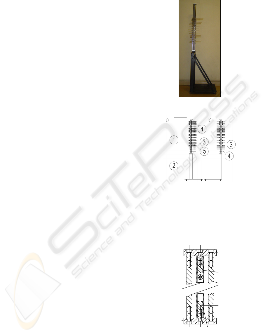
The components of the apparatus (Figure 2) are
the “body” (1) and the “support” (2).
The “body” has a vertical piece (5), mounted in
the support (2), with 18 horizontal pieces (4) called
“2D Positioner”. Two pieces, one vertical, where
the body of the instrument fits, and a stand where the
person to be evaluated stands up, constitute the
“support”.
Figure 1: Image of Vertebral Metrics.
Figure 2: Diagram of Vertebral Metrics.
Figure 2 presents two views of the whole
instrument, with (a) and (b) showing the frontal view
and the left lateral view, respectively.
The Vertical piece of the body (Figure 3)
consists of a rectangular profile with two plates at
the ends, which provide rigidity and the fixation to
the system.
Figure 3: Vertical piece of the body of Vertebral Metrics.
A detail of the instrument, is presented in Figure
4. The “2D Positioner” (4) slides along the scaled
ruler attached on the vertical piece (3).
DEVELOPMENT OF A MECHANICAL INSTRUMENT TO EVALUATE BIOMECHANICALLY THE SPINAL
COLUMN IN PREGNANT WOMEN
311
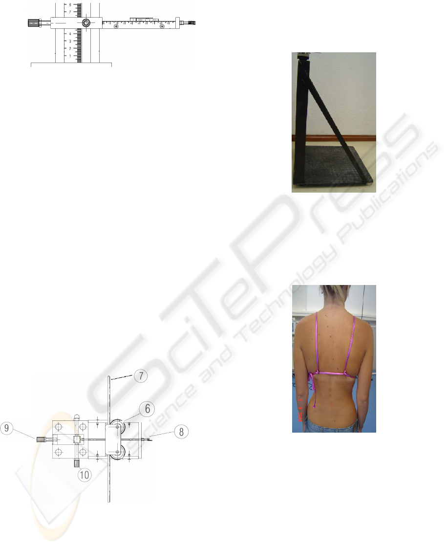
Figure 4: One of the “2D Positioner” and the vertical
piece of the body of the instrument.
Figure 5 shows components, 6, 7, 8, 9 and 10 of
the “2D Positioner. The component 7 is a square
sectioned rod, with a cone shaped tip, which is
where it makes contact with each vertebra apophysis
contact point. This rod is manoeuvred by two
pinions, (6), placed horizontally and touching the
rod via a neoprene O-ring surrounding the pinions. It
fixes the y position of the vertebral apophysis. This
coordinate is calculated through the distance from
the conical end, (5), to each “2D Positioner”, (4), of
the body of the instrument.
The pieces 6 and 7, moved by the horizontal
threaded rod (8), are fixed at the top by a plate and
two bolts. These bolts are inserted in two vertical
threaded holes, passing through the rotational axis of
the pinions 6.
At the fixed end of the rod 8 there is a small handle
which rotation enabling rotational movement of the
rod, which, in turn, allows movement of piece 7.
Measurements of the x position (see Figure 4) of the
vertebral apophysis of each of the vertebrae are then
possible.
Figure 5: Top view of “2D Positioner”.
The component 9 is a piece that fixes the “2D
Positioner” which moves upwards/downwards on
the vertical piece (3), as component 10 rotates.
Finally, coordinate z is obtained from the
measurement of the position of the horizontal part in
piece 4 (see Figure 1).
The support of Vertebral Metrics has two pieces:
one horizontal plate supported on four feet, where
the person to be evaluated stands, and one vertical
piece, shown in Figure 6, where the vertical part of
the body of the instrument (see Figure 2) fits.
Figure 6: The support of Vertebral Metrics.
After marking the vertebral apophyses (Figure 7),
with a washable pen, each pregnant woman stands
up on the stand of the support of the instrument with
the posterior face of the trunk facing the body of
Vertebral Metrics.
Figure 7: Marking the vertebral apophyses with a
washable pen.
All the 18 “2D Positioners” of the body of
Vertebral Metrics are identical and adjustable,
allowing the identification of the x, y and z positions
of the vertebral apophyses, from the first cervical
vertebra to the first sacral vertebra.
Three of the “2D Positioner” pieces are used
distinctively. The first is placed in the occipital
region and is used, during the data collection, as a
reference point. The second piece collects the data of
the cervical vertebra and the piece number fifteen
collects the data from the first, second and third
BIODEVICES 2009 - International Conference on Biomedical Electronics and Devices
312
lumbar vertebra. The remaining horizontal pieces
will identify the position of all other vertebral
apophyses of the spinal column (Fig 8).
The x, y and z positions of each “2D Positioner” are
then obtained.
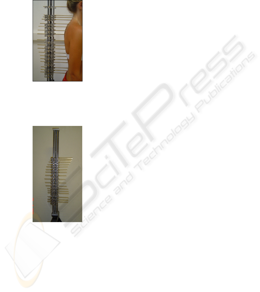
Figure 8: Example of application of Vertebral Metrics.
Each data collection lasts seven minutes. Figure 9
shows the position of the “2D Positioner” after
application of Vertebral Metrics.
Figure 9: After the application of Vertebral Metrics.
The collected data are then recorded and
transferred to a specific data basis with correction
factors associated with the instrument included. The
final data will then be inserted into the previously
mentioned mathematical model.
3 CONCLUSIONS
Vertebral Metrics is a non-invasive mechanical
instrument, which assesses the curvatures and lateral
deviations of the spine in a standing position. The
patent was registered and the study with pregnant
women was accepted by the Ethics Committees of
the Maternidade Dr Alfredo da Costa and Regional
Health Administration of Lisbon and Vale do Tejo.
The validation process for the instrument,
including the calculation of the correction factors
and uncertainties associated with it was
accomplished.
This instrument allows a global assessment of the
spine. Thus, identification of dysfunctions and / or
diseases of the spinal column in pregnant women,
will be shown on a thorough diagnosis. Intervention
programs, directly connected to specific problems of
each person, may then be elaborated and
implemented.
Vertebral Metrics was originally planned and
built to be applied to pregnant women. However, it
can be applied to any type of population after
redefining the dimensions of the different parts.
REFERENCES
Alexandre, N.; Moraes, M. (2001) Modelo de avaliação
fisio-funcional da coluna vertebral. Rev. Latino-am
Enfermagem Março, 9 (2); 67-75.
Knoplich, J. (2003) Enfermidades da coluna vertebral..,
Robe Editorial. São Paulo, 3ªed
Harrison et al (2005) Sagittal skin contour of the cervical
spine: interexaminer and intraexaminer reability of the
Flexicurve instrument. Journal of Manipulative and
Physiological Therapeutics. Vol 28, 7; 516-519
Harlick, J. Milosavljevic, S. (2007) Palpation
identification os spinus processes in the lumbar spine.
Manual Therapy 12, 56-62
Hinman, M. (2004) Comparison of thoracic kiphosis and
postural stiffness in younger and older women. Spine
Journal 4; 413-417.
Lee, Y.; Chen, Y. (2000) Regressionally determined
vertebral inclination angles of the lumbar spine in
static lifts. Clinical Biomechanics 15; 672-677.
Norton, BJ; Ellison, JB. (1993) Realiability and concurrent
validity os the Metrecom for length measurements on
inanimate. Phys. Ther. 73; 266-274.
Teixeira, FA; Caravalho, GA( 2007) Confiabilidade e
validade das medidas da cifose torácica através do
Método Flexicurva. Revista Brasileira de Fisioterapia.
Vol.11, 3; 199-204.
Pinel-Giroux, F.; Mac-Thiong, J.; Guise, J.; Labelle, H.
(2006) Computerized assessment of sagittal curvatures
of spine – Comparison between Cobb and tangent
circles techiques. J. Spinal Disord. Tech, vol.
19,7;507-512
Walsh e Breen (1995) Reability and validity of the
Metrecom Skeletal Analysis in the assessment os
sagittal plane lumbar angles. Clinical Biomechanics
vol. 10 nº4; 222-223
DEVELOPMENT OF A MECHANICAL INSTRUMENT TO EVALUATE BIOMECHANICALLY THE SPINAL
COLUMN IN PREGNANT WOMEN
313
