
TREATMENT OF MITRAL VALVE INSUFFICIENCY BY SHAPE
MEMORY POLYMER BASED ACTIVE ANNULOPLASTY
Pilar Lafont Morgado, Andrés Díaz Lantada, Héctor Lorenzo-Yustos, Julio Muñoz-García
División de Ingeniería de Máquinas – E.T.S.I. Industriales – Universidad Politécnica de Madrid
C/ José Gutiérrez Abascal, nº 2. 28006 – Madrid, Spain
Ignacio Rada Martínez, Antonio Jiménez Ramos, José Luis Hernández Riesco
Hospital Gómez Ulla
Glorieta del Ejército s.n. 28047 – Madrid, Spain
Keywords: Shape Memory Polymers (SMP), Mitral valve Insufficiency, Annuloplasty ring, Laser Stereolithography,
Silicone mould vacuum casting, Biomaterials.
Abstract: Active materials are capable of responding in a controlled way to different external physical or chemical
stimuli by changing some of their properties. These materials can be used to design and develop sensors,
actuators and multifunctional systems with a large number of applications for developing medical devices.
(for both surgery and implants).
Shape memory polymers are active materials with thermo-mechanical coupling (changes in temperature
induce shape changes) and a capacity to recover from high levels of distortion, (much greater than that
shown by shape memory alloys), which combined with a lower density and cost has favoured the
appearance of numerous applications. In many cases, these materials are of medical grade, which increases
the chances of obtaining biocompatible devices.
This paper presents the design, manufacture, “shape memory” programming process and in vitro trials of an
active annuloplasty ring for the treatment of mitral valve insufficiency, developed by using shape memory
polymers. This has been done with the collaboration betweeen researchers from Universidad Politécnica de
Madrid and doctors from the Hospital General Central de la Defensa.
1 MITRAL INSUFFICIENCY AND
POSSIBLE IMPROVEMENTS
IN ANNULOPLASTY
TREATMENT
1.1 Mitral Valve Insufficiency
The mitral valve is made up of two components
whose mission is to channel the blood from the left
auricle to the left ventricle. Firstly, there is the so-
called “mitral valve complex” comprising the mitral
annulus, the valve leaflets, and the commissures
joining both valves. Apart from the mitral valve
complex itself, this valve has the so-called “tensor
complex”, which in turn comprises the chordae
tendineae which continue with the papillary muscles
attached to the left ventricle.
A failure of any of these elements leads to
functional changes in the mitral apparatus, such as
mitral insufficiency, explained below, and
hemodynamic repercussions.
Mitral insufficiency (or regurgitation) is defined
as the systolic regurgitation of blood from the left
ventricle to the left auricle, due to incompetence in
mitral valve closing. This can arise for three main
reasons: a) primary disease of the mitral valve; b) an
anatomic or functional alteration in the chordae
tendineae or in the papillary muscles, and c) a
disorder in the correct function of the left auricle and
the left ventricle (Díaz Rubio, 1994).
Valve reconstruction is currently the preferred
treatment for mitral insufficiency provided this is
possible. With the aid of preoperative transesophagic
echocardiography lesions can be located and their
extent seen, so a surgeon can evaluate if valve repair
is possible and thus design an exact plan for any
17
Lafont Morgado P., Díaz Lantada A., Lorenzo-Yustos H., Muñoz-García J., Rada Martínez I., Jiménez Ramos A. and Luis Hernández Riesco J. (2008).
TREATMENT OF MITRAL VALVE INSUFFICIENCY BY SHAPE MEMORY POLYMER BASED ACTIVE ANNULOPLASTY.
In Proceedings of the First International Conference on Biomedical Electronics and Devices, pages 17-22
DOI: 10.5220/0001046400170022
Copyright
c
SciTePress

operation required. Nowadays, the object of this
surgery is not simply limited to eliminating mitral
insufficiency but in many cases to reconstructing the
geometry of the entire mitral valve apparatus to
ensure a durable repair.
Surgically restoring the geometry to normal
conditions consists in: a) augmenting or reducing the
abnormal leaflets; b) replacing broken or short
chordae tendineae using “Goretex” type sutures, and
c) annuloplasty.
1.2 Treating Mitral Insufficiency with
Annuloplasty
Carpentier’s description of a rigid prosthetic ring to
allow a selective reduction of the entire mitral
annulus opened the way to modern mitral repair.
Annuloplasty consists in inserting the said ring-
shaped device into the coronary sinus and after
applying traction, retraction or heat, it reduces its
perimeter, thereby reducing the mitral annulus and
improving the contact between the valve leaflets,
which leads to a reduction in the patient’s degree of
mitral insufficiency (Hernández, 2005).
Since then, a series of implants have been
developed that can be basically classified as rigid or
flexible and total or partial. Rigid monoplane
implants have been displaced due to the large
number of experimental and clinical works showing
that the perimeter of the mitral annulus constantly
changes in size and shape during the heart cycle.
The recent findings showing that these changes are
produced in a three-dimensional way with a
hyperbolic paraboloid shaped ring has given rise to
new rigid three-dimensional prosthesis. Duran
proposes replacing the most conventional devices
for other flexible or semi-rigid designs that
reproduce the three-dimensional shape, such as the
one marketed by Medtronic Inc..
1.3 Desirable Improvements
Employing Progressive Procedures
However, inserting a device to close the mitral valve
means making additional demands on the heart that
may lead to postoperatory problems. It would be
ideal to insert a ring with the same shape as the
patient’s mitral annulus and, when they have
recovered from the operation, progressively act on
this ring (in several stages) and remotely. This seeks
to maintain a balanced situation and not excessively
overload the patient’s heart during the operation.
In this way, the progressive closing of the
patient’s mitral annulus can be controlled and, by
using non-invasive inspection technologies, the
improvement in the patient’s mitral insufficiency
can be evaluated after each stage of the ring
actuation.
2 SOLVING MECHANICAL
OPERATION USING SHAPE
MEMORY POLYMER BASED
DEVICES
2.1 Shape Memory Polymers in
Medical Devices
Shape memory polymers (SMPs) are materials that
give a mechanical response to temperature changes.
When these materials are heated above their
“activation” temperature, there is a radical change
from rigid polymer to an elastic state that will allow
deformations of up to 300%. If the material is cooled
after manipulation it retains the imposed shape; it
“freezes”, the said structure returning to a rigid but
“non-equilibrium” state. When the material is heated
above its activation temperature, it recovers its
initial undeformed state.
The cycle can be repeated numerous times
without degrading the polymer and most suppliers
can formulate different materials with activation
temperatures ranging between –30 ºC and 260 ºC,
depending on the application required.
They are therefore active materials that present
thermomechanical coupling and
a capacity to recover
from high levels of distortion
, (much greater than
shown by shape memory alloys), which combined
with a lower density and cost has favoured the
appearance of numerous applications. Their
properties allow applications for manufacturing
sensors and actuators, especially for the aeronautic,
automobile and medical industry (Lendlein, Kelch,
2002).
The main problem associated with the use of
shape memory polymers is the lack of structured
processes for developing devices based on these
materials. The design process for these devices and
the transformation processes for these materials need
to be more thoroughly investigated.
The main advantages of shape memory polymers
are:
• They are new materials with the ability to
change their geometry from an initial
deformed shape to a second shape
predetermined during the manufacturing
process.
BIODEVICES 2008 - International Conference on Biomedical Electronics and Devices
18
• They are more economical than shape
memory alloys.
• Different additives can be used to change
their properties “a la carte”, to better adapt
them to the end application.
• The levels of deformation are much greater
than those obtainable using shape memory
alloys.
• They can also be more easily processed and
allow the use of “Rapid Prototyping
Technologies”, which speeds up the
production of devices.
• More complex geometries and actuators
can be obtained than with developments
based on shape memory alloy.
However, due to their recent appearance, in
many cases their mechanical and thermomechanical
properties are still not completely typified, which
gives rise to doubts concerning how devices based
on these materials will react. One of the basic aims
of current research is to increase knowledge of the
properties of these polymers by improving
characterization processes.
Regarding the development of medical devices,
both surgical and implantable ones, they have
additional advantages to those mentioned above:
• They are frequently medical grade
materials which increases the chances of
obtaining biocompatible devices.
• The combined use of preoperative
inspection technologies and CAD-CAE-
CAM technologies means that prostheses
and customised devices can be tailored for
patients.
• Their activation temperature and properties
can be adapted to the application, thanks to
the amount of copolymers employed and
the use of additives.
Among the medical devices developed that take
benefit from the advantages of these polymers, the
most notable are self-expanding stents, drug release
devices, thrombectomy devices, intelligent sutures
and active catheters (Lendlein, 2002, 2005, Wilson,
2006).
2.2 Shape Memory Polymers for Active
Annuloplasty
Commercial annuloplasty rings based on shape
memory polymers have been patented but not yet
developed.
The Sorin Group’s Memo 3D manages to reduce
its shape by using a shape memory alloy (Nitinol
type, similar to those used in the manufacture of
self-expanding stents). However, the change of
shape is produced during the operation itself on
making contact with human body temperature,
which means that no postoperative measures are
possible.
Besides, the capacity of shape memory polymers
to recover their shape against efforts of up to around
7 MPa means that a 3 mm thick annuloplasty ring,
similar to devices currently in use, manufactured
with these materials will be able to overcome a
circumferential force of between 4 N to 12 N that is
imposed by the patient’s mitral annulus.
In accordance with the above, what is proposed
is a ring made of shape memory polymer and
electrical resistances or heaters distributed inside to
activate the “shape memory effect” and therefore the
required shape change.
Firstly, the ring adapts to the end size required
(that needed to eliminate the mitral insufficiency)
and with the resistances already in place. The ring is
then uniformly heated to a temperature higher than
the transition temperature (situated for the end
product between 41 ºC and 43 ºC) and is forced to
take on the expanded transitory shape (to do this
cone-shaped tools can be used with a cross section
similar to that of the mitral annulus), letting it cool
down to room temperature. The device also consists
of a battery to power the resistances and heat them.
The rise in temperature of the resistances causes a
local rise in temperature, which, if suitably
controlled leads to a change in phase of the SMP and
the associated size reduction.
Using an associated electronic control enables
the resistances to be operated in pairs and at
different times, in order to carryout the progressive
or “step by step” operation required on the ring.
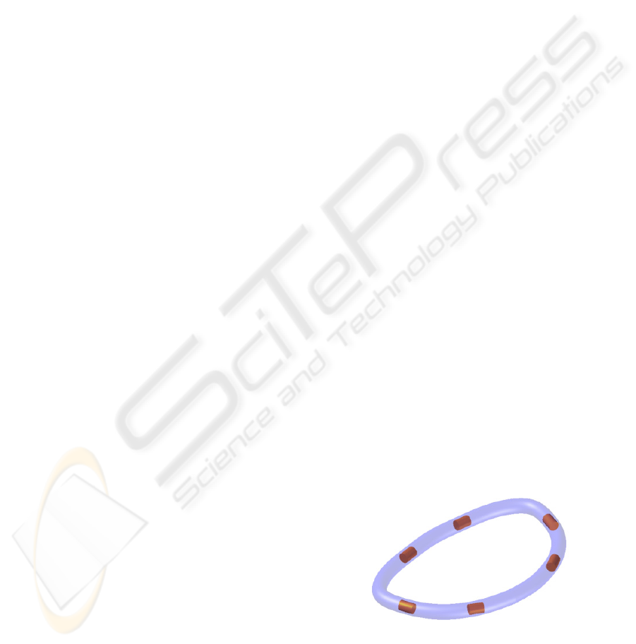
Figure 1 shows a preliminary design.
Figure 1: Preliminary active annuloplasty ring design.
SMP with internal resistances for heating.
A patent for this device was applied for by the
authors under the title of “Active annuloplasty
system for the progressive treatment of valve
insufficiencies and other cardiovascular pathologies”
TREATMENT OF MITRAL VALVE INSUFFICIENCY BY SHAPE MEMORY POLYMER BASED ACTIVE
ANNULOPLASTY
19
on 13 December 2006 with Application Number
P200603149 and is currently being evaluated by the
Spanish Patents and Trade Marks Office.
The following sections present the design
alternatives and the prototypes obtained, as well as
the first “in vitro” trials performed, the results, and
future recommendations for optimising the results.
The development has been carried out in
collaboration between researchers from from
Universidad Politécnica de Madrid and doctors from
the Hospital General Central de la Defensa.
3 DESIGN AND MANUFACTURE
OF PROTOTYPES
Computer aided design and calculation technologies,
(CAD – Computer Aided Design and CAE –
Computer Aided Engineering), have become an
essential tool for developing medical devices. They
enable alternative shapes and designs to be obtained
quickly, as well as making it easier to evaluate their
advantages by being able to analyse stress,
deformations, ergonomics or dynamic response.
They are also highly valuable for comparing and
selecting the different materials that can be used. In
addition, when combined with preoperative
inspection techniques, they serve to design
implantable devices tailored for the patient
measurements and simulate their implantation.
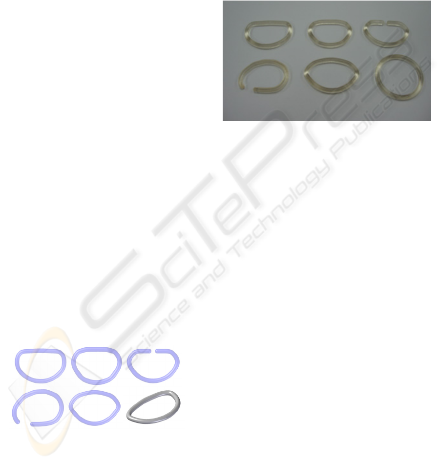
Figure 2 shows alternative designs for
annuloplasty rings made by using the “Solid Edge
v.18” computer design package. With the help of
these programmes it is very simple to change the
parameters of a design, which enables a shape to be
adapted to the size of a particular patient’s mitral
annulus or change the thickness of rings depending
on how long the device is required to last.
Figure 2: Alternative designs for annuloplasty rings
produced with CAD technologies.
Bellow is explained how prototypes are
manufactured from the designs shown and the
advantages of using rapid prototyping and rapid
tooling technologies.
Figure 3 shows the physical models obtained in
epoxy resin by laser stereolithography using an
SLA-350 machine available at the Product
Development Laboratory of Universidad Politécnica
de Madrid, from the designs shown in Figure 2.
Together with the annuloplasty designs, also shown
is a 3 mm thick, 30 mm outer diameter toroidal ring
to give the image an idea of scale.
Figure 3: Models obtained by laser stereolithography from
files containing the 3D part geometry.
The parts obtained by stereolithography are
particularly suitable for checking sizes, shapes and
appearance. They can also be used as models for
obtaining silicone moulds, which are subsequently
used to obtain polyurethane resin replicas, more
resistant and suited to working trials, and which also
possess shape memory properties. With the vacuum
casting process different types of bicomponent
resins can be used, with a wide range of properties,
and the prototypes obtained reproduce the mould
cavities with great precision (roughnesses of up to
50 μm) (Lafont, 2000).
The chosen material is a polyurethane resin from
MCP Iberia company with reference 3174 which is
supplied in bicomponent form, which means it can
be cast (after mixing the two components) in
silicone moulds to obtain the prototype shape
required.
It must be pointed out that shape change
temperature of the polyurethane resin used is not
suited to the “in vivo” end trials, nor fits the initial
specifications which required a range of 41 to 43 ºC
to activate the shape memory effect.
However, this polyurethane resin has been used
because it is easies to manipulate and cast in silicone
moulds, which enables prototypes to be made in less
than 5 days from computer file to end material.
Other mould manufacture technologies are
currently being used for casting alternative shape
memory polymers which do not attack the silicone
moulds and whose transition temperature can be set
from 4 to 6 ºC above that of the human body, suited
to the “in vivo”.
BIODEVICES 2008 - International Conference on Biomedical Electronics and Devices
20
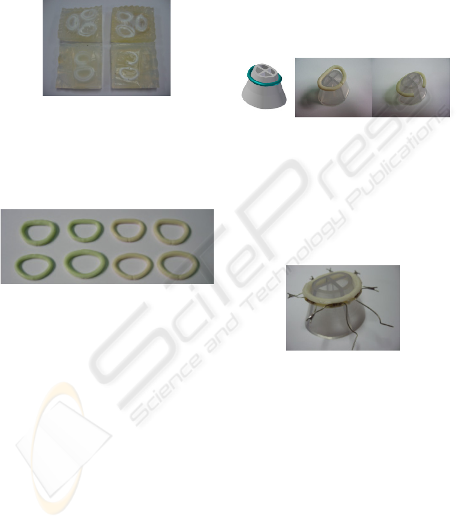
Figure 4 shows silicone moulds obtained from
the physical epoxy resin models displayed in Figure
3. These moulds enable prototypes to be obtained
from the material with shape memory properties.
Figure 4: Silicone moulds obtained from laser
stereolithographic models.
Enhanced design models have led to the
construction of new silicone moulds and the
obtaining of improved prototype annuloplasty rings,
both solid ones and with circumferential grooves for
housing the heating resistances. These are shown in
Figure 5.
Figure 5: Different polyurethane resin prototypes obtained
under vacuum casting in silicone moulds. Both open and
closed rings were made to analyse alternative
performances.
4 PROGRAMMING PROCESS OF
THE SHAPE MEMORY
When the annuloplasty rings have been shaped to
ensure the mitral valve closes properly, they need to
undergo heat deformation at 80 ºC in the case of
polyurethane resin, (higher temperature than that
needed to activate the shape memory effect), to
increase their cross section until it coincides with the
patient’s mitral valve annulus in the initial state of
insufficiency.
By doing this, a temporary shape is obtained and
the ring can be implanted without submitting the
patient’s heart to an additional overload due to a
sudden reduction in the section of the mitral valve.
After the surgical operation the recovery effect
of the original shape is activated, which produces a
gradual, controllable closure of the valve and a
controlled recovery of mitral regurgitation.
To perform this “shape memory programming
process”, tools were used that were obtained by laser
stereolithography in the form of a cone base with a
similar cross section to that of the patient´s mitral
annulus.
Figures 6 shows the tool and deformations
caused to ring prototypes thanks to the use of a
counter-shape that acts as a press on the tool and the
prototype.
Figure 6: Design and prototype of a tool for programming
shape memory effect. Deformation caused at 80 ºC to
obtain a temporary shape.
Figure 7 shows an annuloplasty ring with the
temporary shape already applied and prepared for
implant and the first “in vitro” trials. With the aid of
a cone base a 15% increase in cross section was
produced (maximum inner diameter ring size pass
from 26 to 28 mm), which will be used to evaluate
the subsequent shape memory recovery in “in vitro”
trials.
Figure 7: Active ring with heating resistances with the
temporary shape already applied. It is ready to be
implanted and subsequently activated.
5 RESULTS OF “IN VITRO”
TRIALS
For performing the first “in vitro trials” two pig
hearts were used because of their similarity to
human ones, as is demonstrated by their being used
for biological valve replacement operations.
Figure 8 shows the process for activating the
shape memory process in the ring and reducing the
associated mitral ring cross section.
The 4,7 Ω resistances (a total of 7 resistances
with serial connection) were supplied with power by
a 12 V transformer. Thus an intensity of 364 mA
was obtained, similar to what can be supplied by
TREATMENT OF MITRAL VALVE INSUFFICIENCY BY SHAPE MEMORY POLYMER BASED ACTIVE
ANNULOPLASTY
21

implantable commercial devices. The images (left to
right) show a 10.7% reduction in cross section
during an operating period of 150 seconds. This
means a 71%, recovery compared to effort since the
increase in cross section induced was 15%.
By interrupting heating the temperature
decreases and the recovery process is halted, which
means the required effect can be obtained step by
step. By recommencing the heating process the
recovery will continue, although in these first “in
vitro” trials heating was done continuously in order
to evaluate the maximum recovery that could be
obtained and the duration of the entire process.
Temperature was continuoustly measured using a
thermocouple.
Error!
Figure 8: Activating the shape memory effect using
heating resistances.
Despite it being desirable a cross section
reduction of 15% to 20%, it is very important to
point out the material’s capacity for recovery while
overcoming the forces imposed by the mitral
annulus of the hearts used.
6 FUTURE IMPROVEMENTS
AND CONCLUSIONS
For the postoperative and progressive treatment of
mitral insufficiency the use of an annuloplasty
device made of shape memory polymer has been
proposed. It has electrical resistances distributed
inside it to activate the “shape memory” effect, so
that the required change in shape to reduce mitral
regurgitation can be progressively induced. This
provides an alternative to current devices, that do not
permit any change of shape after implantation, and
therefore any errors committed during the operation
cannot be corrected.
The design, manufacturing, “shape memory”
effect programming and “in vitro” trials of such an
annuloplasty ring for treating mitral insufficiency,
developed by using shape memory polymers, have
been presented.
This has been done in collaboration between
researchers from from Universidad Politécnica de
Madrid and doctors from the Hospital General
Central de la Defensa.
Using computer aided manufacture and design
technologies has enabled different designs and
prototypes to be produced in parallel, as well as
rapid improvements to obtain the devices that were
used in the in vitro trials.
Future actions regarding improvements in the
shape memory programming process should lead to
optimising the reduction in mitral ring cross section
up to the required 15% to 20%. Using alternative
shape memory polymers with a lower activation
temperature will also result in more suitable devices,
since they will require a smaller size heating system
and will be easier to manufacture.
However, it is very important to point out the
material’s capacity for recovery against the forces
imposed by the mitral annulus of the hearts used,
which shows the feasibility of developing an active
annuloplasty system based on the use of shape
memory polymers.
REFERENCES
Díaz Rubio, M., Espinós, D., 1994. Tratado de Medicina
Interna. Editorial Médica Panamericana.
Carpentier, A., 1983. Cardiac Valve Surgery – The French
Correction. Journal of Thoracic and Cardiovascular
Surgery.
Duran, C., 1992. Duran Flexible Annuloplasty Repair of
the Mitral and Tricuspid Valves: Indications, Patient
Selection, and Surgical Techniques Using the Duran
Flexible Annuloplasty Ring. Medtronic Inc..
Hernández, J.M., 2005. Manual de Cardiología
Intervencionista. Sociedad Española de Cardiología.
Sección de Hemodinámica y Cardiología
Intervencionista.
Lendlein, A., S. Kelch, 2002. Shape-Memory Polymers.
Angewandte Chemie International.
Lendlein, A., Langer, R., 2002. Biodegradable, elastic
shape-memory polymers for potential biomedical
applications. Science.
Lendlein, et al., 2005. Light-induced shape-memory
polymers. Nature.
Wilson,T., et al., 2006. Shape Memory Polymer
Therapeutic Devices for Stroke. Lawrence Livermore
National Laboratory.
Lafont, P., Lorenzo, H. et al., 2000. Rapid Tooling:
moldes rápidos a partir de estereolitografía. Revista de
plásticos modernos.
BIODEVICES 2008 - International Conference on Biomedical Electronics and Devices
22
