
A STUDY OF TWO COLOR SYSTEMS USED IN
CONTENT-BASED IMAGE QUERY ON MEDICAL IMAGERY
Liana Stanescu, Dumitru Burdescu, Cosmin Stoica and Marius Brezovan
University of Craiova, Faculty of Automation, Computers and Electronics
Keywords: Image feature extraction, image processing, content-based visual quer
y, color, histogram, HSV color space,
l color space, medical images.
Abstract: The article presents a comparative study over two methods used in content-based visual query. The two
methods refer at two different color systems used for representing color information from images: HSV
quantized to 166 colors and l1l2l3 quantized to 64 colors. The originality of the study comes from the fact
that it was made on a database with medical images from digestive tract area, captured by an endoscope.
The scope was to check the quality of the content-based visual query on images representing five different
diagnoses (colitis, ulcer, esophagitis, polyps and ulcerous tumor) and taking into consideration that some
parameters were modified during the capturing process: viewing direction, intensity and direction of the
illumination, parameters that affect mostly the medical images captured during the diagnosis process.
1 INTRODUCTION
As the world is in the middle of the digital era, the
quantity of visual information is increasing (Sebe
and Lew, 2001). More than 2700 digital pictures are
made in every second (in total 85 billion images
yearly). For example, PhotoWorks includes tens of
millions of images on its web site. The Internet
allows us to have access to a big part of these
images. The common images are completed by
images with special purpose, like medical images
with an estimation of 2 billion per year. Because of
the tendency for digital (television, movies) and
because everybody will have access to everything,
the number of images will be increasing. The world
production of digital information in 2007 is
estimated to be more than 10
9
GB (250 MB for each
man on the planet, ignoring his technological
development). It is estimated that in the next 10
years, each of us will manipulate terabytes of
information (video, static images, music, photos and
documents) every day.
These image databases are associated with the
problem of the content-based retrieval, solved in two
steps (Sebe and Lew, 2001).
In the first step, when inserting a new image, it
will be
pre-processed and some features will be
automatically extracted: color, texture and shape.
The result will be a characteristics vector that will be
stored in the database.
In the second step the content based retrieval is
made, by choosing a query
image, calculating the
characteristics vector, comparing this vector with
each vector of the images stored in the database and
viewing the most similar images.
The color is one of the base image properties. In
content based retrieval on color feature it is desired
to find the images from the database having the
color composition closest to the color composition
of the query image (Del Bimbo, 2001, Gevers and
Smeulders, 1999).
The color content of an image is best represented
by
color histograms.
Comparing color histograms of the query image
and
target image is done by histograms intersection
or by the quadratic distance between histograms that
takes into consideration the conceptual similitude
between two colors (Sebe and Lew, 2001, Smith,
1997).
As was said before, one of the domains where a
large numb
er of images are accumulated is the
medical domain. The advantages of using content-
based visual query on medical images are on the
following directions (Muller et al, 2004):
• Medical teaching
• Medical resea
r
ch
• Diagnostic aid
• Electronic patient records
337
Stanescu L., Burdescu D., Stoica C. and Brezovan M. (2007).
A STUDY OF TWO COLOR SYSTEMS USED IN CONTENT-BASED IMAGE QUERY ON MEDICAL IMAGERY.
In Proceedings of the Fourth International Conference on Informatics in Control, Automation and Robotics, pages 337-340
DOI: 10.5220/0001621303370340
Copyright
c
SciTePress

The medical images are being produced directly
by medical equipment used in patient diagnosis, or
by digitalizing the images stored on other devices.
In each of these methods some changes can
occur:
• Changes in viewing direction
• Changes in direction of the illumination
• Changes in the intensity of the illumination
As a result, the purpose of the paper is to make a
comparative study of the content-based query results
effectuated on medical images database where the
color information is represented by HSV and l1l2l3
color systems.
The originality of the study is given by the fact
that the experiments are made on medical images
from digestive area produced by an endoscope. The
ill area is seen from different directions and in
different illumination intensity. This study, unlike
the others made on CorelDraw images, uses images
produced in real condition, in patient diagnosis.
The paper has the following structure: In section
2 the two color systems are presented. In section 3
the conditions and the results of the experiments are
presented, and in section 4 the conclusions of the
comparative study are discussed.
2 CONTENT-BASED IMAGE
QUERY ON COLOR FEATURE
The color is the visual feature that is immediately
perceived on an image. The color space used for
representing color information in an image has a
great importance in content-based image query, so
this direction of research was intensely studied (Del
Bimbo, 2001).
There is no color system that it is universal used,
because the notion of color can be modeled and
interpreted in different ways. Each system has its
own color models that represent the system
parameters (Gevers, 2004).
There were created several color spaces, for
different purposes: RGB (for displaying process),
XYZ (for color standardization), rgb, xyz (for color
normalization and representation), CieLuv, CieLab
(for perceptual uniformity), HSV (intuitive
description) (Gevers, 2001, Gevers, 2004). The color
systems were studied taking into consideration
different criteria imposed by content-based visual
query (Gevers and Smeulders, 1999):
• The independence of the imaging device
• Perceptual uniformity
• Linear transformation
• Intuitive for user
• Robust against imaging conditions: invariant to a
change in viewing direction, invariant to a
change in object geometry, invariant to a change
in direction and intensity of the illumination and
invariant to a change in the spectral power
distribution of the illumination.
The studies have shown that two of these color
systems can be used, with good results in a content-
based visual query process, namely HSV and l1l2l3
(Gevers et al, 2006).
It was proved that the HSV color system has the
following properties (Gevers, 2004):
• It is close to the human perception of colors
• It is intuitive
• It is invariant to illumination intensity and
camera direction
The studies made on nature and medical images
have shown that in the case of the HSV, RGB and
CieLuv color systems, the HSV color space
produces the best results in content based retrieval
(Stanescu et al, 2006).
Still, the HSV color space has several problems
(Gevers, 2004) :
• Nonlinear (but still simple) transformation from
RGB to HSV
• Device dependent
• the H component becomes instable when S is
close to 0
• the H component is dependent of the
illumination color
Gevers and Smeulders have proposed a new
color system, named l, whose components are
defined using the equations (Gevers and Smeulders,
1999):
(1)
Where R, G, B are the color values in the RGB
color space. They also showed that the l color
system is invariant to viewing direction, illumination
direction and intensity. In this case it is also a
nonlinear, but simple, transforming from RGB space
to l space.
In case of medical images the main problems are
regarding changing illumination intensity and
viewing direction. That is why the two color spaces
presented above are chosen.
222
2
)()()(
)(
),,(1
BGBRGR
GR
BGR
−+−+−
−
=
l
222
2
)()()(
)(
),,(2
BGBRGR
BR
BGR
−+−+−
−
=
l
222
2
)()()(
)(
),,(3
BGBRGR
BG
BGR
−+−+−
−
=
l
ICINCO 2007 - International Conference on Informatics in Control, Automation and Robotics
338

The operation of color system quantization is
needed in order to reduce the number of colors used
in content-based visual query: from millions to tens.
The quantization of the HSV color space to 166
colors, solution proposed by J.R. Smith, is the idea
used in this study (Smith, 1997). For the color space
l1l2l3 the solution of quantization to 64 colors is
chosen, keeping 4 values for each component of the
system. The fact that a color system is quantized to
166 colors and the other to 64 colors does not
influence the quality of the content-based image
query process, the research studies showing clearly
this aspect (Stanescu et al, 2006). The color
histograms represent the traditional method of
describing the color properties of the images. They
have the advantages of easy computation and up to
certain point are insensitive to camera rotating,
zooming, and changes in image resolution (Del
Bimbo, 2001). In case of both color systems, to
compute the distance between the color histograms
of the query image and the target image, the
intersection of the histograms is used (Smith, 1997).
The studies have also shown that using this metric in
content-based visual query gives very good results
as quadratic distance between histograms that is
more difficult to calculate (Smith, 1997, Stanescu et
al, 2006).
3 EXPERIMENTS
The experiments were performed in the following
conditions.
A database with 520 color images from the field
of the digestive apparatus was created. The images
are from patients with the following diagnosis:
polyps, ulcer, esophagitis, ulcerous tumors and
colitis. For each image there are several images with
affected area captured from 3 or 4 viewing
directions. For each image in the database there is
another identical image, but having the illumination
intensity changed.
A software tool that permits the processing of
each image was created. The software tool executes
the following steps:
1. the transformation of image from RGB
color space to HSV color space and the
quantization to 166 colors
2. the transformation of image from RGB
color space to l1l2l3 color space and the
quantization to 64 colors
3. calculation of the two color histograms
with 166, respectively 64 values, that
represent the characteristics vectors and
storing them in the database
In order to make the query the procedure is:
• a query image is chosen
• the dissimilitude between the query image
and every target image from the database is
computed, for each two specified criteria
(color histograms with 166 colors and the
color histogram with 64 colors);
• the images are displayed on 2 columns
corresponding to the 2 methods in
ascending order of the computed distance.
For each query, the relevant images have been
established. Each of the relevant images has become
in turn a query image, and the final results for a
query are an average of these individual results.
The experimental results are summarized in table
1. Method 1 represents the query using the HSV
color space quantized at 166 colors and Method 2
represents the query on color using the l1l2l3 color
space quantized at 64 colors. The values in the table
represent the number of relevant images of the first 5
images retrieved for each query and each of the
methods, as an average of the values obtained on
each executed query.
Table 1: Experimental results.
It must be mentioned that the queries were made
for each of the 5 diagnostics in part. The notion of
relevant image was strictly defined. The images
from the same patient captured at different
illumination intensity and from different points of
view were considered relevant for a query, and not
the ones with the same diagnosis. The quality of the
content-based image query process was strictly
analyzed. In figure 1 there is an example of content-
based image query considering the two specified
methods for images categorized as colitis. The first
column contains 5 images retrieved by Method1 and
the second contains the images retrieved using
Method2. In the first case there are 5 relevant
images and in the second case, 4 relevant images.
4 CONCLUSION
The paper presents the condition in which the
quality of the content-based visual query process
was studied, using a collection of medical images
from digestive tract. The quality was measured
Query Method 1 Method 2
Polyps 3.6 3.2
Colitis 3.5 3.1
Ulcer 3.2 2.9
Ulcerous Tumor 3.5 3.1
Esophagitis 3.4 3.1
A STUDY OF TWO COLOR SYSTEMS USED IN CONTENT-BASED IMAGE QUERY ON MEDICAL IMAGERY
339
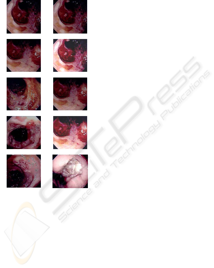
Figure 1: The retrieved images using the two specified
methods.
calculating the precision and recall parameters. HSV
system, quantized to 166 colors and l1l2l3 color
system quantized to 64 colors were considered
highlighting the way they influence the process of
content-based visual query if some important
parameters that often affects medical images are
modified: viewing direction, direction and intensity
of the illumination.
Several conclusions can be formulated after the
experimental results were analyzed:
1. to find images representing the same ill area,
that were captured by an endoscope from
several viewing directions, the solution that
uses HSV color system quantized to 166
colors gives the best results
2. for images representing the same ill area,
captured to different illumination intensities,
the solution that uses l1l2l3l color system
quantized to 64 colors, gives the best results
in querying process
3. globally, the solution that uses HSV color
space gives most satisfying results, because
the database includes both types of images
In general, for medical images, the first case,
with images representing ill area captured from
different angles is the most frequent case. So, that is
why the use of HSV color space, quantized to 166
colors, is recommended. The situation in the
database that was studied was the same, namely, the
number of images captured from different angles
was higher than the number of images where only
the illumination intensity was different.
In the future the study will be extended by using
a bigger database with much more images in order to
see if this conclusion will be also confirmed. New
experiments with images from other parts of the
human body or images produced by other medical
devices will be effectuated.
REFERENCES
Del Bimbo, A., 2001. Visual Information Retrieval,
Morgan Kaufmann Publishers. San Francisco USA.
Gevers, T., Smeulders, W.M., 1999. Color-based object
recognition. Pattern Recognition. 32, 453-464
Gevers, T., 2001. Color in Image Search Engines. In
Principles of Visual Information Retrieval. Springer-
Verlag, London.
Gevers, T., 2004. Image Search Engines: An Overview. In
Emerging Topics in Computer Vision. Prentice Hall.
Gevers, T., Van de Weijer. J., Stokman, H., 2006. Color
Feature Detection. In Color Image Processing:
Methods and Applications. CRC Press.
Muller, H., Michoux, N., Bandon, D., Geissbuhler, A.,
2004. A Review of Content_based Image Retrieval
Systems in Medical Application – Clinical Benefits
and Future Directions. Int J Med Inform. 73(1)
Sebe, N., Lew, M., 2001. Color-based retrieval. Pattern
Recognition Letters. 22, 223-230
Smith, J.R., 1997. Integrated Spatial and Feature Image
Systems: Retrieval, Compression and Analysis, Ph.D.
thesis, Graduate School of Arts and Sciences.
Columbia University.
Stanescu, L., Burdescu, D.D., Ion, A., Brezovan, M.,
2006. Content-Based Image Query on Color Feature in
the Image Databases Obtained from DICOM Files. In :
International Multi-Conference on Computing in the
Global Information Technology. Bucharest. Romania
ICINCO 2007 - International Conference on Informatics in Control, Automation and Robotics
340
