
A SIMPLE SCHEME FOR CONTOUR DETECTION
Gopal Datt Joshi
Center for Visual Information Technology, IIIT Hyderabad
Gachibowli, Hyderabad, India-500032
Jayanthi Sivaswamy
Center for Visual Information Technology, IIIT Hyderabad
Gachibowli, Hyderabad, India-500032
Keywords:
Contour detection, Surround suppression, Primary visual cortex, Human visual system.
Abstract:
We present a computationally simple and general purpose scheme for the detection of all salient object con-
tours in real images. The scheme is inspired by the mechanism of surround influence that is exhibited in
80% of neurons in the primary visual cortex of primates. It is based on the observation that the local context
of a contour significantly affects the global saliency of the contour. The proposed scheme consists of two
steps: first find the edge response at all points in an image using gradient computation and in the second step
modulate the edge response at a point by the response in its surround. In this paper, we present the results of
implementing this scheme using a Sobel edge operator followed by a mask operation for the surround influ-
ence. The proposed scheme has been tested successfully on a large set of images. The performance of the
proposed detector compares favourably both computationally and qualitatively, in comparison with another
contour detector which is also based on surround influence. Hence, the proposed scheme can serve as a low
cost preprocessing step for high level tasks such shape based recognition and image retrieval.
1 INTRODUCTION
Contour detection in real images is a fundamental
problem in many computer vision tasks. Contours are
distinguished from edges as follows. Edges are vari-
ations in intensity level in a gray level image whereas
contours are salient coarse edges that belong to ob-
jects and region boundaries in the image. By salient
is meant that the contour map drawn by human ob-
servers include these edges as they are considered to
be salient. However, the contours produced by differ-
ent humans for a given image are not identical when
the images are of complex, natural scenes. In such
images, multiple cues are available for the human vi-
sual system (HVS) - low level cues such as coherence
of brightness, texture or continuity of edges, interme-
diate level cues such as symmetry and convexity, as
well as high level cues based on recognition of fa-
miliar objects. Even if two observers have exactly
the same set cues, they may choose contours at vary-
ing levels of granularity. Thus saliency of an edge
is a subjective matter and varies accordingly. Nev-
ertheless, the fact remains that a contour map drawn
by human observers is sparser than an edge map de-
rived by processing the digital image. This can be
seen from Fig 1(a) which shows a test image and the
corresponding ground truth data (Fig. 1(b))indicating
the contours considered relevant by a human observer
(dat, 2003). If we compare this with the edge maps
in Fig. 1(c), (d) extracted by a Canny detector we can
observe that the contour map is sparse. This is de-
spite selecting a low scale (to capture gross informa-
tion) and using two different thresholds for the edge
detection. In general, a contour map is an efficient
representation of an image since it retains only salient
information and hence is more valuable for high level
computer vision tasks. The design of a detector that
can extract all contours from a wide range of images
is therefore of interest.
The key to extracting contours appears, from the
ground truth, to be the ability to assess what is
relevant and what is not in a local neighbourhood.
For instance, the grassy texture has been rejected in
the ground truth while the edges defining the ele-
phants’ feet have been retained. An assessment-based
strategy has been attempted to contour detection us-
ing local information around an edge such as im-
age statistics, topology, texture, colors, edge con-
tinuity, density, etc. Specifically, these approaches
have used statistical analysis of gradient field (Meer
236
Datt Joshi G. and Sivaswamy J. (2006).
A SIMPLE SCHEME FOR CONTOUR DETECTION.
In Proceedings of the First International Conference on Computer Vision Theory and Applications, pages 236-242
DOI: 10.5220/0001374702360242
Copyright
c
SciTePress

(a) (b)
(c) (d)
Figure 1: Demonstration of texture as a problem in contour
detection process. (a) Image of elephants (b) Ground truth
image. Canny edge map using low scale and low threshold,
(c) low scale and high threshold.
and Georgescu, 2001), anisotropic diffusion (Perona
and Malik, 1990; Black et al., 1998), complementary
analysis of boundaries and regions (Ma and Manju-
nath, 2000) and edge density information (Dubuc and
Zucker, 2001). These approaches, by design, are very
extensive in computation.
The HVS is capable of extracting all important
contour information in its early stages of processing.
Some attempts have also been made to model con-
tour detection in the HVS. One such model assumes
that saliency of contours arise from long-range in-
teraction between orientation-selective cortical cells
(Yen and Finkel, 1998). This model accounts for a
number of experimental findings from psychophysics
but is computationally intensive and its performance
is unsatisfactory on real images. Another model also
emphasises the role of local information and focuses
on cortical cells which are tuned to bar type features
(Grigorescu et al., 2003). This scheme computes ori-
ented Gabor energy at a single scale and over twelve
different orientations followed by a non-classical re-
ceptive field (non-CRF)
1
inhibition. Results of testing
1
The classical receptive field (CRF) is, by definition,
the area within which one can activate an individual neu-
ron. The region beyond this area which can modulate the
response of the concerned neuron is called a non-classical
receptive field.
of this scheme on images of animals in their natural
habitat, are reasonably good. However, the scheme
is computationally expensive and produces a contour
map which is quite sparser than an edge map though
not as sparse as the ground truth (contour map). In
this paper, we seek to find a solution to contour detec-
tion which is computationally low in cost as well as
effective on a wide range of images including natural
images. The paper is organized as follows: section 2
presents the development and details of the scheme;
section 3 proposes an implementation of the scheme;
section 4 summarises the results and section 5 draws
some conclusions based on the performance of the
proposed scheme.
2 PROPOSED SCHEME
The human visual system, in its early stages of
processing, differentiates between isolated bound-
aries such as object contours and region boundaries,
on the one hand, and edge in group, such as those
in texture, on the other hand. This is accomplished
in a series of processing stages. At the retinal level,
the ganglion cells process the visual input from rods
and cones to produce an image similar to that of
an edge detector used in computer vision (Marr and
Hildreth, 1980). The ganglion cells signal the spa-
tial differences in the light intensity falling upon the
retina. Their receptive field is organized into a center-
surround fashion, in which the excitatory and in-
hibitory subfields are integrated into circularly sym-
metric regions. The classical work in (Marr and
Hildreth, 1980) modelled this receptive field with a
Laplacian of Gaussian function. At the output of this
stage (retina), the visual system provides an efficient
representation of the image by removing redundant
information such as uniform light intensity on adja-
cent retinal locations. The axons of the ganglion cells
project to an area in the brain called the Lateral Genic-
ulate Nucleus (LGN). This area has no known filter
function but serves mainly to project binocular visual
input to various sites, especially to the visual cortex.
In the visual cortex, Hubel and Wiesel (Hubel and
Wiesel, 1962) found simple and complex cells in cat
primary visual cortex (area V1) that are selective
to intensity changes in specific orientation. These
orientation-selective cells are organized in columns,
in which all cells in a column have the same pre-
ferred orientation, and adjacent columns have incre-
mental change in orientations (Hubel and Wiesel,
1962). The orientation selectivity of cells is accom-
plished through a spatial summation of the inputs
from LGN. This leads to interesting features. For
example, complex cells differ from simple cells by
showing less specificity regarding the position of the
A SIMPLE SCHEME FOR CONTOUR DETECTION
237
stimulus. Computationally, this functionality can be
modeled using input from the simple cells.
Figure 2: Contour detection scheme in (Grigorescu et al.,
2003).
Other classes of cells besides the simple and com-
plex cells also exist in V1. These are the end stopped
cells (Dobbins et al., 1987), bar (contour) cells (Bau-
mann et al., 1997), and grating cells (von der Heydt
et al., 1991). Neurophysiological measurements on
cells have showed that the response of an orientation-
selective neuron to an optimal stimulus in its recep-
tive field is reduced if the stimulus extends to the
surround. This effect is termed as surround inhibi-
tion and it is exhibited in a majority (80%) of the
orientation-selective cells in the visual cortex of pri-
mates(Knierim and van Essen, 1992). In general,
an orientation-selective cell with surround- inhibition
will respond most strongly to a single bar, line, or
edge in its receptive field and will show reduced re-
sponse when more bars are added to the surroundings
like sine wave gratings. These type of cells was called
the bar cell, referring to the preference of the cell for
bars versus gratings. These cells were the source of
inspiration for the contour detector reported in (Grig-
orescu et al., 2003). The detection scheme is based
on fitering the input image with a Gabor filter bank;
summing the Gabor energy output at 12 different ori-
entations and then applying surround inhibition. This
scheme is shown in Fig. 2. The Gabor filtering stage
is essentially a local energy computation stage (Mor-
rone and Burr, 1988). Edge and line features are
signalled by local maximas in the local energy map.
Hence, the input to the surround inhibition stage in
Fig. 2 is edge information.
Edge information can also be derived using a gra-
dient computation. The difference between edge de-
tection using gradients as opposed to local energy is
that the latter is capabile of detecting step/impulse
discontinuities as well as ramps and other luminance
profiles in the image. Most of the edges in natural
images have step profiles which can be effectively
picked by the gradient computation. A drawback of
Gabor based commutation is that it leads to a poor
localization since the window over which it is com-
puted needs to be wide enough to attain a good fit to
the Gabor profile. Furthermore, local energy compu-
tation is far too expensive (12×2=24 filters) compared
to gradient computation which needs to be done only
in two orthogonal directions (2 filters) to determine
edges at various orientation. Hence, we argue that
a simpler scheme for contour detection would be to
derive the edge information from a gradient compu-
tation followed by an assessment based on the local
context.
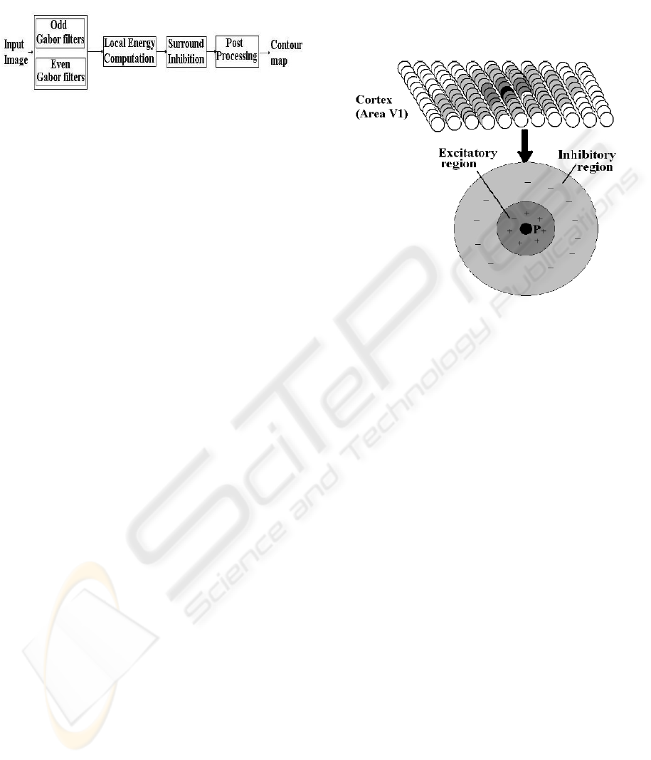
Figure 3: Neighboring cells profile of a cortical cell in area
V1.
Next, we turn our attention to the second part of
the contour detection scheme, namely assessment of
the edge information. This assessment needs to be
done based on the local context and a surround in-
hibition mechanism has been used for this purpose
in (Grigorescu et al., 2003). A recent neurophysi-
ological study (Cavanaugh et al., 2002) of cells in
V1 has however shown that the surround region actu-
ally can have an excitatory influence in addition to an
inhibitory influence. Specifically, the findings about
neuronal behaviour can be summarised as follows: (i)
Every cell responds to a stimulus if it falls on its cen-
tral region (CRF). This is shown by the circle P in
Figure 3; (ii) Besides the CRF, a neuron has a sur-
round region which is made up of two parts, namely
an inner annular shaped region (shown in dark gray
in Figure 3) which is excitatory and an outer annu-
lar shaped region (shown in light grey in Figure 3)
which is inhibitory; (iii) The surround region which
can influence the response of a neuron is limited in
extent. The behavior of these neurons give us a clue
about the role of local context in the visual perception
of a stimulus which is obtained by combining exci-
tatory and inhibitory influences. We draw inspiration
from this study and propose a surround mechanism
with excitatory and inhibitory components. An con-
tour detection scheme which computes the gradient
information first and then employs this second step
will not respond (sufficiently) to edges which belong
to texture regions. Such a scheme is easy to imple-
ment as well.
VISAPP 2006 - IMAGE ANALYSIS
238
3 IMPLEMENTATION OF
SCHEME
The proposed scheme for contour detection can be
implemented in a simple manner as follows. First,
compute a gradient map. Next, incorporate the sur-
round influence on the gradient map using a mask
operation. Finally, binarise the output of the second
stage using a standard procedure.
3.1 Gradient Estimation in the
Discrete Domain
As mentioned earlier , it suffice to perform the gra-
dient computation in two orthogonal directions using
Sobel or Prewitt masks.
3.2 Surround Interaction
The surround influence can be implemented by ei-
ther taking into account the direction of the gradient
or ignoring the same. The former will lead to less
amount of surround supression in natural images be-
cause they generally contain texture edges at arbitrary
orientations. In other words, derive contour map will
be noisy. An surround influence operation which ig-
nore the edge orientation can on the other hand, im-
prove the level of supression. Hence, the edge ass-
esment based on the surround influence can be imple-
mented as a convolution operation with an appropriate
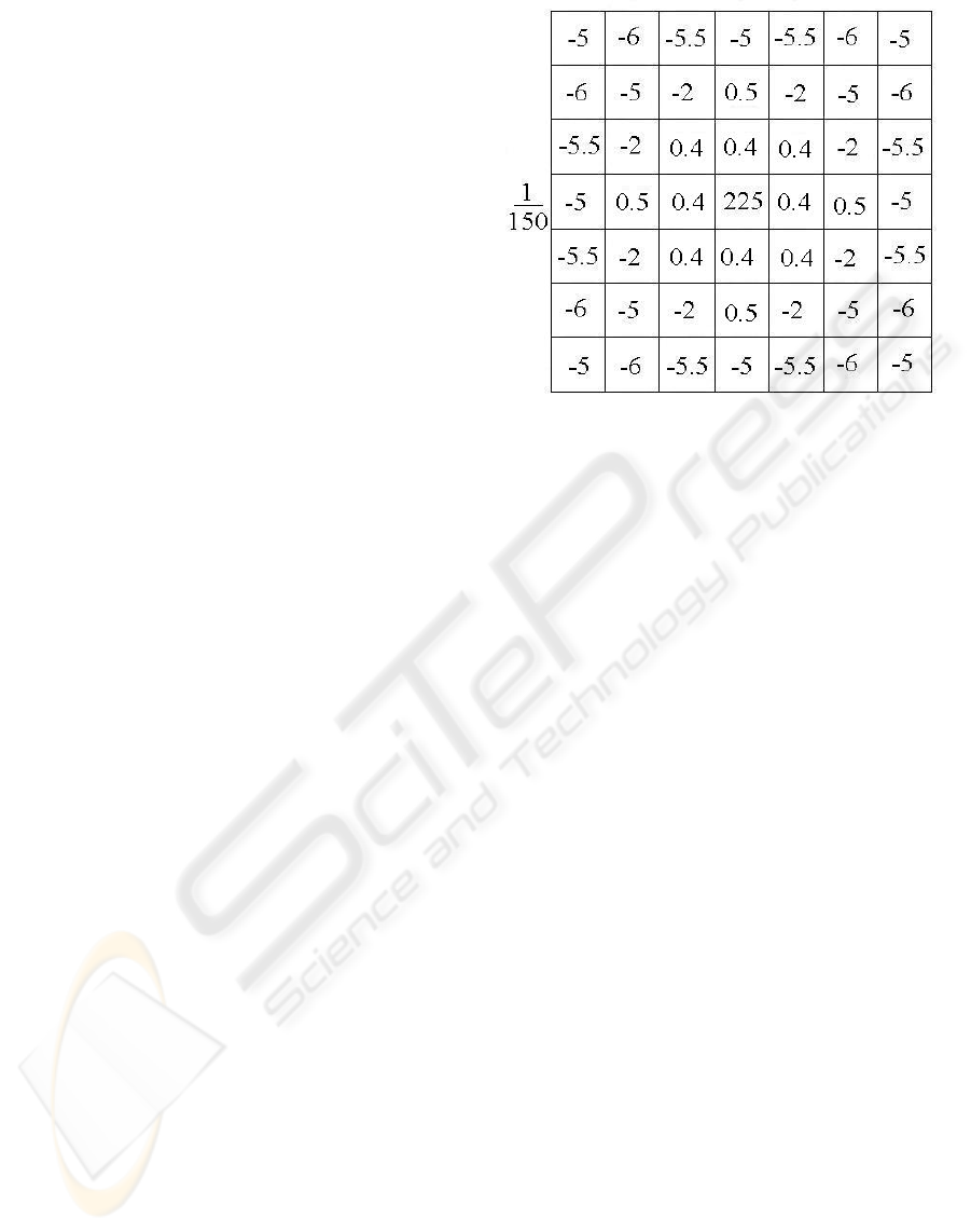
isotropic mask. We will now explain how this mask
can be designed. As explained in the previous section,
this surround consists of two annular regions, the in-
ner being excitatory and the outer being inhibitory in
nature. Hence, in the mask, we will define two an-
nular regions surrounding the central pixel. The inner
one is assigned positive weights (excitatory) while the
outer one is assigned negative weights (inhibitory).
The magnitude of the weights is chosen based on the
following neurophysiological findings: the influence
of a point in the surround on the cell response is de-
pendent on its distance from the centre and this is
roughly Gaussian in profile. Furthermore, the excita-
tory influence is weaker than the inhibitory influence
(Series et al., 2003). The size of the required mask
was determined by varying it from 7 × 7 to 15 × 15.
It was found that a mask of size 7 × 7 is optimal to
achieve the best results. Hence, we propose a 7 × 7
mask as shown in Figure. 4. The weights in the outer
inhibitory region of the optimal mask was found by
sampling a Gaussian profile of standard deviation 1.4.
The sum of the weights inside this mask is set at 0.52
to help enhance a contour pixel.
Figure 4: A 7 × 7 surround mask.
3.3 Binarisation
A binary contour map can be constructed by using
a standard procedure such as nonmaxima supression
followed by hysteresis thresholding (Canny, 1986).
Let the gradient magnitude M (x, y) and orientation
map Θ(x, y) specify the local strength and local edge
direction respectively. Nonmaxima suppression seeks
to thin regions where M(x, y) is non-zero, to gener-
ate candidate contours as follows: two virtual neigh-
bors are defined at the intersections of the gradient
direction with a 3 × 3 sampling grid and the gradi-
ent magnitude for these neighbors is interpolated from
adjacent pixels. The central pixel is retained for fur-
ther processing only if its gradient magnitude is the
largest of the three values. The final contour map is
computed from the candidates by hysteresis thresh-
olding. This process involves two threshold values t
l
and t
h
, t
l
<t
h
. All the pixels with M (x, y) ≥ t
h
are
retained for the final contour map, while all the pix-
els with M(x, y) ≤ t
l
are discarded. The pixels with
t
l
<M(x, y) <t
h
are retained only if they already
have at least one neighbor in the final contour map.
4 EXPERIMENTAL RESULTS
4.1 Ground Truth Image Data
Most of the methods for the evaluation of edge and
contour detectors use natural images with associated
desired output that is subjectively specified by the
observer [(Martin et al., 2004), (Grigorescu et al.,
2003)]. Some recent studies (Shin et al., 2001) show
that the performance of such an operator must be
A SIMPLE SCHEME FOR CONTOUR DETECTION
239

considered task dependent. For object recognition,
for example, some operators may perform better than
others despite similar performance on synthetic im-
ages. The proposed surround interaction mechanisms
is aimed at a better detection of objects contours in
natural scenes.
We tested the performance of the proposed scheme
on 40 natural images from a database designed to
evaluate the performance of contour detection (dat,
2003). For each test image, an associated desired out-
put binary contour map that was drawn by human is
given. It should be noted that the ground truth data in-
cludes more than one type of pixels: (i) pixels which
are parts of a contour of an object (ii) pixels which
are part of a boundary between two (textured) regions.
Our proposed scheme on the other hand, is designed
to extract only the first type of contour pixels.
4.2 Performance Measure
In order to have a quantitative comparison between
the contour detector proposed in (Grigorescu et al.,
2003) we use the performance measure introduced in
the same. Let f
p
and f
n
are number of false positive
and false negative pixels detected in the final contour
map, respectively. The performance measure is de-
fined as follows:
P =
t
p
t
p
+ f
p
+ f
n
(1)
where, t
p
is the number of correctly detected con-
tour pixels (True positive). The performance measure
P is a scalar taking values in the interval [0, 1].If
the desired output pixels are correctly detected and no
background pixels are falsely detected as contour pix-
els, then P =1. For all other cases, P takes values
smaller than one, being closer to zero as more con-
tour pixels are falsely detected and/or missed by the
operator.
For computing the performance measure, we must
determine which true positives are correctly detected,
and which detection is false. The binary contour map
in the ground truth data can be used for this pur-
pose. Let us consider how to compute P of a out-
put contour image given a ground truth contour map.
One could simply correspond coincident contour pix-
els and declare all unmatched pixels either as false
positives or misses. However, this approach would
not tolerate any localization error and result in a poor
performance measure. For robustness, it is desirable
that the correspondence of output contour pixels to
ground truth tolerate localization errors since ground
truth data is not accurately localized. The approach
proposed in (Grigorescu et al., 2003) considers a con-
tour pixel as correctly detected if a corresponding
ground truth contour pixel is present in a 5×5 (empir-
ically find) square neighborhood (window) centered
Table 1: Performance of proposed scheme and reported
by Cosmin (Grigorescu et al., 2003) on 3 natural im-
ages(elephant, goat and hyena).
Image Method Performance
Goat (Grigorescu et al., 2003) 0.34
Proposed Scheme 0.51
Elephant (Grigorescu et al., 2003) 0.42
Proposed Scheme 0.61
Hyena (Grigorescu et al., 2003) 0.55
Proposed Scheme 0.76
at the respective pixel coordinate. A window based
approach leads to a less robust performance measure,
as different sizes of the window can be shown to af-
fect the performance value significantly, which is a
undesirable. A large window will boost the number
of true positive. However, an explicit correspondence
of the detected contour and ground truth contour pix-
els is the only way to robustly count the hits, misses
and false positives that we need to compute P .We
have used the algorithm presented in (Martin et al.,
2004) for the correspondence between output contour
map and a ground truth contour map. The algorithm
converts the corresponding problem into a minimum
cost bipartite assignment problem, where the weight
between a output contour pixel and ground truth con-
tour pixel is proportional to their relative distance in
the image plane. One can then declare all contour
pixels matched beyond some threshold to be non-hits.
The correspondence computation is detailed in (Mar-
tin et al., 2004).
4.3 Results
The proposed contour detection scheme was tested on
40 images from a database reported in (Grigorescu
et al., 2003). Of these, we present results on 3 im-
ages in Fig 5 for illustrative purposes. A qualita-
tive comparison between the results of our contour
detection scheme and the contour reported in (Grig-
orescu et al., 2003) can be made by observing the re-
sults in this figure. The Canny edge detector outputs
are also included for reference. The first and second
columns show the input images and the corresponding
ground truth images, respectively; the third column
shows the best results of the Canny edge detector; the
fourth column shows the results of the contour detec-
tion reported by (Grigorescu et al., 2003); and finally,
the fifth column shows the results of the proposed
VISAPP 2006 - IMAGE ANALYSIS
240
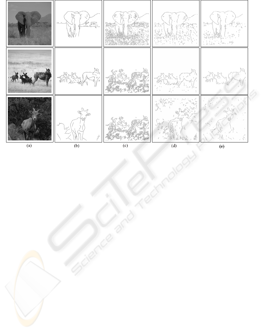
Figure 5: Results of contour detection on test images. (a) & (b) show input images and associated ground truth images
respectively (c) Canny edge map (d) Best contour map reported in (Grigorescu et al., 2003) (e) Best results obtained by the
proposed contour detection scheme.
scheme. The first point to note is that the obtained
contours in the fourth and fifth columns are closer to
the ground truth than the Canny output which is jus-
tifiably very ’noisy’. Furthermore, it can be seen that
the results of the proposed scheme are closer to the
ground truth. For instance, the grassy region in the
elephant image (top row) is suppressed well. The best
result of the contour map can be seen from the hyena
images (in the bottom row) where the output is very
close to the ground truth image.
A quantitative comparison of the contour and edge
detectors is shown in Table 1. The figures in the ta-
ble show that the proposed scheme outperforms the
scheme in (Grigorescu et al., 2003) in all 3 images
shown in Fig 5. This is consistent with our visual
analysis of the results.
Fig. 6 shows statistical Box- and - Whisker plots
for ten of the images used in our experiments. These
plots are helpful in interpreting the distribution of per-
formance value over ten different images. The aver-
age obtained value is above 0.5 which is encouraging
since the measure is computed using ground truth im-
ages which included contour pixels belonging to ob-
ject as well as texture region boundaries, whereas our
scheme is designed for extracting only the former.
5 CONCLUSION
Though centre-surround receptive fields have been
explored as a possible solution for good edge detec-
tion, a centre-surround mechanism is also applica-
ble at a wider level (summation across neighbour-
ing cells) to achieve contour detection. The proposed
scheme for contour detection is motivated from such
center-surround influence in the cortical cells of pri-
mates. It contributes to better contour detection not by
enhancing responses to contours, but by selectively
suppressing edges based on the surround. Specifi-
cally, an edge (signalled by a strong gradient) qual-
ifies to be a contour only if it is salient in a local con-
text where saliency implies that either the surround
has no edges or the surround has weaker edges. Thus,
the proposed scheme is not dependent on any prior
knowledge which makes it a general preprocessing
step for high order tasks. A further attractive feature
of the scheme is that it is also computationally low in
cost compared with many of the earlier approaches to
general purpose contour detection.
In practice, contour detection is an intermediate
level operation in computer vision with its output of-
ten used as input for further stages performing higher
level processing. It is hence of interest to know the
A SIMPLE SCHEME FOR CONTOUR DETECTION
241
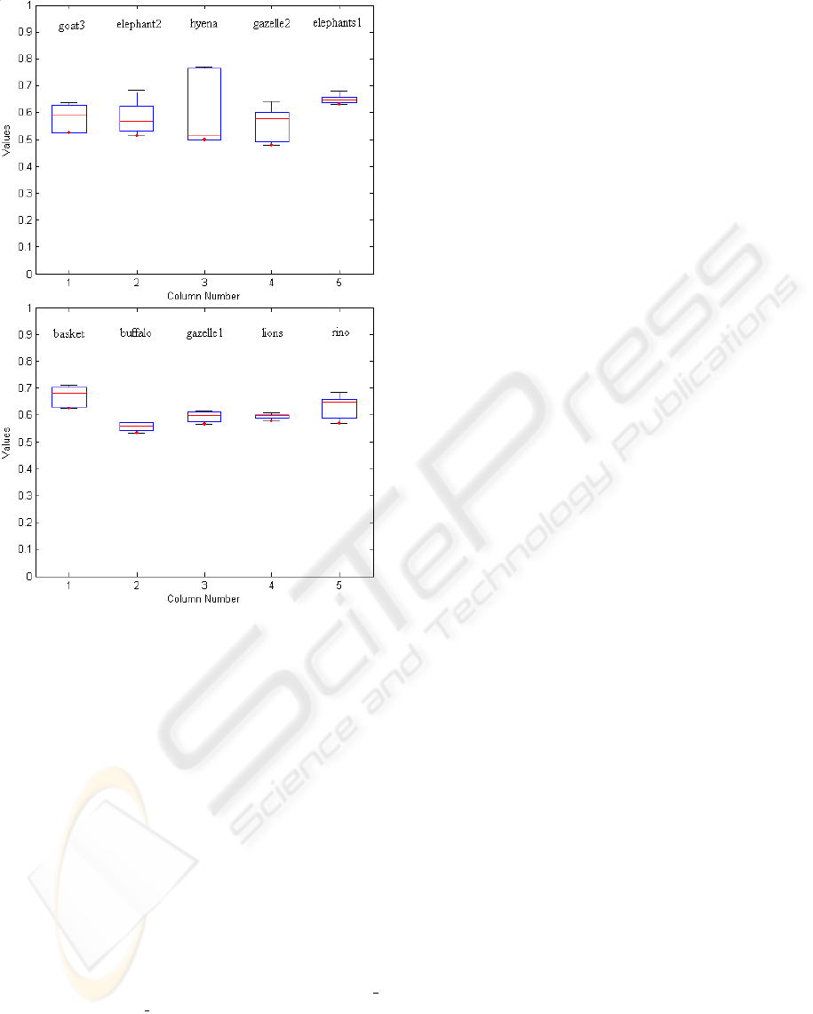
Figure 6: The distribution of performance value over ten
different images.
appropriateness of its use given a specific high level
task. As can be seen from the results, the proposed
contour scheme largely suppresses the local back-
ground information and hence it is not appropriate
to deploy it in tasks where the background informa-
tion is essential, e.g. texture classification or region
based segmentation. In other high-level tasks such as
shape-based recognition and image retrieval, the pro-
posed scheme can play a very useful role in their per-
formance improvement.
REFERENCES
(2003). http://www.cs.rug.nl/∼imaging/databases/contour
database/contour database.html.
Baumann, R., van der Zwan, R., and Peterhans, E. (1997).
Figure-ground segregation at contours: a neural mech-
anism in the visual cortex of the alert monkey. In Eu-
ropean Journal of Neuroscience.
Black, M., Sapiro, G., Marimont, D., and Heeger, D.
(1998). Robust anisotropic diffusion. In IEEE Trans-
action on Image Processing.
Canny, J. (1986). A computational approach to edge detec-
tion. In IEEE Transactions on Pattern Analysis and
Machine Intelligence.
Cavanaugh, J., Bair, W., and Movshon, J. (2002). Nature
and interaction of signals from the receptive field cen-
ter and surround in macaque v1 neurons. In Journal
of Neurophysiology.
Dobbins, A., Zucker, S. W., and Cynader, M. S. (1987).
Endstopped neurons in the visual cortex as a substrate
for calculating curvature. In Nature.
Dubuc, B. and Zucker, S. (2001). Complexity, confusion
and perceptual grouping. part ii: mapping complexity.
In International Journal on Computer Vision.
Grigorescu, C., Petkov, N., and Westenberg, M. (2003).
Contour detection based on nonclassical receptive
field inhibition. In IEEE Transactions on Image
Processing.
Hubel, D. H. and Wiesel, T. N. (1962). Receptive fields,
binocular interaction and functional architecture in the
cats visual cortex. In Journal of Psychology.
Knierim, J. and van Essen, D. (1992). Neuronal re-
sponses to static texture patterns in area v1 of the alert
macaque monkey. In Journal of Neurophysiology.
Ma, W.-Y. and Manjunath, B. (2000). Edgeflow: A tech-
nique for boundary detection and image segmentation.
In IEEE Transactions on Image Processing.
Marr, D. and Hildreth, E. (1980). Theory of edge detection.
In Proceedings of the Royal Society.
Martin, D. R., Fowlkes, C. C., and Malik, J. (2004). Learn-
ing to detect natural image boundaries using local
brightness, color, and texture cues. In IEEE Trans-
actions on Pattern Analysis and Machine Intelligence.
Meer, P. and Georgescu, B. (2001). Edge detection with em-
bedded confidence. In IEEE Transactions on Pattern
Analysis and Machine Intelligence.
Morrone, M. C. and Burr, D. C. (1988). Feature detection in
human vision: A phase-dependent energy model. In
Proceedings of the Royal Society, London Series B.
Perona, P. and Malik, J. (1990). Scale-space and edge detec-
tion using anisotropic diffusion. In IEEE Transactions
on Pattern Analysis and Machine Intelligence.
Series, P., Lorenceau, J., and Fregnac, Y. (2003). The silent
surround of v1 receptive fields: theory and experi-
ments. In Journal of Physiology Paris.
Shin, M. C., Glodgof, D. B., and Bowyer, K. (2001). Com-
parision of edge detectors using an object recognition
task. In Computer Vision and Image Understanding.
von der Heydt, R., Peterhans, E., and Drsteler, M. R. (1991).
Grating cells in monkey visual cortex: coding texture?
In Channels in the Visual Nervous System: Neuro-
physiology, Psychophysics and Models (Blum B, ed).
Yen, S. and Finkel, L. (1998). Extraction of perceptually
salient contours by striate cortical networks. In Vision
Research.
VISAPP 2006 - IMAGE ANALYSIS
242
