
A Self-adaptation Method for Human Skin Segmentation based on Seed
Growing
Anderson Carlos Sousa e Santos and Helio Pedrini
Institute of Computing, University of Campinas, Campinas-SP, 13083-852, Brazil
Keywords:
Skin Detection, Image Analysis, Face Detection, Color Models.
Abstract:
Human skin segmentation has several applications in image and video processing fields, whose main purpose
is to distinguish image portions between skin and non-skin regions. Despite the large number of methods
available in the literature, accurate skin segmentation is still a challenging task. Many methods rely on color
information, which does not completely discriminate the image regions due to variations in lighting conditions
and ambiguity between skin and background color. Therefore, there is still need to adapt the segmentation to
particular conditions of the images. In contrast to the methods that rely on faces, hands or any other body
content detector, we describe a self-contained method for adaptive skin segmentation that makes use of spatial
analysis to produce regions from which the overall skin can be estimated. A comparison with state-of-the-art
methods using a well known challenging data set shows that our method provides significant improvement on
the skin segmentation.
1 INTRODUCTION
Human skin detection in digital images is a crucial
part in several applications, such as face detection,
gesture analysis, content-based image retrieval, nu-
dity detection and consequent adult content filtering.
Skin detection can be seen as a classification prob-
lem, whose purpose is to determine which image pix-
els belong to the skin or non-skin classes. Such task
presents many challenges. For instance, variation in
scene illumination interferes with the appearance of
the skin, different cameras produce distinct colors and
ethnic diversity promotes various skin tones. These
factors make the skin detection process quite com-
plex.
Most studies (Kawulok et al., 2014; Kakumanu
et al., 2007; Phung et al., 2005) use color information
as evidence to detect skin since this property is able
to provide computational efficiency while it demon-
strates to be robust to occlusions and rotation and
scaling transformations (Kakumanu et al., 2007).
The main obstacle is the existence of an overlap
between skin and non-skin colors that occurs inde-
pendently of the color space. To minimize that, some
segmentation methods (Kawulok et al., 2014) attempt
to perform a color separability dependent on the skin
color that appears in the image. The most significant
improvements are performed with the aid of face de-
tection, which provides an estimation of the remain-
ing skin.
This work presents an adaptive segmentation
method with no need for face or any other body
content detection. It is based on an estimation from
regions found through spatial analysis performed
with a skin probability map. Experiments conducted
on a large well known data set show that our method
outperforms other skin segmentation approaches
available in the literature.
The text is organized as follows. Section 2
presents the main concepts and work related to
skin detection. Section 3 describes the proposed
method for self-adaptive segmentation of human
skin. Experimental results are shown in Section 4.
Finally, Section 5 concludes the paper with final
remarks and directions for future work.
2 BACKGROUND
Many approaches have been proposed to address de-
tection and segmentation of human skin. The simplest
strategy is based on static decision rules defined in
some color space or even in a combination of them.
Various different color spaces have been explored,
such as RGB (Fleck et al., 1996), normalized RG (So-
riano et al., 2000), HSV (Sobottka and Pitas, 1998;
Wang and Yuan, 2001), YCbCr (Hsu et al., 2002). A
transformation of RGB into a single-channel has been
proposed by Cheddad et al. (Cheddad et al., 2009),
specifically for the purpose of skin detection.
455
Sousa e Santos A. and Pedrini H..
A Self-adaptation Method for Human Skin Segmentation based on Seed Growing.
DOI: 10.5220/0005295204550462
In Proceedings of the 10th International Conference on Computer Vision Theory and Applications (VISAPP-2015), pages 455-462
ISBN: 978-989-758-089-5
Copyright
c
2015 SCITEPRESS (Science and Technology Publications, Lda.)

A more sophisticated strategy relies on modeling
the statistical distribution of color. There are two main
approaches to that: parametric and non-parametric
techniques. Both approaches calculate the probability
of a given color (c) to be skin (P(skin|c)), which gen-
erates a probability map such that segmentation can
be performed through a threshold. However, para-
metric approaches assume that the skin distribution
fits some explicit model.
Most of the methods available in the literature rely
on mixture of Gaussians (Yang and Ahuja, 1999), al-
though there are also methods based on single Gaus-
sian (Subban and Mishra, 2014) or elliptical boundary
model (Lee and Yoo, 2002).
On the other hand, non-parametric models esti-
mate the probabilities directly from the training data
without any assumptions on its distribution shape. To
do so, histograms for skin (H
skin
(c)) and non-skin
(H
¬skin
(c)) are built over an annotated data set. The
conditional probabilities are obtained as
P(c|skin) =
H
skin
(c)
∑
(H
skin
(i))
(1)
P(c|¬skin) =
H
¬skin
(c)
∑
(H
¬skin
(i))
(2)
From these probabilities, Bayes rule can be ap-
plied to produce the desired posterior probability as
P(skin|c) =
P(c|skin)P(skin)
P(c|skin)P(skin) + P(c|¬skin)P(¬skin)
(3)
where P(skin) and P(¬skin) are the prior probabili-
ties and usually are set to 0.5. Vezhnevets et al. (Vezh-
nevets et al., 2003) demonstrated that the choice of
the prior probabilities does not influence the over-
all result. Jones and Rehg (Jones and Rehg, 2002)
conducted an extensive evaluation of the method and
showed that it outperforms Gaussian mixture models
for a sufficient large data set.
There is usually an overlap between skin and non-
skin colors when the models become more accu-
rate (Kawulok et al., 2014). Taking this fact into con-
sideration, many researchers have adapted the men-
tioned methods according to the context. For in-
stance, Kovac et al. (Kovac et al., 2003) defined differ-
ent rules depending on lighting conditions, whereas
Phung et al. (Phung et al., 2003) created an iterative
method for determining an optimal threshold for the
probability map of a particular image.
Nevertheless, the most significant results
are obtained by content-based adaptation, more
specifically for face detection. The first of such
approaches (Fritsch et al., 2002) uses the region
acquired by a face detector to update a unimodal
Gaussian previously determined. Taylor and Mor-
ris (Taylor and Morris, 2014) recently proposed to
only use the skin of the face in normalized RG to
build a Gaussian model, discarding any previous
training. A more robust technique (Kawulok, 2010)
uses the face region to build a local skin histogram
and, consequently, a P
f ace
(skin|c) is derived and
combined with the general probability for the final
map.
Another strategy, namely spatial analysis, consid-
ers the structural alignment in the neighborhood of
pixels classified as skin - generally with a probability
map - such that it refines the segmentation process by
removing false positives.
Most of these methods perform an expansion of
seeds found by a high threshold. The expansion
can be performed through different criteria: thresh-
old hysteresis (Ruiz-del Solar and Verschae, 2004),
energy accumulation (Kawulok, 2010) or cost prop-
agation (Kawulok, 2013). The latter one is the more
complex and provides superior results, where the Di-
jkstra’s algorithm (Dijkstra, 1959) is used to calculate
shortest routes in a combined domain composed of
luminance, hue and skin probability.
Some of the methods described in this section will
be used for comparison in our experiments, described
in Section 4. For a more extensive review of the state-
of-the-art methods, refer to Kawulok et al. (Kawulok
et al., 2014).
3 PROPOSED METHODOLOGY
We propose a method for skin segmentation that com-
bines spatial analysis and adaptive models for better
skin probability estimation. The methodology can be
divided into three main steps. First, seeds are ex-
tracted from the probability map through a precise
and systematic strategy for spreading them over the
images. Second, a controlled propagation method is
applied to grow the seeds into skin blobs. Finally,
these blobs are used to estimate the skin color present
in the images and such that can optimize the probabil-
ity map. The main steps of our method are presented
in the diagram shown in Figure 1.
Figure 2 illustrates the application of the proposed
method to an input image, where the general probabil-
ity map, the extracted seeds, the blobs after propaga-
tion, the final probability map and the resulting seg-
mentation are shown.
Algorithm 1 describes in details the main steps
performed by the proposed skin segmentation
method.
VISAPP2015-InternationalConferenceonComputerVisionTheoryandApplications
456

Color image
Seed extraction
Propagation
Skin general
probability
map
Local skin
probability
map
Adaptive detection
Final probability map
Figure 1: Main stages of the skin detection process.
3.1 Seed Extraction
The most important step in a region growing algo-
rithm is the proper choice of the seeds. Since seeds
can correspond to false positives, the propagation pro-
cess does not guarantee that inadequate seeds do not
occur in the images, which can compromise the final
estimation.
As described in Section 2, skin region propaga-
tion methods usually rely on a fixed high threshold
and size based analysis for producing the seeds. Such
methods do not take different characteristics of each
image into account, as well as its respective proba-
bility map, once the same threshold is applied to all
images. Another disadvantage is the assumption that
the resulting skin-like seeds (false positives) are very
small, which is not always true. Taking these factors
into consideration, we propose an adaptive seed ex-
traction with a homogeneity-based analysis.
In order to obtain the best high threshold for a par-
ticular image, we first apply a median filter (Gonzalez
et al., 2009) to the probability map and then take the
maximum probability (Line 3, Algorithm 1). To allow
for images with no skin, if the maximum value is
Algorithm 1: Proposed skin segmentation method
based on seed growing.
input : color image I, histogram of skin (H
skin
)
and non-skin (H
nonskin
) colors.
output: Final probability map M
f inal
1 Build general probability map (M
global
)
according to Equations (1), (2) (3), using H
skin
and H
nonskin
2 M
blur
← blur(M
global
, size)
3 T
seed
← max(M
blur
)
4 if T
seed
≤ 0.5 then
5 return M
6 end
7 edges ← edgeDetector(I)
8 for x ∈ I do
9 if M
global
(x) ≥ T
seed
∧ x /∈ edges then
10 Seeds ← x
11 end
12 end
13 Q ← {Seeds} {where Q is a priority queue}
14 for x ∈ I do
15 if x ∈ Seeds then
16 C(x) = 0
17 else
18 C(x) = −1
19 end
20 end
21 while Q 6=
/
0 do
22 q = pop(Q)
23 for s ∈ Neighbors(q) do {8 -
neighborhood}
24 c = C(q) +ρ(q → s)
25 if (c < C(s) ∨C(s) < 0) ∧ s /∈ edges
then
26 C(s) = c
27 Q ← s
28 end
29 end
30 end
31 Normalize C by scaling the costs from 0 for the
maximal cost to 1 for a zero cost
32 for x ∈ I do
33 if C(x) > 0 then
34 H
skin local
(color(x))++
35 end
36 end
37 Build final probability map (M
f inal
) according
to Equation (5) using H
skin local
38 return M
f inal
smaller than a minimum threshold (T
seed
min
), it is dis-
carded, otherwise it is assigned as the seed threshold
for the original probability map. Therefore, we obtain
ASelf-adaptationMethodforHumanSkinSegmentationbasedonSeedGrowing
457

(a) Original (b) General probability map
(c) Seeds (d) Blobs after propagation
(e) Final probability map (f) Segmentation result
Figure 2: Examples of images obtained by applying each
stage of our method to an input image.
seeds with high probability by considering the con-
text.
To prevent the occurrence of false positives, we
exclude the choice of seeds located in edge regions,
since skin is usually a smooth and homogeneous re-
gion. The used edge detector is described in the fol-
lowing section.
3.2 Propagation
The objective here is to expand the seeds into skin
blobs. The main drawback of propagation methods
is the “leakages”, that is, the seed growth to a region
of non skin. In order to avoid those, we establish a
strict control of the propagation. We modified the cost
propagation proposed in Kawulok (Kawulok, 2013)
by adding a constraint in which the propagation can-
not flow out the image edges. As a consequence, an
increase in the false negative rate is concerned with a
reduction of the false positive rate. The next step will
address these undetected skin regions.
Prior to the actual propagation, an edge detector is
applied to the images. In order to benefit from color
information, we utilize a color edge detection tech-
nique that combines (through logical or operator) the
results of Canny detector (Gonzalez et al., 2009) for
each of the three channels in HSV color space. Fol-
lowing that, a morphological dilation operation is per-
formed, such that small gaps can be closed.
The propagation process is similar to the work
by Kawulok (Kawulok, 2013) except that the process
stops when an edge is reached for a certain direction
(Line 25, Algorithm 1). Besides preventing false posi-
tives, this also speeds up the algorithm, once the orig-
inal approach calculates the costs from the seeds to
every other pixel in the image.
3.3 Adaptive Detection
Once we have generated the skin blobs, we use them
to build a local statistical model (Line 34, Algo-
rithm 1) that adapts to the particular conditions of the
image. From the histogram of these resulting skin re-
gions, we obtain a P
local
(c|skin). As for non-skin, we
assume that the local distribution follows the global
one, which gives
P
local
(c|¬skin) = P
global
(c|¬skin) (4)
The final probability is defined as
P(skin|c) = γP
local
(skin|c) + (1 − γ)P
global
(skin|c) (5)
where P
local
(skin|c) and P
global
(skin|c) are both cal-
culated as in Equation (3), differentiating by using
local and global data, respectively. The parameter γ
controls the importance of the local model.
From Equation (5), we generate the final skin
probability map, in which the detection can be per-
formed through a fixed threshold or generated by
more complex techniques developed for the general
probability map. However, the description of such
methods is beyond the scope of this paper.
4 EXPERIMENTS
The experiments were evaluated through two different
data sets. To train the Bayes classifier, we used 8963
non-skin images and 4666 skin images from the Com-
paq database (Jones and Rehg, 2002), which contains
images acquired from the Internet in a diverse vari-
ety of settings and its approximately 1 billion pixels
makes it sufficient large for non-parametric estima-
tion of skin color distribution.
For evaluation and comparison purposes, we used
the ECU database (Phung et al., 2005) divided into a
1000 images for validation and 3000 images for test.
This data set ensures a diversity in terms of back-
ground scenes, lighting conditions and skin types.
Both data sets provide a ground-truth that makes
possible identify the pixel class (skin or non-skin) for
the training and quantitatively evaluate the detection
output.
VISAPP2015-InternationalConferenceonComputerVisionTheoryandApplications
458
The performance of the skin detection was mea-
sured through a number of metrics: true positive rate
(η
t p
- percentage of skin correctly classified as skin);
false positive rate (δ
f p
- percentage of non-skin clas-
sified as skin); precision (η
prec
- percentage of cor-
rectly classified pixels out of all the pixels classi-
fied as skin); F
score
(harmonic mean between η
prec
and η
t p
) and detection error (δ
min
= (1 − η
t p
) + δ
f p
).
Additionally for non-binary classification, the ROC
(receiver operating characteristics) and the respective
area under curve (AUC) are applied.
All the following experiments were conducted on
an Intel Core i7 3.50GHz with 32GB RAM running
64 bits Ubuntu 12.04 operating system.
In order to determine the bin size of the his-
togram, we experimented a number of different sizes,
as shown in Table 1. Since 32 bins per channel pro-
duced the highest value of AUC, this value was used
in the proposed method both for local and general
models.
Table 1: Bin size evaluation for validation data set.
Bin size AUC
8
3
0.892348
16
3
0.918403
32
3
0.934036
64
3
0.923436
128
3
0.917958
256
3
0.914159
Furthermore, the seed detection process demon-
strated to be sensitive to the bin size, performing bet-
ter for more quantized color values. Another factor
that influenced the seeds is the kernel size for the me-
dian filter. We empirically observed that the amount
of seeds found is directly related to it. If it is too
small, then very few seeds are found; otherwise, if it
is too large, several false positives are placed as seeds.
A 15 × 15 median filter was used to generate the re-
ported results.
A comparison between our seed extraction
method and different fixed thresholds are presented
in Table 2. Since an important issue in the seed
extraction is to avoid false positives while retaining
some true positives, the precision (η
prec
) seems
appropriate for the evaluation. As it can be noticed,
our method provides superior results with a large
difference.
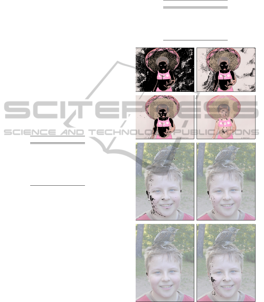
Another desired quality for seeds is that they
should be spread over the skin regions to prevent from
missing any isolated region. Therefore, we present
a qualitative comparison in Figure 3. The seeds ac-
quired with fixed thresholds are displayed along with
the seeds collected by our method and the T
seed
found
Table 2: Evaluation of seed extraction for validation data
set.
Method η
prec
(%)
T
seed
= 0.70 68.32
T
seed
= 0.80 73.39
T
seed
= 0.90 82.64
T
seed
= 0.95 88.70
Our seeds 94.66
(a) T seed = 0.8 (b) T seed = 0.9
(c) T seed = 0.95 (d) Our seeds [0.983012]
(e) T seed = 0.7 (f) T seed = 0.8
(g) T seed = 0.9 (h) Our seeds [0.722982]
Figure 3: Examples of seeds detected through different
thresholds for our method.
by it are placed in brackets.
It is possible to observe that our method not only
avoids the non-skin regions but also maintains the dis-
ASelf-adaptationMethodforHumanSkinSegmentationbasedonSeedGrowing
459

persal. It is also noticeable that a high threshold is
required in the first image in order to avoid misclassi-
fication, while the second will not produce significant
seeds for the same value. Thus, our seed extraction
method overcomes such problem with a choice of a
proper threshold for each image.
The edge detection is an important stage of our
method since it supports both seed extraction and
propagation. Some experiments were conducted in
the edge detector for different color spaces: only lu-
minance, HSV, RGB and YCbCr. The HSV model
better captured people’s contours in images under ab-
normal lighting and, therefore, was employed in the
experiments. Canny detector was applied with both
low and high thresholds equal 100 and a dilation pro-
cess was performed with a 3 × 3 structuring kernel.
For comparison, we selected some state-of-the
art methods available in the literature: Cheddad’s
decision rule (Cheddad et al., 2009), statistical
model (Jones and Rehg, 2002), face-based adap-
tation (Kawulok, 2010) built with Viola-Jones
face-detector (Viola and Jones, 2004) and cost prop-
agation (Kawulok, 2013). Our method was tested
with γ = 0.8 and T
seed
min
= 0.5, whereas original
parameters were employed in the other approaches.
Figure 4 presents a comparison of the ROC
curves. The points in the curves were obtained
with different thresholds, except for Cheddad’s rule,
whose output is binary.
False positive rate
True positive rate
0% 10% 20% 30% 40%
50% 60% 70% 80% 90% 100%
Cheddad’s rule
Statistical model − AUC = 0.9323
Cost Propagation − AUC = 0.9132
Face−based adaptation − AUC = 0.9487
Proposed Method − AUC = 0.9520
Figure 4: ROC curves for comparison of the tested methods.
To present quantitative values, Table 3 shows the
results for a fixed threshold obtained through maximal
Youden’s index (Youden, 1950), which represents the
closest point to the optimum value (0,1). Best values
are highlighted in bold.
Cheddad’s rule, as expected, holds the worst re-
Table 3: Detection results for different methods.
Method η
t p
δ
f p
F
score
δ
min
(%) (%) (%) (%)
Cheddad 89.33 19.51 64.78 30.18
Statistical model 87.90 14.51 69.71 26.61
Face-based adaptation 86.83 11.79 72.63 24.96
Cost propagation 90.40 14.46 71.05 24.06
Proposed method 89.78 11.24 74.95 21.46
sults, which demonstrates the inadequacy of such re-
strict and biased method. Statistical model performs a
little better, however, it still has a higher false positive
rate.
From the compared methods, the cost propagation
has the lowest δ
min
whereas the face-based adaptation
the highest F
score
. In fact, their values are very similar,
considered as a tie. The first has δ
f p
very similar to
the statistical model, what suggests that its improve-
ment is accomplished by an increase in the true pos-
itive rate. Oppositely, the second maintains the η
t p
,
performing a reduction in the number of false pos-
itives. Our proposed method, which holds the best
results, improves on both metrics.
Table 4 gives the true positive rate for fixed false
positive rate values, where only methods of proba-
bilistic output were considered. It shows the behavior
of the methods in different tolerance settings.
Table 4: True positive rates for fixed values of false positive
rate.
Method η
t p
δ
f p
η
t p
δ
f p
(%) (%) (%) (%)
Statistical model 81 10 92 20
Face-based adaptation 85 10 92 20
Cost propagation 84 10 94 20
Proposed method 88 10 95 20
Figure 5 illustrates the results for applying the
evaluated methods on four different image samples
from the test set. In the first row, it is possible to
observe that the face-based adaptation misclassified a
piece of blue shirt possibly due to the girl’s blue eyes.
Furthermore, in the second row shows the detection of
several different false positives, which suggests that
the face was not correctly detected.
The main drawback of the cost propagation ap-
proach is that it detects part of the background as skin,
as illustrated in the third row of Figure 5. This occurs
because, in some points, there is a smooth transition
between skin and false skin regions, such that a “leak-
age” occurs.
Our method overcomes such problems since it
tends to use more than just one region for the esti-
mation. Thus, our local model is an accurate repre-
sentation in cases of variation of skin through differ-
VISAPP2015-InternationalConferenceonComputerVisionTheoryandApplications
460
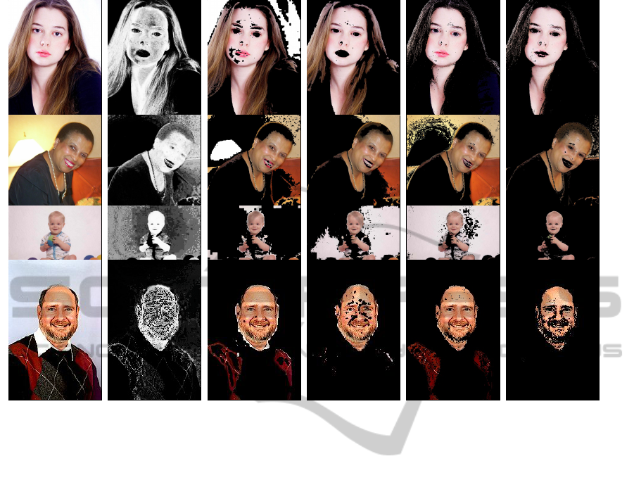
(s) Original (t) Probability map (u) Cheddad (v) Cost propagation (w) Face-based (x) Proposed
Figure 5: Examples of skin detected with different methods.
ent locations. Furthermore, seeds were always found
in our tests, while no faces were detected in 12% of
the images. Although “leakages” can still occur, they
are significantly reduced as demonstrated through the
results.
It is also important to highlight the viability of our
method in real-time applications, since the average
time per image in the test set was 282ms in an un-
optimized version of our code.
5 CONCLUSIONS AND FUTURE
WORK
This work presented a new adaptive human skin seg-
mentation method that makes use of skin probability
map and eliminates the need for object detection. The
main contributions of our approach include: a new
method for seed extraction based on spatial analysis
and a self-contained adaptation.
Experimental results demonstrated that the pro-
posed technique outperforms state-of-the-art skin seg-
mentation methods for a large and well-known test
set. Nevertheless, additional improvements can be
made. False positives generated in the propagation
stage of our method has a large contribution to the
overall accuracy, what makes us conjecture that even
little enhancements in the propagation control, such
as better edge detection, will significantly decrease
the error rates.
Our method could also be combined with face-
based adaptive methods through two strategies: use
of detected faces to improve seed extraction and use
as an alternative when a face is not present in the im-
age or has not been correctly detected.
As future directions, we intend to investigate more
powerful features, such as textural information, to dis-
card incorrect seeds, as well as new strategies for con-
trolling the propagation.
ACKNOWLEDGEMENTS
The authors are grateful to FAPESP, CNPq and
CAPES for the financial support.
REFERENCES
Cheddad, A., Condell, J., Curran, K., and Mc Kevitt, P.
(2009). A Skin Tone Detection Algorithm for an
ASelf-adaptationMethodforHumanSkinSegmentationbasedonSeedGrowing
461

Adaptive Approach to Steganography. Signal Pro-
cessing, 89(12):2465–2478.
Dijkstra, E. W. (1959). A Note on Two Problems in Con-
nection with Graphs. Numerische Mathematik, 1:269–
271.
Fleck, M. M., Forsyth, D. A., and Bregler, C. (1996). Find-
ing Naked People. In European Conference on Com-
puter Vision, pages 593–602. Springer.
Fritsch, J., Lang, S., Kleinehagenbrock, M., Fink, G. A.,
and Sagerer, G. (2002). Improving Adaptive Skin
Color Segmentation by Incorporating Results from
Face Detection. In 11th IEEE International Work-
shop on Robot and Human Interactive Communica-
tion, pages 337–343.
Gonzalez, R., Woods, R., and Eddins, S. (2009). Digital
Image Processing Using MATLAB. Gatesmark Pub-
lishing.
Hsu, R.-L., Abdel-Mottaleb, M., and Jain, A. K. (2002).
Face Detection in Color Images. IEEE Transac-
tions on Pattern Analysis and Machine Intelligence,
24(5):696–706.
Jones, M. J. and Rehg, J. M. (2002). Statistical Color Mod-
els with Application to Skin Detection. International
Journal of Computer Vision, 46(1):81–96.
Kakumanu, P., Makrogiannis, S., and Bourbakis, N. (2007).
A Survey of Skin-Color Modeling and Detection
Methods. Pattern Recognition, 40(3):1106–1122.
Kawulok, M. (2010). Energy-based Blob Analysis for Im-
proving Precision of Skin Segmentation. Multimedia
Tools and Applications, 49(3):463–481.
Kawulok, M. (2013). Fast Propagation-based Skin Regions
Segmentation in Color Images. In 10th IEEE Interna-
tional Conference and Workshops on Automatic Face
and Gesture Recognition, pages 1–7.
Kawulok, M., Nalepa, J., and Kawulok, J. (2014). Skin
Detection and Segmentation in Color Images. In Ad-
vances in Low-Level Color Image Processing, pages
329–366. Springer.
Kovac, J., Peer, P., and Solina, F. (2003). Human Skin Color
Clustering for Face Detection. In EUROCON - Com-
puter as a Tool, volume 2, pages 144–148. IEEE.
Lee, J.-Y. and Yoo, S. I. (2002). An Elliptical Boundary
model for Skin Color Detection. In International Con-
ference on Imaging Science, Systems, and Technology.
Citeseer.
Phung, S. L., Bouzerdoum, A., and Chai Sr, D. (2005). Skin
segmentation using color pixel classification: Analy-
sis and comparison. IEEE Transactions on Pattern
Analysis and Machine Intelligence, 27(1):148–154.
Phung, S. L., Chai, D., and Bouzerdoum, A. (2003). Adap-
tive Skin Segmentation in Color Images. In Interna-
tional Conference on Multimedia and Expo, volume 3,
pages III–173.
Ruiz-del Solar, J. and Verschae, R. (2004). Skin Detection
using Neighborhood Information. In Sixth IEEE Inter-
national Conference on Automatic Face and Gesture
Recognition, pages 463–468. IEEE.
Sobottka, K. and Pitas, I. (1998). A Novel Method for Au-
tomatic Face Segmentation, Facial Feature Sxtraction
and Tracking. Signal Processing: Image Communica-
tion, 12(3):263–281.
Soriano, M., Martinkauppi, B., Huovinen, S., and Laak-
sonen, M. (2000). Skin Detection in Video under
Changing Illumination Conditions. In 15th Interna-
tional Conference on Pattern Recognition, volume 1,
pages 839–842. IEEE.
Subban, R. and Mishra, R. (2014). Human skin segmenta-
tion in color images using gaussian color model. In
Recent Advances in Intelligent Informatics, pages 13–
21. Springer.
Taylor, M. J. and Morris, T. (2014). Adaptive Skin Segmen-
tation via Feature-based Face Detection. In SPIE Pho-
tonics Europe, pages 91390P–91390P. International
Society for Optics and Photonics.
Vezhnevets, V., Sazonov, V., and Andreeva, A. (2003). A
Survey on Pixel-based Skin Color Detection Tech-
niques. In Graphicon, volume 3, pages 85–92.
Moscow, Russia.
Viola, P. and Jones, M. J. (2004). Robust Real-Time Face
Detection. International Journal of Computer Vision,
57(2):137–154.
Wang, Y. and Yuan, B. (2001). A Novel Approach for Hu-
man Face Detection from Color Images under Com-
plex Background. Pattern Recognition, 34(10):1983–
1992.
Yang, M.-H. and Ahuja, N. (1999). Gaussian Mixture
Model for Human Skin Color and its Application in
Image and Video Databases. In SPIE: Storage and
Retrieval for Image and Video Databases VII, volume
3656, pages 458–466.
Youden, W. J. (1950). Index for Rating Diagnostic Tests.
Cancer, 3(1):32–35.
VISAPP2015-InternationalConferenceonComputerVisionTheoryandApplications
462
