
Microwave Radiation as Interface to the Brain Functional State
V. S. Kublanov
Ural Federal University, 19 Mira street, Ekaterinburg, Russia
Keywords: Brain, Microwave Radiation, Interface, Absorption Coefficient, Thermodynamic Temperature Permittivity,
Brightness Temperature, Fluctuations, Wavelet Analysis, Frequency Range, Metabolic and Humoral
Processes.
Abstract: The paper deals with investigations of spectral characteristics of fluctuations of brain microwave radiation
changes in metabolism production in the brain tissue. In the frequency band from 0.02 Hz to 0.013 Hz, these
fluctuations mainly show liquid circulation of products in the intercellular and intracellular spaces of brain
tissue. In the frequency band below 0.013 Hz, these fluctuations show thermodynamic changes in tissues.
This conclusion is related to radiation in the frequency band from 650 to 850 MHz and is based on the
results of anaysis of the phenomenological models of brain tissue radiation and thermodynamic processes in
it, and, also, on the experimental data received by means of the measuring radiophysical system.
1 INTRODUCTION
The brain is the most complicated biological
structure, in which functioning the interconnected
dynamic systems participate (Haken, 2006). Those
are: neural networks, glia, brain covers, and system
of liquor and blood circulation. Investigations of a
brain, behavior, and cognitive activity have a large
number of peculiarities. The brain plays a special
role in the organization of life sustenance of human
being. Its tissues possess high intensity of metabolic
processes, have no internal stocks neither oxidable
substrate, nor an oxidizer and, consequently, demand
intensive and very reliable blood circulation.
Therefore, it is necessary to search for models
adequately reflecting features of regulation
mechanisms of processes in brain tissue and to work
out apparatus complexes for functional researches of
brain including the investigation of a homeostasis
function in its tissues.
It is known that metabolism processes in brain
tissues are accompanied by fluctuations of
thermodynamic temperature and variations of the
liquid circulation (Godik and Gulyaev, 1991). The
same parameters define characteristics of the brain
microwave radiation. Note that the brain brightness
temperature T
br
(t) depends on two parameters:
absorption coefficient
(t) of medium and its
thermodynamic temperature Т(t)
T
br
tTt
k
t
2
2
,
(1)
where k is the Boltshman constant, is the
wavelength of radiation.
It is important to search for such conditions when
the brightness temperature Tbr(t) of brain can be
mainly defined by one of these parameters. The
problem can be solved by researching the
phenomenological models of microwave radiation of
brain tissues and thermodynamic processes in them.
In our investigations, methods of
phenomenological simulation of these processes and
physiological verification of their features were
applied to verification of the experimental data
received by means of contact microwave
radiothermography. For formating certain
physiological factors in an investigated organism,
the loading tests for their stimulation were used.
2 MODELS OF BRAIN
PROCESSES
According to equation (1), the brightness
temperature Т
br
depends on two values
and Т.
However, search of such conditions is of interest
when the brightness temperature Т
br
of the brain can
318
Kublanov V..
Microwave Radiation as Interface to the Brain Functional State.
DOI: 10.5220/0004371703180322
In Proceedings of the International Conference on Biomedical Electronics and Devices (MHGInterf-2013), pages 318-322
ISBN: 978-989-8565-34-1
Copyright
c
2013 SCITEPRESS (Science and Technology Publications, Lda.)

be mainly defined by one of them. Such search is
based on investigation of phenomenological models
of microwave radiation of brain tissues and
thermodynamic processes in them.
The results of the phenomenological modelling
of the microwave radiation and thermodynamic
processes on brain tissue revealed the following
facts.
It shows that fluctuations of own microwave
radiation of a brain in the band of frequencies from
650 to 850 MHz are mainly defined by changes of
an absorption coefficient of white and grey
substances of a brain. On the one hand, it is so since
the basic contribution to radiation is brought by
partial radiations of deep structures of a brain (grey
and white substances). Moreover, it is because in
these layers on depth more than 15 mm, a
thermoneutral zone is formed, in which changes of
thermodynamic temperature can be neglected.
The estimations received by modelling
characterize quasistatic processes in a brain and
allow us to obtain recept of the changes domain of
the absorption coefficient
and the thermodynamic
temperature Т in its tissues, but not time dynamics
of these changes.
There are few data about the changes of
thermodynamic temperature in brain tissues under
the functional-loading tests received by direct
methods of measurement. So, according to contact
measurements, for thermoneutral zones of a brain of
the human, the thermodynamic temperature can
change not more, than (0.05 – 0.06)
0
С, and their
period makes tens minutes (Ivanov, 1990). At the
same time, it is shown that under functional-loading
testes change of a blood flow happens rather quickly
within several seconds. But temperature changes of
a cerebral tissue are significantly slow, they depend
on the rate of transfer of heat defined by a blood
flow thermal conduction
i
.
According to (Yablonskiy et al., 2000), any
temperature inhomogenities in the brain have a
characteristic length depending on the blood
flow that are described as follows:
Fp
bb
i
,
(2)
where р
b
is the blood density,
b
and
i
are the
specific heat capacity of tissues of a brain and blood,
accordingly.
Typically, is approximately of several
millimeters. For large animals that have head
diameter of several centimeters or higher (this
includes adult humans with the head diameter
of 15 cm; neonates with the head diameter of 6
cm; most primates, etc.), the temperature distribution
near the brain surface can be treated as a one-
dimensional problem with temperature depending
only on the distance from the brain surface.
The temperature relaxation time constant is
described:
,
tissue
rCBFс
с
bb
(3)
where c
tissue
is the heat capacity of the
tissue,
rCBF is the local cerebral blood flow.
For the human and higher animals, this constant
of time makes some tens seconds. These data were
proved to be true by the experimental results:
temperature reaction to visual stimulation by
analogy «on-off» is formed in (50 – 80) sec
(McElligott and Melzack, 1967). The similar result
has been received in experiment on laboratory rats,
in which the maximum of the temperature response
was reached in 60 sec after including the stimulation
(Trubel, 2006).
Results of indirect estimation of the temperature
response in brain tissues at glucose test are presented
in (Guyton, 2010): skin rise in temperature in a
projection of one of veins in the head (v.
retromandibularus) is observed in (150 – 200) sec.
In the obtained experimental data (Ivanov, 1990),
the time constant of thermodynamic changes in a
brain coincides with theoretical estimations, that is,
it is at least not less than (50 – 80) sec. Therefore, it
is possible to suppose that the spectrum of
fluctuations of the brain own microwave radiation
(defined by thermodynamic processes in its tissues)
is in range of frequencies below 0.013 Hz.
3 EXPERIMENTAL
RESEARCHES
During experimental researches of own radiation, a
complex of works was performed. Metabolic or
hydrodynamic processes in brain tissues were
mainly activated by means of special functional-
loading tests.
In the first case for research of the contribution
of thermodynamic temperature in changes of the
own microwave radiation, the provocative influence
by a glucose is chosen. Remind that metabolic
processes of a human body entirely depend on a
metabolism of a glucose, which is the basic power
MicrowaveRadiationasInterfacetotheBrainFunctionalState
319
resources of a human body, and some organs and
tissues (the brain, erythrocytes) use exclusively it as
power raw materials. So, the change of a metabolism
in organism tissues is accompanied by the change of
their local temperature.
In the second case the passive antiorthostatic
load was applied. The main objective of these
researches is formation in the experimental model of
influence of weightlessness on a human brain. Thus,
the head inclination angle relatively the feet was
minus 15
0
. In antiorthostatic position, a blood
redistribution in vessels of the top half of the body
or shift of a hydrostatic vector of liquid motion on
vascular spaces are caused by moving «gravitation
indifferent point» of the body towards a brain. Thus,
the condition of long counteraction to the
antigravitational vascular mechanisms is created in
system of cerebral circulation. This state is formed
by erect walking of a human that leads to raised
filling the brain with blood and to shift filter-
absorptive equilibriums in capillaries towards
augmentation of degree of hydration of intercellular
spaces, i.e., to formation of physiological brain
edema.
Researches were implemented with use of the
measuring radiophysical system (Kublanov, 2009)
on a group of 46 healthy volunteers with age from
18 to 25 years.
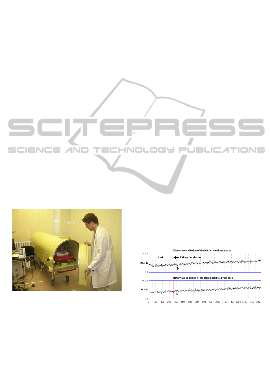
The general view of the measuring radiophysical
system is given on Fig. 1.
Figure 1: General view of the measuring radiophysical
system.
Except monitoring the brain own microwave
radiation by measuring, the radiophysical system
allows one to record simultaneously the heart rate
variability and to assess changes in the autonomic
nervous system in the real time mode. The choice of
this system is stipulated by the fact that the
autonomic nervous system is a part of nervous
system and coordinates activity of the systems
participating in conservation of dynamic balance of
vital signs, regulating a metabolism, excitability, and
automatism of an internal and the central nervous
system. Cooperating with the somatic nervous
system and endocrine system, it provides
maintenance of constancy of a homeostasis and
adaptation in varying environmental conditions. But
in this paper, these topics are not discussed.
During experiments, the patient was inside
screened cabins of the measuring radiophysical
system. Procedure of measurements included two
stages: functional rest and loading test. Time
intervals of staying the patient in each of these
stages were chosen long enough to exclude influence
of transient processes onto results of the research.
In analysis of dynamic parameters of time-and-
frequency characteristics of signals of the brain
microwave radiation, wavelet analysis was applied.
Analysis was implemented in the following three
frequency bands: (0.1 – 0.05) Hz, (0.02-0.013) and
less 0.013 Hz.
3.1 The Glucose Test
During experiments, the following regimes got out:
patient was in condition of functional rest within 300
sec. After that through gastrointestinal tract, the
fixed dose of an aqueous solution of glucose was
entered from calculation 0.2 gr glucose per 1 kg of
mass of the patient body. After the receiving the
glucose the time interval of observation was more
than 1500 sec.
In Figure 2, graphs of the target microwave
radiation signals from the measuring radiophysical
system MRTHR are shown during the test with the
glucose.
Figure 2: Signals on an exit of the measuring
radiophysical system during the test with the glucose of
healthy patient X. (the instant of taking the glucose is
marked by a horizontal arrow, and the beginning of
reaction of radiation is marked by a vertical arrow).
In Table 1, values of the mean intensities of the
microwave radiation fluctuations in four frequency
bands in various phases of functional research are
given.
BIODEVICES2013-InternationalConferenceonBiomedicalElectronicsandDevices
320
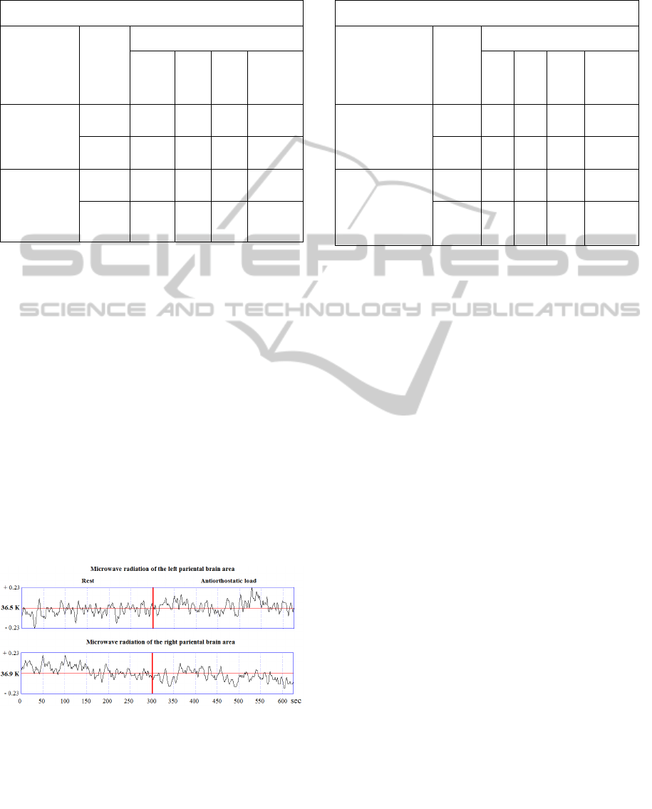
Table 1: Values of the mean intensities of the microwave
radiation fluctuations in various phases of functional
research.
Intensity of microwave radiation, К
Functional
status /
[time], sec
Area
Frequency bands, Hz
0.10,
0.05
0.05
,
0.02
0.02,
0.013
Below
0.013
Rest /
0, 300
Left 0,16 0.00 0,14 0,03
Right
0,18 0.00 0,18 0,12
Glucose
load /
300,1600
Left 0,21 0.00 0,19
0,46
Right
0,15 0.00 0,15
0,43
Taking the glucose, the trend of the brain own
microwave radiation is formed by (20 – 50) sec. The
largest changes of intensity are observed in the band
of frequencies below 0.013 Hz.
In Table 2, the values of these fluctuations are in
bold.
3.2 The Antiorthostatic Test
In these experiments three functional conditions of
the patient were formed: functional rest, an
antiorthostatic load and clinostatic load. The patient
was in each of these conditions within 300 sec.
In Figure 3, the target signals of microwave
radiation of the measuring radiophysical system
MRTHR are presented during the functional
researches with an antiorthostatic load of the healthy
patient G.
Figure 3: Signals on an exit of the measuring
radiophysical system during functional researches with an
antiorthostatic load of the healthy patient G.
In Table 2, values of the mean intensities of the
microwave radiation fluctuations in four frequency
bands in various phases of functional research are
given.
Table 2: Values of the mean intensities of the microwave
radiation fluctuations in various phases of functional
research.
Intensity of microwave radiation, К
Functional
status /
[time], sec
Area
Frequency bands, Hz
0.10
,
0.05
0.05
,
0.02
0.02,
0.013
Below
0.013
Rest /
0, 300
Left 0.14 0.00 0.14 0.06
Right
0.09 0.00 0.18 0.1
Antiorthostatic
load /
300, 600
Left 0.14 0.00
0.25
0.00
Right
0.14 0.00
0.27
0.01
After passage from the condition of the functional
rest into the condition defined by an antiorthostatic
load, the trend of the microwave radiation is formed
during (5 – 10) sec. The largest changes of
microwave radiation are observed in frequency
bands from 0.02 to 0.013 Hz. This corresponds to
range (50 – 80) sec of fluctuation period of
microwave radiation.
In Table 3, the values of these fluctuations are in
bold.
4 CONCLUSIONS
Results of theoretical and experimental researches of
the brain own microwave radiation in a band of
frequencies from 650 to 850 MHz show that its
fluctuations of the microwave radiation are objective
reflexion of the physiological changes in brain
tissues. Spectrum of these fluctuations in the
frequency range from 0.02 to 0.013 Hz mainly
reflects changes of dielectric permeability in tissues
in depth more than 10 mm and is a consequence of
the humoral processes. In the field of frequencies
below 0.013 Hz, intensity of fluctuations of brain
microwave radiation is defined by thermodynamic
changes in its tissues that is stipulated by metabolic
processes. These conclusions confirm legitimacy of
the hypothesis offered by us before about the
mechanism of transport of a liquid in intercellular
and intracellular spaces of a nervous tissue.
Note, also, that in our researches the
radiophysical system was applied. This is non-
invasive, diagnostic, and meets the concept of
development of the modern public health.
MicrowaveRadiationasInterfacetotheBrainFunctionalState
321

This allows us to regard described approach as a
method for observation of functional changes in the
brain at early stages of formation of various
pathologies.
REFERENCES
Haken, H., 2006. A macroscopic approach to complex
system, Springer-Verlag. Berlin, Heidelberg, New
York, 3
th
edition.
Godik, E. E., Gulyaev, Y.V., 1991. Functional imaging
of the human body. IEEE Engineering in Medicine
and Biology Society, V. 10, Issue 4: 21-29.
Ivanov, K. P., 1990. Foundations of body energetics. The
total energy, heat transfer, and temperature control,
Nauka, Leningrad Branch. Leningrad.
Yablonskiy, D. A., Ackerman, J. J., Raichle, M. E., 2000.
Coupling between changes in human brain
temperature and oxidative metabolism during
prolonged visual stimulation. Proceedings of the
National Academy of Sciences of the USA, V.97, Issue
13: 7603–7608.
McElligott, J. C., Melzack, R., 1967. Localized thermal
changes evoked in the brain by visual and auditory
stimulation. Experimental Neurology, V.17, Issue 3:
293-312.
Trubel, H. K., Sacolick, L. I., Hyder, F., 2006. Regional
temperature changes in the brain during
somatosensory stimulation. Journal of Cerebral Blood
Flow & Metabolism, V. 26, Issue 1: 68-78.
Guyton, F. C., Hall, J. E., 2010. Textbook of Medical
Physiology, Elsevier Saunders. Philadelphia, 12
th
edition.
Kublanov, V. S., 2009. Radiophysical system for
examining functional state of a patient’s brain.
Biomedical Engineering, Vol. 43, № 3: 114-119.
BIODEVICES2013-InternationalConferenceonBiomedicalElectronicsandDevices
322
