
Efficacy of Beam Computer Tomography (CBCT) in Diagnosis of
Disease Lesions in Paranasal Sinuses
Edward Kijak
Department of Prosthetic Dentistry, Faculty of Medicine and Dentistry, Pomeranian Medical University,
Rybacka 1, 70-204 Szczecin, Poland
Keywords: Volumetric Tomography, TMJ Dysfunction, Sinus Disease.
Abstract: Different techniques of X-ray imaging often confirm the suspected diagnosis, but not infrequently redirect the
diagnostic process in other areas. A modern ultrahigh-resolution volumetric tomography, called a cone beam
computed tomography (CBCT) also, is one of the most innovative technique and able to visualise these ana-
tomical structures that conventional techniques are not. A differential diagnosis of TMJ dysfunction is partic-
ularly difficult due to the quantity of factors that influence the generation of symptoms. Laying on of symp-
toms that mask the main disease means that frequently, without additional examinations, it is not possible, in
an univocal way, to describe the type and extent of this disease. The study assesses the usefulness of volu-
metric tomography ( CBCT ) in an accidental detecting in the maxillary lesions sinuses of a temporomandib-
ular joints dysfunction. The analysis was performed on the base of 249 studies of volumetric tomography.
The face part of the skull was made by the a camera with a large imaging field (FOV) 17 cm x 23 cm i-CAT
Next Generation (ISI). It was found that a significant number of patients (almost half) with TMD have the
changes in paranasal sinuses. Based on the observations, the relevance and legitimacy of tested technique
helping in stomatognathic system diseases diagnosis was analysed.
1 INTRODUCTION
Different X-ray imaging techniques used in dentistry
have different accuracy of image of anatomical de-
tails of a diagnosed area (Różyło-Kalinowska, 2011).
The diagnostic value of conventional static and func-
tional panoramic imaging (including digital) largely
depends on human factors, mainly the skills and con-
sistency of a radiologist who performs the examina-
tion. An incorrect positioning of the examined pa-
tient, an incorrect angle of incidence during exposure
to radiation and instability of above factors can have
a negative influence on a detailed, correct analysis. In
addition, the conventional radiographic techniques
provide a two-dimensional back projection image
with overlapping various anatomical details. A tech-
nological progress in this area forces physicians to
change their approach to diagnostic algorithms.
Radiological examination using cone beam com-
puted tomography (CBTC) allows for the imaging of
hard and soft anatomical structures in three projec-
tions. CBTC provides an undistorted real-size image
allowing for accurate measurements. The accuracy of
images reconstructed by volumetric tomography is so
superior that all anatomical details in the area under
investigation do not require additional testing; a
three-dimensional image reconstruction gives a detail
insight into the topography of skeletal structures (Ró-
żyło-Kalinowska, 2009, Szkutnik, 2001). CBCT is
used also successfully in laryngology. Because of the
accuracy of this imaging technique it is possible to
diagnose and plan treatment for paranasal sinusitis
simultaneously. Importantly, the technique is safe for
patients since the dose of ionizing radiation is re-
duced. The average dose of radiation in CBCT is in
the range of 20-650 μSv depending on the size of an
imaging field and an image resolution (Jager, 2001,
Kijak, 2012, Loubele M, 2009, Bargan, 2010, Anjos,
2012) and it is about 20-30-fold lower than in con-
ventional multi-slice CT CBCT is increasingly being
used for diagnosis of patients with suspected dysfunc-
tion of the tempormandibular joints (TMD), suffering
from pain, located in the vicinity of the front of the
ear and middle part of the face. Differential diagnosis
of TMJ dysfunction is particularly difficult due to the
quantity of factors that could infuence the generation
of symptoms. The laying on of symptoms that mask
the basic disease means that frequently, without
148
Kijak, E.
Efficacy of Beam Computer Tomography (CBCT) in Diagnosis of Disease Lesions in Paranasal Sinuses.
DOI: 10.5220/0006657101480152
In Proceedings of the 11th International Joint Conference on Biomedical Engineering Systems and Technologies (BIOSTEC 2018) - Volume 2: BIOIMAGING, pages 148-152
ISBN: 978-989-758-278-3
Copyright © 2018 by SCITEPRESS – Science and Technology Publications, Lda. All rights reserved
additional examinations, it is not possible, in a univo-
cal way, to describe the type and extent of the disease.
Very often, the result of specialized laryngologi-
cal examination is negative and the patient returns to
a dentist. With non-specific subjective symptoms,
limited only to painful problems, the study of volu-
metric tomography leads to accidental detection of le-
sions in the maxillary sinuses. Often the patient is re-
ferred to a laryngologist or maxillofacial surgeon. It
was provided that these are not individual cases.
The aim of the study was the analysis of imagines
obtained by CBCT with a medium and large imaging
field in diagnosis of the lesions in paranasal sinuses
and commissioned causing dysfunction of temporo-
mandibular joints.
2 MATERIALS AND METHODS
2.1 Materials
The material consisted of 249 studies of volumetric
tomography. The face part of the skull was made by
a the camera with a large imaging field (FOV) 17 cm
x 23 cm i-CAT Next Generation (ISI).
Figure 1: Size and gender of the study group.
A total of 249 subjects (182 F and 67 M) aged 13
to 88 years were enrolled, who reported to the Depart-
ment of Prosthetic Dentistry Pomeranian Medical
University due to TMD-related ailments.
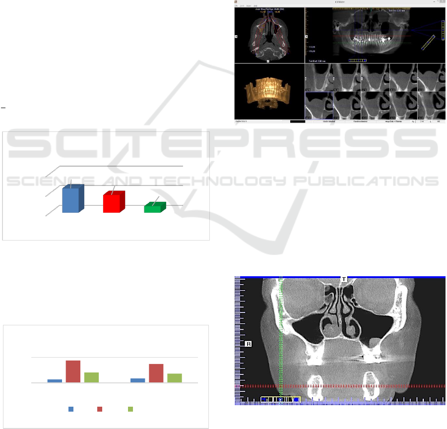
Figure 2: Age of tested people.
2.2 Methods
An important feature of the device is a short scanning
time from 5 seconds at low volumes up to 27 seconds
at the largest imaging field that can be adjusted by
varying the test resolution in the range of 0.125 to 0.4.
Patient’ sitting position with correctly setting head
minimizes the presence of motion artefacts. Using
Quantum IQ software, soft-tissue imagines were ob-
tained that were much better than other types of ima-
gines (Photo 1). The exposure parameters were 110
kV and 5 mAs. The exposure time was 14 seconds
and 0.3 voxel resolution was used the most com-
monly.
Photo 1: CBCT - large imaging field.
3 RESULTS
Volumetric tomography allows the diagnosis of even
the smallest pathological lesions located in the max-
illary sinuses (Photo 2) beginning from the most com-
monly diagnosed inflammatory conditions (edema)
of the sinus lining (Photo 3), through various types
and extent of hyperplasia (Photo 4) until the total air
loss of one of the bays – Photo 5.
Photo 2: Limited pathological change in the bottom of the
left maxillary sinus.
0
200
400
F + M F M
249
182
67
Study group - sex
13
17
88
74
40,53
36
0
100
F M
Group structure - age
MIN MAX MID
Efficacy of Beam Computer Tomography (CBCT) in Diagnosis of Disease Lesions in Paranasal Sinuses
149
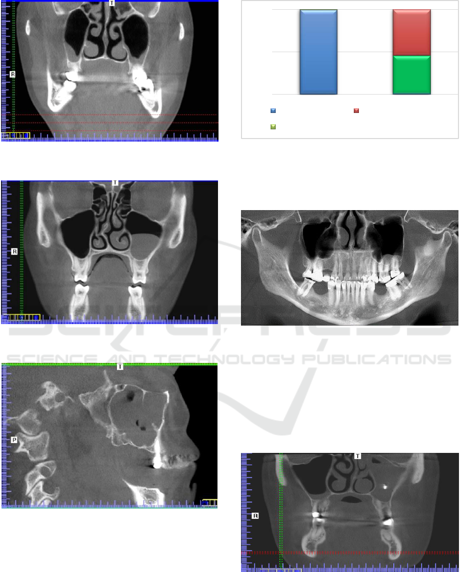
Photo 3: Oedematous mucosa of alveolar recess of the two
maxillary sinuses.
Photo 4: The disease process includes the entire light of the
right maxillary sinus.
Photo 5: The disease process includes the entire light of the
right maxillary sinus.
Figure 3: The incidence of lesions disease of maxillary si-
nuses detected by CBCT.
The studies have shown that the incidence of le-
sions is relatively high at almost 50%.
Photo 6: The root appex of the teeth 25, 26, 27 in the max-
illary sinus.
From the point of view of the therapeutic possibil-
ities available to the dentist, we are interested in teeth
related changes that are located in the alveolar recess
of the maxillary sinuses: roots in the sinuses (Photo
6),pushed through the endodontic treatment of the
material (Photo 7), and unerupted tooth located in the
immediate vicinity of the bay.
Photo 7: The material stuffed as a result of endodontic treat-
ment in the bottom of the left sinus.
CBCT and the capabilities of computer programs
that support this imaging method greatly extend the
249
114
135
0%
50%
100%
Number of studies No changes disease
Study with changes
45.78%
BIOIMAGING 2018 - 5th International Conference on Bioimaging
150
potential for the effective topological evaluation of
pathological lesions "accidentally" detected. Even
when they are diagnosed in a traditional pantomo-
graphic examination, a lesion derived from the alve-
olar recess as observed in a volumetric study, the di-
agnosis has to be change.
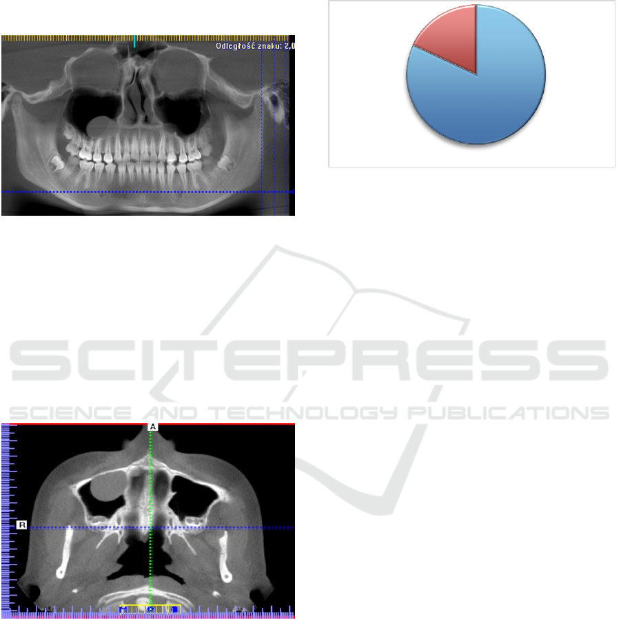
Photo 8: Disease lesion in the right maxillary sinus cavity -
image in volumetric tomography.
Photo 8 shows a classic pantomographic imaging
performed by CBTC, which was helpful in the diag-
nosis of a polyp lesion that originates from the alveo-
lar sinus of the right maxillary sinus. Further evalua-
tion of the same image diametrically changed our
view of this pathology (Photo 9). The disease is not
associated with the teeth because it is clearly visible
in the axial projection that it originates from the an-
tero-medial walls of the sinus.
Photo 9: The same lesion (Figure 8) - CBCT image in axial
projection.
The precise identification of the source of a dis-
ease process directs the further course of diagnostic
and therapeutic procedures. Random detected patho-
logical changes are not necessarily a direct cause of
ailments. It may reflect an asymptomatic condition
that is accompanied by a main disease. This should
not, however, slow down the doctor from further
diagnosis, which is becoming more targeted and eas-
ier. The disease lesions detected in both bays in the
study population recorded in more than one-fifth of
cases.
Figure 4: Disease lesions in both sinuses.
The CBCT images obtained, evaluated in a multi-
faceted image reconstruction, allow for the precise
definition of a disease process extention. Frequently,
a study of volumetric tomography, with a large field
of imaging, is a diagnostic procedure that is suffi-
ciently adequate to an implement proper treatment in
the therapeutic process.
4 DISCUSSION
Detection of various pathological processes occurring
in the sinus of the paranasal sinuses with the diagnosis
of dysfunction of the chewing organ is not uncommon
(Phothikhun, 2012). The accuracy of CBCT studies is
so significant that it is impossible to compare such
tests with traditional and routine radiology proce-
dures used in dentistry (Różyło- Kalinowska, 2009,
2011). The ability to choose the size of the imaging
(cone), gives the possibility of the radiological pro-
tection by lowering the dose of ionizing radiation. In
the case of functional abnormalities of the chewing
organs, the multitude of anatomical and physiological
details cause a multi-faceted pain. Symptoms often
correlate with the clinical picture of the stomatog-
nathic system functional evaluation (Anjos, 2012).
Often, a dentist dealing with the problem faces a di-
lemma: adhere to radiological protection rules and
commission CBCT examinations limited to the tem-
poral and temporal joints, or extend the examination
to adjoining areas. The choice is difficult especially
in the absence of pathognomonic symptoms. Radio-
logical protection should be superior. Adherence to
the principles developed by the European Academy
of DentoMaxilloFacial Radiology (EADMFR)
should not, however, limit diagnostic possibilities
Zmiany w obu zatokach
(55)
(249)
22.43%
77.56%
Efficacy of Beam Computer Tomography (CBCT) in Diagnosis of Disease Lesions in Paranasal Sinuses
151

(Scarfe, 2008)). In case of maxillofacial dysfunction
there are unfortunately no uniform procedures. The
difference in dose of ionizing radiation between pan-
tomographic imaging supplemented with conven-
tional sinus radiographs and CBCT with medium to
large imaging with modern imaging is not as signifi-
cant (Loubbele, 2008). The benefit of the evaluation
of paranasal sinuses in volumetric thomography is un-
doubtedly greater (Rege, 2012), so it should account
for the balance of gains and losses.
5 CONCLUSIONS
Volumetric Tomography is a valuable x-ray imaging
technique useful in the differential diagnosis of TM
dysfunction. The laying on of symptoms that mask
the main disease means that frequently, without addi-
tional examinations, it is not possible, in a univocal
way, to describe the type and extent of a disease. The
diagnosis of functional disorders of the muscular of
the chewing systems should take into account the dis-
ease lesions in the maxillary sinus. Detection of vari-
ous pathological processes occurring in the sinuses
paranasal, during of the diagnosis of dysfunction of
the chewing organ is not uncommon.
Limiting the imaging field in a volumetric tomog-
raphy study significantly reduces the diagnostic po-
tential of the study. The advantage of the possibility
of additional evaluation of the collateral sinuses and
the diagnosis of the underlying disease process is un-
doubtedly greater.
REFERENCES
Anjos Pontual M.L., Freire J.S., Barbosa J.M. et al., 2012.
Evaluation of bone changes in the temporomandibular
joint using cone beam CT. Dentomaxillofacial Ra-
diol.;41(1):24–29.
Bargan S., Merrill R., Tetradis S. 2010. Cone beam com-
puted tomography imaging in the evaluation of the tem-
poromandibular joint. J Calif Dent Assoc, 1;38(1):33–
39.
Howerton-W.B.Jr., Mora-M.A., 2008. Advancements in
digital imaging. What is new and on the horizon? J.
Am. Dent. Assoc., 139, Suppl. 3, 20S ‑24 Jager L.,
Rammelsberg P., Reiser M., 2001. Bildgebene Diagnostik
der Normalanatomie des Temporomandibulargelenks,
Radiologe, (41), 734-740.
Kijak E., Lietz-Kijak D., Frączak B., Cieślińska-Wilk G.,
2012. Use of x-ray and electronic functional tests in the
diagnosis of temporal and temporal joint dysfunction.
Journal Stomatolog (22), 28-33.
Loubbele-M et al. 2008. Image quality vs radiation dose of
four cone beam computed tomography scaners.
Dentoxillofac. Radiol., 37, 6, 309-319.
Loubbele M.,Bogaerts R., Van Dijck E., Pauwels R.,
Vanheusden S., Suetens P., Marchal G., Sanderink G.,
Jacobs R.: 2009. Comparison between effective radia-
tion dose of CBCT and MSCT scanners for dentomaxil-
lofacialis applications. Eur. J. Radiol., (71), 461-468.
Phothikhun S., Suphanantachat S., Chuenchompoonut V. et
al., 2012. Cone beam computed tomographic evidence
of the association between periodontal bone loss and
mucosal thickening of the maxillary sinus. J
Periodontol.; 83(5):557–564.
Rege I. C., Sousa T. O., Leles C. R. et al., 2012. Occur-
rence of maxillary sinus abnormalities detected by cone
beam ct in asymptomatic patients. BMC Oral Health.
12, 30.
Różyło-Kalinowska I., 2009. The standards of the Euro-
pean Academy of Radiology Oral and Maxillofacial on
volumetric imaging (CBCT). . Journal Stomatolog, 6,
12-16.
Roberts J. A., Drage N. A., Davies J., Thomas D. W., 2009.
Effective dose from cone beam CT examinations in
dentistry. Brit. J. Radiol., 82, 35-40.
Różyło-Kalinowska I., Różyło T. K., 2009. Volumetric im-
aging options for a dental patient. Journal Stomatolog
(5), 18-23.
Różyło-Kalinowska I., Różyło T.K., Taras M., 2009, Use
of volumetric imaging in general dental diagnosis.
Your Stomat Review (YSR), (3), 77 ‑80.
Różyło-Kalinowska I., Różyło T. K., 2011 The book, Volu-
metric tomography in dental practice. The publishing
company Czelej. 1
st
edition.
Scarfe W. C., Farman A. G., 2008. What is cone beam and
how does it work? Clin. North AM., 52, 4, 707-730.
Sztuk S., Urbanik A., Stypułkowska J., 2001. Functional
imaging of temporal and temporomandibular joints us-
ing 3D computer tomography reconstruction. Polish
Radiology Review, (65), 21-24.
Rege I. C., Sousa T. O., Leles C. R. et al.: Occurrence of
maxillary sinus abnormalities detected by cone beam ct
in asymptomatic patients. BMC Oral Health. 2012, 12,
30.
BIOIMAGING 2018 - 5th International Conference on Bioimaging
152
