
Feasibility of Eye-tracking based Glasses-free 3D Autostereoscopic
Display Systems for Medical 3D Images
Dongwoo Kang, Seok Lee, Hyoseok Hwang, Juyong Park, Jingu Heo, Byongmin Kang,
Jin-Ho Lee, Yoonsun Choi, Kyuhwan Choi and Dongkyung Nam
Multimedia Processing Lab, Samsung Advanced Institute of Technology, Suwon-si, South Korea
Keywords: Three-dimension, 3D, Glasses-free 3D Autostereoscopy, Eye-tracking, Medical 3D, Cardiac CT, Coronary
CTA, 3D Heart, Display.
Abstract: Medical image diagnosis processes with stereoscopic depth by 3D display have not been developed widely
yet and remain understudied Many stereoscopic displays require glasses that are inappropriate for use in
clinical diagnosis/explanation/operating processes in hospitals. An eye-tracking based glasses-free three-
dimensional autostereoscopic display monitor system has been developed, and its feasibility for medical 3D
images was investigated, as a cardiac CT 3D navigator. Our autostereoscopic system uses slit-barrier with
BLU, and it is combined with our vision-based eye tracking system to display 3D images. Dynamic light field
rendering technique is applied with the 3D coordinates calculated by the eye-tracker, in order to provide a
single viewer the best 3D images with less x-talk. To investigate the feasibility of our autostereoscopic system,
3D volume was rendered from 3D coronary CTA images (512 by 512 by 400). One expert reader identified
the three main artery structures (LAD, LCX and RCA) in shorter time than existing 2D display. The reader
did not report any eye fatigue or discomfort. In conclusion, we proposed a 3D cardiac CT navigator system
with a new glasses-free 3D autostereoscopy, which may improve diagnosis accuracy and fasten diagnosis
process.
1 INTRODUCTION
Three-dimensional (3D) medical imaging techniques
are increasingly employed for evaluation of not only
identifying complex organ structures but also
diagnosing abnormalities. Recent advanced 3D
imaging techniques such as MR, CT and Ultrasound
showed the usefulness and evoked the demand of 3D
medical imaging displaying system. Also 3D
graphics techniques have been developed fast, which
enables high quality 3D medical volume rendering
(Chan et al., 2013, Ferroli et al., 2013, Langdon et al.,
2014). However, the advanced and complex 3D
medical images are displayed with 2D monitors,
where 3D objects are projected into 2D plane.
3D displays have become available these days.
Especially, three-dimensional movies presented via
stereoscopic displays have become more popular in
recent years aiming at a more engaging viewing
experience. However, medical image diagnosis
processes with stereoscopic depth by 3D display have
not been developed widely yet and remain
understudied.
Additionally, many stereoscopic displays require
glasses that are inappropriate for use in clinical
diagnosis/explanation/operating processes in
hospitals. A few studies of 3D autostereoscopy in
medical image analysis area exist (Narita et al., 2014).
A glass-free 3D autostereoscopic display monitor
has been developed by Samsung Advanced Institute
of Technology (SAIT) (Park and Nam, 2012, Park et
al., 2013) which provides almost same 3d quality as 3D
display that requires 3D glasses: ≤5% crosstalk.
We investigated "3D display needs" from medical
doctors in various medical departments from
Samsung Medical Center (SMC). Based on the
collected opinions, we developed a new application
of glass-free medical 3D: Cardiac CT 3D Navigator.
One of the medical imaging systems that require
3D display system is cardiac CT due to complex
anatomy of a heart and isotropic 3D volume of
cardiac CT. 3D display for cardiac CT diagnosis
without glasses techniques may enable physicians
and medical doctors to make a detailed/accurate
134
Kang, D., Lee, S., Hwang, H., Park, J., Heo, J., Kang, B., Lee, J-H., Choi, Y., Choi, K. and Nam, D.
Feasibility of Eye-tracking based Glasses-free 3D Autostereoscopic Display Systems for Medical 3D Images.
DOI: 10.5220/0005826901340138
In Proceedings of the 9th International Joint Conference on Biomedical Engineering Systems and Technologies (BIOSTEC 2016) - Volume 2: BIOIMAGING, pages 134-138
ISBN: 978-989-758-170-0
Copyright
c
2016 by SCITEPRESS – Science and Technology Publications, Lda. All rights reserved
diagnosis and facilitate diagnosis processes.
In this paper, we introduce our glasses-free
autostereoscopic display to medical society, and
discuss the feasibility as a medical 3D display, which
may help improved diagnosis accuracy and fastened
diagnosis process, with an application with cardiac
CT data.
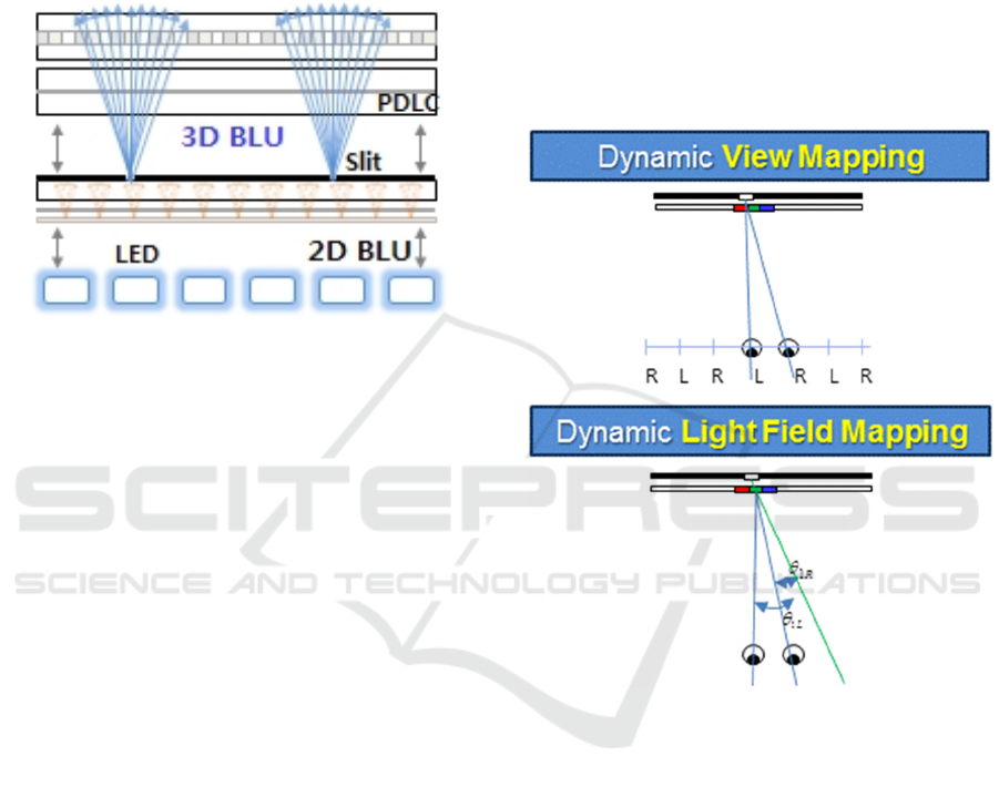
Figure 1: Slit-barrier with BLU composition.
2 METHOD
We developed a new 3D diagnosis system with a new
glasses-free display technique. The new glasses-free
display system is based on eye-tracking technique and
slit-barrier with back light unit (BLU) device
technique. The new system was applied to 3D cardiac
CT, for a new glass-free cardiac CT 3D navigator
system.
2.1 Eye-tracking based Glasses-free 3D
Autostereoscopic Display
Our 3D display consists of display panel, optical
element for 3D and camera. The 4K liquid crystal
display (LCD) panel was used. The optical element
for 3D device is a device that controls the direction of
lights which pass through the panel, where our
display system is based on slit-barrier with BLU
technique. The slits in the barrier allow only left
image pixels to pass into left eye position, right image
pixel to pass into right eye position. Our slit-barrier
locates behind LCD panel and in front of BLU
(Figure 1).
The camera is used for eye-tracking, where
machine learning based eye-tracking algorithm was
applied. With this eye-tracking algorithm, the viewer
doesn’t have to find the good position to see the 3D
properly, but can see the 3D in any position that
tracking is allowed. The eye-tracker identifies the
viewer’s the 3D coordinates of the pupil center, the
subpixel values for the left and right views in display
panel are adjusted to be seen correctly by viewer's
eyes.
The real-time machine learning based eye-
tracking algorithm starts from face detection using the
AdaBoost learning algorithm (Freund and Schapire,
1997). From the detected face region, subregions with
eyes are extracted by shape alignments using
Supervised Descent Method (SDM) (Xiong et al.,
2013). 23 landmark points of eyes, nose and mouth
were used for SDM shape alignments.
Figure 2: 3D rendering techniques. Our autostereoscopy
uses dynamic light field mapping method.
3D Light ray image is generated by a 3D
rendering algorithm, where each pixel’s light ray
direction was determined by the slit-barrier.
Especially, based on eye tracking algorithm,
Dynamic Light Field Mapping (DLFM) is applied for
the 3D rendering (Park and Nam, 2012, Park et al.,
2013). Using the position and direction information
of each light, each light is mapped to the 3D eye
coordinates. While Dynamic View Mapping (DVM)
3D rendering method (Boev et al., 2008), which many
vendors uses currently just switches left/right images
according to the eye position, DLFM maps each light
ray to eye coordinates (Figure 2). Because a DLFM
technique utilizes the photorefractive effect of each
ray, viewer can see the 3D images without the
limitation of viewing distance. Also DLFM can solve
Feasibility of Eye-tracking based Glasses-free 3D Autostereoscopic Display Systems for Medical 3D Images
135
the image degradation problem of large 3D displays.
The overall specification of our 3D
autostereoscopic system is shown in Table 1, and the
prototype of our display is shown in Figure 3.
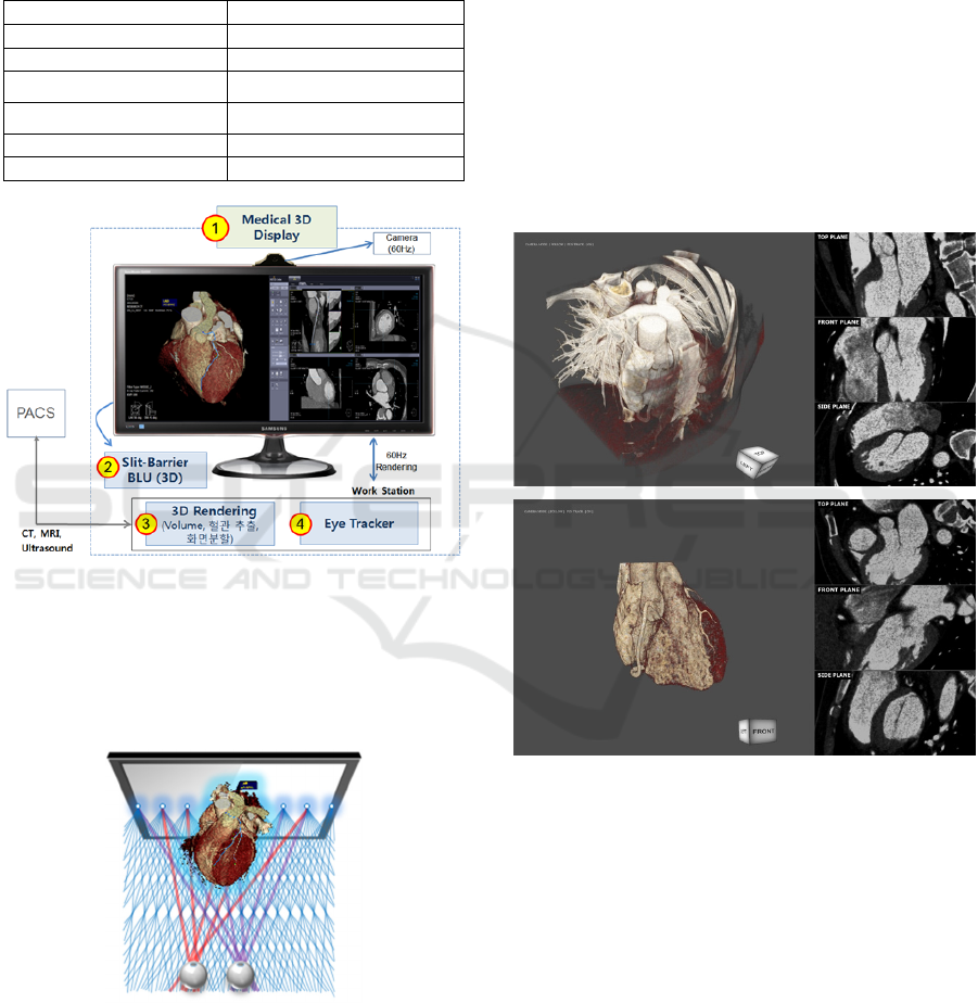
Table 1: Eye-tracking based autostereoscopic display
system specification.
Panel size 31.5 Inch
2D resolution 3840x2160
3D resolution 1536x720
Viewing distance
100cm
±50cm
Viewing Angle
H60
°/V40°
User 1 person only
Frame Rate 60Hz
Figure 3: Prototype of eye-tracking based autostereoscopic
display system for medical 3D.
2.2 3D Cardiac CT Navigator: 3D
Autostereoscopy Visualization
Feasibility
Figure 4: 3D cardiac CT navigator concept.
3D coronary CT angiography (CTA) images were
visualized with our 3D eye-tracking based 3D
autostereoscopic display system. Under advice of
medical doctors at SMC, we made a 3D cardiac CT
navigator software. A 3D CTA anonymized image
data was obtained from SMC. The CTA dataset was
acquired on the dual-source 64-slice CT scanner
(Definition Siemens Medical Solution, Germany)
with a gantry-rotation time of 330mms and standard
collimation of 0.6mm, and had excellent image
quality. 3D CTA scan parameters were 512x512
matrix, voxel size 0.38x0.38x0.3mm
3
, and 412 slices.
The patient did not have any luminal stenosis or
plaque.
The proposed navigator system aims identifying
the 3D structure of the complex organs easily, and we
developed a 3D cardiac CT navigator proto as an
example (Figure 4). With help of enhanced 3D depth
perception in our 3D display, viewers can recognize
the complex 3D structure in depth.
Figure 5: 3D cardiac CT navigator S/W proto. Original CT
volume is rendered without any segmentation (up) and
whole segmentation (down).
For 3D visualization, we followed the standard
cardiac CT image and graphics processing from a
coronary CTA image dataset: (1) whole heart
segmentation, (2) coronary artery segmentation, and
(3) 3D volume rendering. User can adjust the color
and transparency of 3D volumes by option. Also,
multi-planar reconstruction (MPR) is aligned with 3D
volume rendering by side (Figure 5). Further, our
software has an option of converting the volume to
3D mesh, which graphics artists can decorate
manually.
One expert reader from SMC was asked to
BIOIMAGING 2016 - 3rd International Conference on Bioimaging
136
identify the structure of heart with our 3D
autostereoscopy. In a segmented heart only 3D
volume, the expert reader identified main heart
structures including three main coronary artery
structures (LAD, LCX and RCA).
3 RESULTS AND DISCUSSION
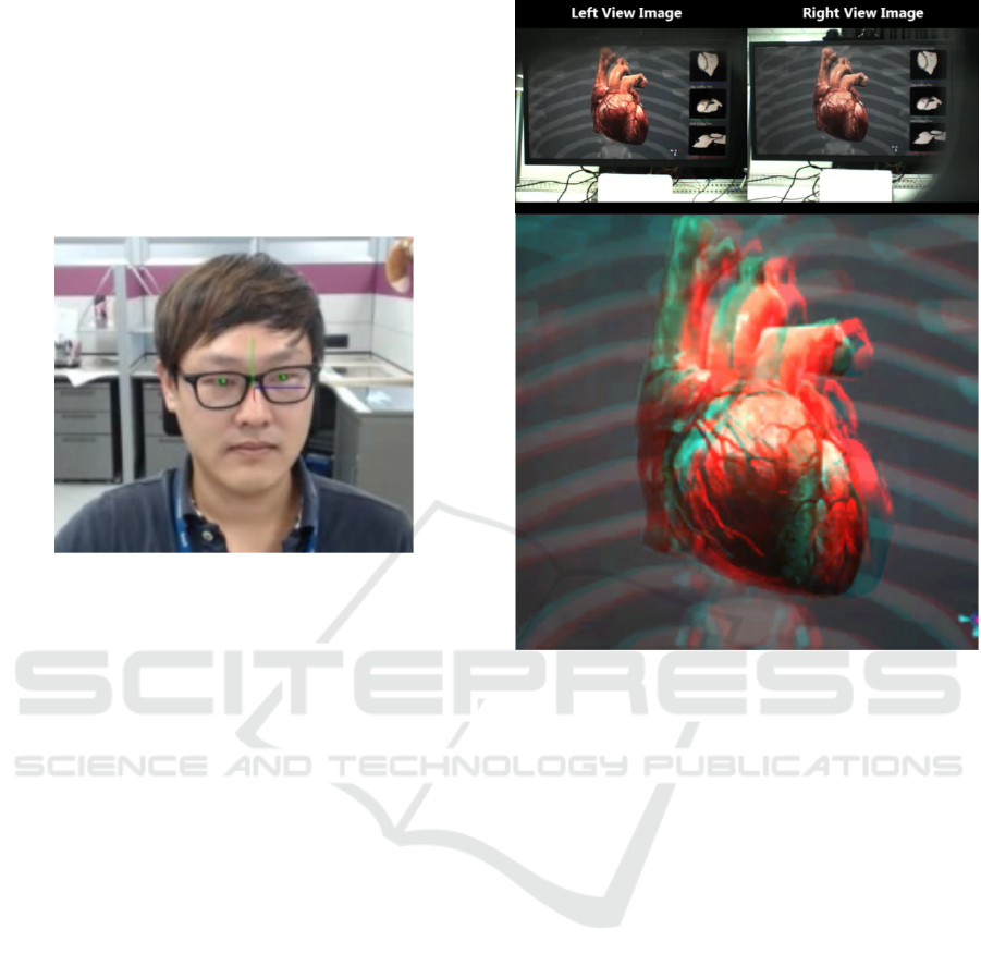
Figure 6: Eye-tracking for 3D autostereoscopy.
A 3D coronary CTA image was tested with our 3D
glasses-free autostereoscopic display system and 3D
cardiac CT navigator software. The average error of
the eye-tracking was 2mm, which was calculated as
Euclidean distance between the center of pupils and
tracked eye coordinates (Figure 6). Also the tracking
time of eyes was 16ms in average. The average
crosstalk of our 3D display was 4.9%, which was
similar 3D quality to that of 3D display with glasses.
With dynamic light field rendering method, 2 stereo
images (left and right images) were generated for 3D
display, making the depth 20cm (Figure 7). A
standard 2.5 GHz personal computer running
windows 7 was used for running the cardiac software.
The rendered 3D heart volume is 3D light field
rendered by dynamic light field mapping method for
displaying in our 3D autostereoscopy. All the
processes ran in the real time.
The expert reader visually assessed the quality of
our 3D autostereoscopy. He did not report any eye
fatigue or discomfort. Also, he identified the 3D heart
structure with our 3D autostereoscopy, including 4
chambers, aorta, main coronary arteries (LAD, LCX
and RCA). He didn’t provide the quantity assessment
but reported he could identify the 3D structure faster
and easier. Further investigation is required with
quantification for testing usefulness of our 3D
autostereoscopy: this is a limitation of our study. Also
number of cases
Figure 7: Stereo images from 3D mesh for a 3D CT
volume(up) and generated anaglyph (down). A graphics
artist decorated the heart manually.
4 CONCLUSIONS
We proposed a 3D cardiac CT navigator system with
our new glasses-free 3D autostereoscopy. We
introduced the feasibility of 3D autostereoscopy for
medical image diagnosis. It may improve diagnosis
accuracy and fasten diagnosis process. Our 3D
autostereoscopic system can be applied any 3D
volumetric medical images.
ACKNOWLEDGEMENTS
We thank Dr. Jinho Choi at Samsung Medical Center
(SMC) for all the advice for the medical 3D navigator
system.
REFERENCES
Chan, S., Conti, F., Salisbury, K. & Blevins, N. H., 2013,
Virtual reality simulation in neurosurgery: technologies
Feasibility of Eye-tracking based Glasses-free 3D Autostereoscopic Display Systems for Medical 3D Images
137

and evolution, Neurosurgery, 72(1), 154-164.
Ferroli P., Tringali, G., Acerbi, F., Schiariti, M., Broggi, M.
Aquino, D. & Broggi, G., 2013, Advanced 3-
dimensional planning in neurosurgery, Neurosurgery,
72(1), 54-62.
Langdon, W. B., Modat, M., Petke, J., Harman, M., 2014,
Improving 3D medical image registration CUDA
software with genetic programming, Proceedings of the
2014 Annual Conference on Genetic and Evolutionary
Computation, 951-958.
Narita, Y., Tsukagoshi, S., Suzuki, M., Miyakita, Y., Ohno,
M., Arita, H., Saito, Y., Kokojima, Y., Watanabe, N.,
Moriyama, N. & Shibui, S., 2014, Usefulness of a glass-
free medical three-dimensional autostereoscopic
display in neurosurgery, International Journal of
Computer Assisted Radiology and Surgery, 9(5), 905-
911.
Park, J., Nam, D., 2012, Active light field rendering in
multi-view display systems, SID International
Symposium, 36-39.
Park, J., Nam, D., Choi, S. Y., Lee, J., Park, D. S., & Kim,
C. Y., 2013, Light Field Rendering of Multi-view
Contents for High Density Light Field 3D Display, SID
International Symposium, 667-670.
Freund, Y., Schapire, R.E., 1997, A Decision-Theoretic
Generalization of on-Line Learning and an Application
to Boosting, Journal of Computer and System Sciences,
119-139.
Xiong, X., Torre, F. D. L., 2013, Supervised Descent
Method and its Applications to Face Alignment, CVPR,
532-539.
Boev, A., Georgiev, M., Gotchev, A. & Egiazarian, K.,
2008, Optimized Single-viewer Mode of Multiview
Autostereoscopic Display, EUSIPCO, 25-29.
BIOIMAGING 2016 - 3rd International Conference on Bioimaging
138
