Echocardiography Guidance and Evaluation of Myocardial Septal
Myectomy with a Novel Device
Magnus Dencker
1
and Henrik Bjursten
2
1
Department of Medical Imaging and Physiology, Skåne University Hospital, Malmö, Sweden
2
Department of Thoracic Surgery, Skåne University Hospital, Lund, Sweden
Keywords: Hypertrophic Obstructive Cardiomyopathy, Myectomy, Echocardiography.
Abstract: Hypertrophic obstructive cardiomyopathy represents a significant clinical problem. The objective with this
investigation was to address if echocardiography guide and monitor myocardial septal myectomy with a
novel device. An experimental porcine model was used. The findings were that echocardiography can
successfully be used to guide a novel procedure for minimally invasive surgical myectomy. Moreover,
echocardiography can be used to hemodynamically monitor this procedure. Finally, echocardiography can
be used to evaluate the result of the myectomy.
1 INTRODUCTION
Hypertrophic obstructive cardiomyopathy with
dynamic left ventricular outflow tract obstruction
often leads to progressive heart failure, and also
sudden death in some patients (Maron, 2013).
Surgical intervention with myectomy has, in the
past, been the primary strategy for treatment (Maron,
2011). Alcohol septal ablation has in recent years
been introduced as a less invasive alternative (Alam,
2006). The efficacy of this procedure has been
questioned (Yacoub, 2005). We performed the first
test ever of a novel surgical device for minimally
invasive surgical myectomy (Septulus). The
objective was to address following questions; 1)
Could echocardiography guide this procedure? 2)
Could echocardiography be used to
hemodynamically monitor the procedure? 3) Could
echocardiography be used to evaluate the result of
the myectomy?
2 METHODS
An experimental porcine model was used. Two adult
pigs were anesthetized and minimally invasive
surgical myectomy was performed with the
Septulus, introduced into the left ventricle from the
apical approach. Echocardiography examinations
were performed with CX-50 cardiac ultrasound
system with S5-1 probe (Philips Medical Systems,
Best, The Netherlands). The porcine equivalent of
parasternal long-axis was used to visualize septum
and the left ventricular outflow tract.
3 RESULTS
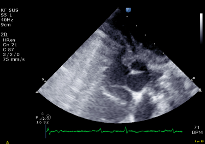
Both the Septulus and the septal portion of the left
ventricle could successfully be imaged in both pigs
(figure 1). The procedure was successfully guided
by the echocardiography images. Moreover, stroke
volume could be calculated from pulsed Doppler
recordings obtained in the left ventricular outflow
tract (figure 2). The results from the myectomy
could be recorded post-operatively (figure 3).
Figure 1: Shows the divice at the septum.
Dencker, M. and Bjursten, H..
Echocardiography Guidance and Evaluation of Myocardial Septal Myectomy with a Novel Device.
Copyright
c
2015 by SCITEPRESS – Science and Technology Publications, Lda. All rights reserved
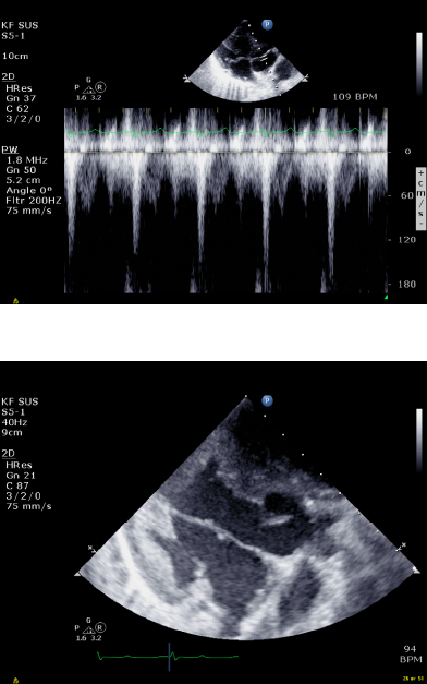
Figure 2: Shows Doppler recordings.
Figure 3: Shows the results from the Myectomy post-
operatively.
4 CONCLUSION
Echocardiography can successfully be used to guide
a novel procedure for minimally invasive surgical
myectomy. Echocardiography can be used to
hemodynamically monitor this procedure.
Echocardiography can be used to evaluate the result
of the myectomy.
REFERENCES
Alam M, Dokainish H, Lakkis N. Alcohol Septal Ablation
for Hypertrophic Obstructive Cardiomyopathy: A
Systematic Review of Published Studies. J Interven
Cardiol. 2006;19:319-27.
Maron BJ, Maron MS. Hypertrophic cardiomyopathy.
Lancet. 2013:381:242-55.
Maron BJ, Yacoub M, Dearani JA. Controversies in
cardiovascular medicine. Benefits of surgery in
obstructive hypertrophic cardiomyopathy: bring septal
myectomy back for European patients. Eur Heart J.
2011:32:1055-8.
