
A Non-linear Finite Element Model for Assessment of Lumbar Spinal
Injury Due to Dynamic Loading
Alexander Tsouknidas
1, 2
, Savvas Savvakis
3
, Nikolaos Tsirelis
3
Antonios Lontos
1
and Nikolaos Michailidis
2
1
Mechanical Engineering Department, Fredereick University, Nicosia, Cyprus
2
Laboratory of Physical Metallurgy, Mechanical Engineering Department, Aristoteles University of Thessaloniki,
Thessaloniki, Greece
3
BETA CAE Systems S.A., Thessaoniki, Greece
Keywords: Lumbar Spine, Non-linear FEA, Critical Stress Development.
Abstract: In this paper a highly detailed model of an adult lumbar spine (L1-L5) was recreated based on Computed
Tomography. Next to the viscoelastic deformation of the intervertebral discs, cortical and cancellous bone
anisotropy was considered, while seven types of ligaments were simulated either by solid or cable elements.
The dynamic behaviour of the spine segment was assessed through stress-strain curves, provoking a non-
linear response of all implicated tissues’ material properties. The model was subjected to dynamic loading
to determine abnormalities in the anatomy’s stress equilibrium that could provoke gait disturbances. Results
indicated the introduced methodology as an effective alternative to in vitro investigations, capable of
providing valuable insight on critical movements and loads of potential patients, as the model can be
employed to optimize therapeutic training or threshold kinematics of any given lumbar spine pathology.
1 INTRODUCTION
The lumbar spine is arguably one of the most
important structural elements of our musculoskeletal
system and as such, dominates complaints received
by orthopaedics. Epidemiologic studies indicate that
lumbar spine pathologies in industrialized
environments have a life-time prevalence of about
70% (Waters et al., 1993), ranging from low back
pain to disc protrusion and spinal fractures, which
can occur during every day activities such as
running.
Finite Element (FE) models of the lumbar spine
become increasingly popular during preoperative
preparation of complex surgeries, customized
implant design and recently optimization of non-
invasive therapeutic intervention. This can be
attributed to the capacity of FE to visualize stress
distributions over the entirety of the examined
anatomy and indicate critical regions.
Linear elastic 3D FE models of spine parts,
simulating their biomechanical response (Little et
al., 2010), (Wang et al., 2006) or investigate trauma
related surgical treatment (Ashish and Pramod,
2009), have been repeatedly introduced over the last
years. To describe however, the biomechanics of
spinal injury, non-linear properties should be
considered (Xiao et al., 2011); (Schmidt et al.,
2006).
Models are conventionally recovered though
Computed Tomography (CT) which is considered as
the golden standard in spine reconstruction (Klinder
et al, 2009) while intervertebral discs were reverse
engineered based on the scanned anatomical
characteristics (Tsouknidas et al., 2012). The
majority of available investigations consider the
remaining connective tissue (ligaments) as cable
elements, able of enduring only tension (Davidson-
Jebaseelan et al., 2010). Even at a non-linear state,
two node link elements are not capable of reflecting
biomechanical alterations of the tissue i.e.
degeneration or fracture. These are however highly
important, as deterioration of ligamentous properties
foster several spine pathogenesies due to increased
range of motion within a spinal unit. El-Rich et al.,
(2009) introduced a two vertebral spine model with
three- and four-nodal elements whereas three
dimensional solid elements were also considered in
studies with simplified geometrical characteristics
292
Tsouknidas A., Savvakis S., Tsirelis N., Lontos A. and Michailidis N..
A Non-linear Finite Element Model for Assessment of Lumbar Spinal Injury Due to Dynamic Loading.
DOI: 10.5220/0004236902920295
In Proceedings of the International Conference on Bioinformatics Models, Methods and Algorithms (BIOINFORMATICS-2013), pages 292-295
ISBN: 978-989-8565-35-8
Copyright
c
2013 SCITEPRESS (Science and Technology Publications, Lda.)

and linear properties (Tsuang et al., 2008).
In this investigation, a bio-realistic model of an
entire lumbar spine with regard to non-linear
material properties and partially solid ligaments, is
introduced, in an effort to allow the quantification of
the effect of mobility and/or loading scenarios on the
occurring spine biomechanics.
2 ANALYTICAL MODEL
During the reconstruction of the lumbar spine (L1-
L5) high resolution CT were the imaging modality
of choice due to their ability to demonstrate high
inherent image contrast between bone and soft
tissue. This enabled relatively unhindered
segmentation of the bone from soft tissue. In order
to achive the reconstruction of the desired bony
tissue, consecutive CT scan slices, were overlayed
(Tsouknidas et al., 2012); (Kobayashi et al., 2009).
Based on this concept, a patients lumbar spine was
scanned in its entirety from below the lower
boundaries of L1 to the upper limit of L5 ensuring
the full 3D representation of the examined area.
Data acquisition was in accordance to DICOM
(Digital Imaging and Communications in Medicine)
and interpolation of the CT information ensured an
isotropic data set. Although this process did not
result in higher resolution of the reconstructed set of
vertebra, it lead to smoother representation allowing
the distinct removal of the remaining soft tissue in
close proximity to the bone.
After the representation of the surfaces the
resulting volumes were generated considering an
outer cortex for each vertebra corresponding to the
cortical bone with a thickness of 0.5mm. The
remaining volume of each vertebra was considered
as cancellous bone.
Due to the severely altering density and the
inhomogeneous tissue of the intervertebral discs,
these were reverse engineered based on the superior
and inferior surface of the connecting vertebral
bodies as described by Tsouknidas et al., (2011).
During the model set up, anterior logitudial
(ALL), posterior logitudial (PLL), intertransverse
ligmante (ITL) and supraspinous ligament (SSL)
were modelled by four uniform layers of hexahedral
elements 0.3mm in thickness each, in order to
encapture their viscoelastic response (Sharma et al.,
1995). The remaining connective tissue, flavum (FL)
and capsular (JC) were considered as two node
tension elements. The simulated geometry,
emphasizing on some of its critical structural
elements, is demonstrated in figure 1.
Figure 1: Meshed lumbar spine model and details of
critical structural elements.
The annulus ground substance of the intervertebral
discs, was meshed by hexahedral elements to
facilitate the implementation of collagen fibers
positioned crosswise within the tetrahedron
structure. The remaining model, nucleus pulposus
and vertebrae, composes of tetrahedral elements and
the unhindered connection at the models contact
areas (hexa - tetrahedral elements interface) was
ensured through the diametrical incision of two
triangles in every rectangle, maintaining the same
nodes throughout the intervertebral disc surface and
the vicinical vertebrae. The same approach was
employed for the solid ligaments (ALL, PLL, ITL
and SSL). These ligaments were modelled through
the stress strain curves illustrated in figure 2
accurate way.
Intrinsic properties were considered for the
remaining ligamentus tissue. Their Young’s module
and poisson ratio as well as the cross sectional area
of each cable element used within the model
originated from literature data (Shirazi-ald et al.,
1984); (Smit et al., 1997).
The annulus fibrosus was considered to exhibit a
incompressible fluid like behaviour in order to
enrapture its hyperelastic response. In order to do so,
the Mooney-Rivlin strain energy density function
was employed as describer by Xiao et al., (2011).
The mechanical properties of bony tissue were
described by the Johnson-Cook elasto-plastic
material law considering strain rate dependent
stress-strain curves.
ANon-linearFiniteElementModelforAssessmentofLumbarSpinalInjuryDuetoDynamicLoading
293
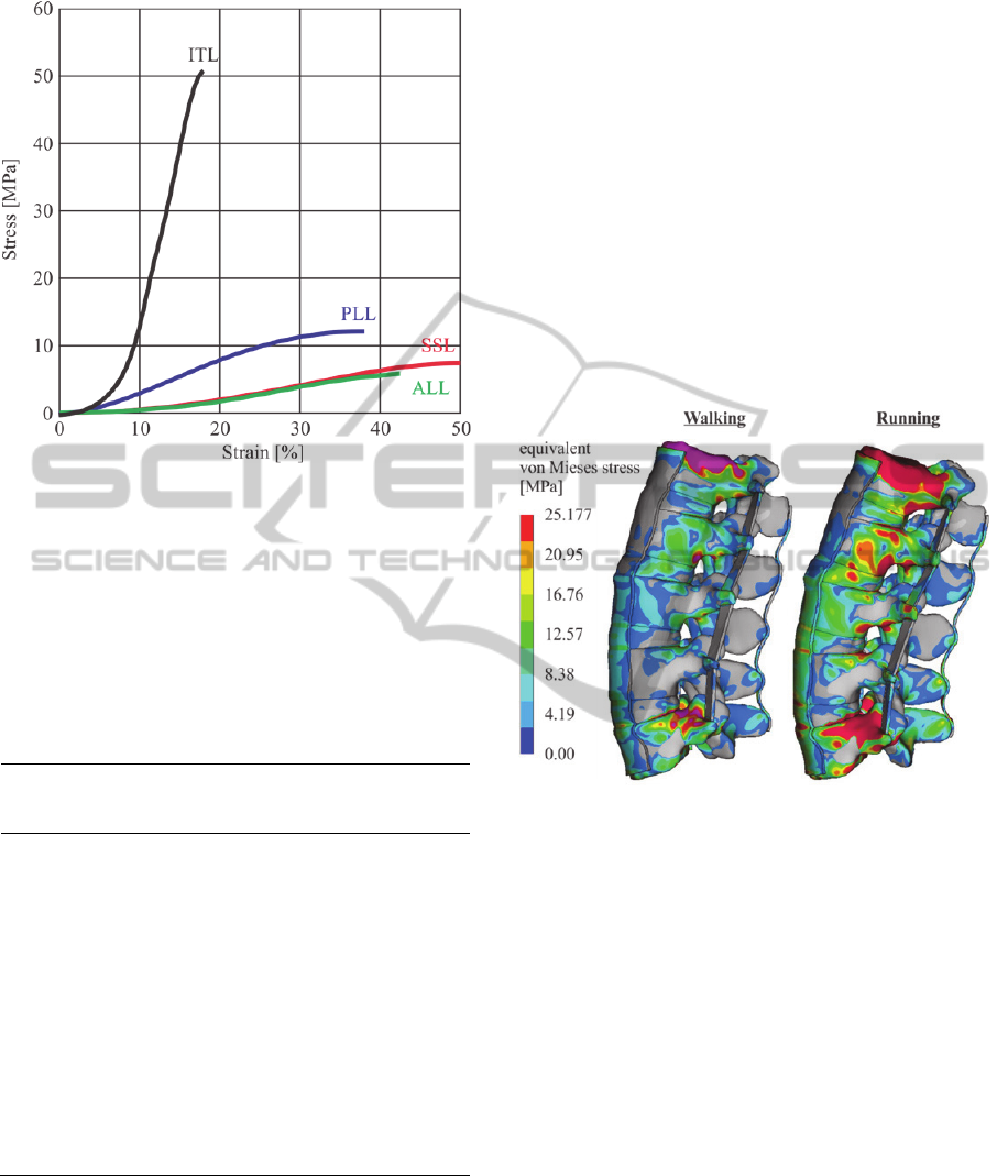
Figure 2: Non-linear properties of solid ligamentus tissue.
To avoid element shear locking and hourglassing
phenomena during the numerical analysis of the
model, second order elements with reduced
integration were employed for all model entities.
Solid ligaments were modelled to comprise of four
layer each, to further supress the appearance of
artificial energy within the model.
Table 1: Mesh related data.
Material type
no. of
Elements
max size
Element
min size
Element
Cortical bone 87.521 1,78 mm 0,08 mm
Cancellous bone 712.361 3,04 mm 0,97 mm
Nucleus pulposus 317.251 2,27 mm 0,72 mm
Annulus 298.657 3,71 mm 1,87 mm
ALL 4260 1,92 mm 1,54 mm
PLL 2100 1,78 mm 1,52 mm
SSL 1420 1,83 mm 1,54 mm
ITL
1380 1,74 mm 1,53 mm
The mesh grid of the spine segment was
generated in ANSA by BETA CAE Systems in order
to ensure anatomic based meshing, leading to a
realistic and isotropic stress transition within the
entire model. Convergence studies were conducted
for every model entity individually, indicating the
optimum mesh density in terms of processing time,
with regard to the results accuracy, related
information are demonstrated in Table 1.
3 RESULTS
Employing the introduced model in for two mobility
scenarios, walking and running, facilitated the
determination of FSU’s biomechanical response to
external stimuli. Corresponding results are presented
in figure 3.
When interpreting these results, it is highly
important to consider that the same scale is used to
indicate stress development of both soft and bony
tissue.
Figure 3: Stress development in the FSU during walking
and running.
Even though stress concentrations are apparent in
the vertebral bodies of both models, these are not
considered as critical as even the highest values
(25.14 and 42.36 respectively) lie below their
strength characteristics. In the running scenario
however, some critical regions can be observed
within intervertebral discs, both qualitative and
quantitative, indicating that health condition of
patients with spinal injuries could be affected
drastically based on everyday activities.
4 CONCLUSIONS
The introduced model facilitates the evaluation of
induced loads on the lumbar spine. Pathological
defects, trauma as well as therapy oriented
intervention can be assessed prior to the actual
practice on the patient. This model may also be a
BIOINFORMATICS2013-InternationalConferenceonBioinformaticsModels,MethodsandAlgorithms
294

valuable tool in preoperative evaluation of the
biomechanical response of the system to a function
specific implant.
ACKNOWLEDGEMENTS
The authors would like to acknowledge the Hellenic
General Secretariat for Research and Technology, as
this work was funded in the frame of the BioSpine
grant (#PE8 3227).
REFERENCES
Waters, T. R. Anderson, V. P., Garg, A., 1993, Revised
NIOSH equation for the design and evaluation of
manual lifting tasks, Ergonomics 36, 749.
Svedmark, P., Tullberg, T., Noz, M. E., Maguire, G. Q. Jr,
Zeleznik, M. P., Weidenhielm, L., Nemeth, G.,
Olivecrona, H., 2012, Three-dimensional movements
of the lumbar spine facet joints and segmental
movements: in vivo examinations of normal subjects
with a new non-invasive method, European Spine
Journal 21(4), 599-605.
Rotter, R., Pflugmacher, R., Kandziora, F., Ewert, A.,
Duda, G., Mittlmeier, T., 2007, Biomechanical in vitro
testing of human osteoporotic lumbar vertebrae
following prophylactic kyphoplasty with different
candidate materials, Spine 32(13), 1400-1405.
Little, J. P., Pearcy, M. J., Tevelen, G., Evans, J. H.,
Pettet, G., Adam, C. J., 2010, The mechanical
response of the ovine lumbar anulus fibrosus to
uniaxial, biaxial and shear loads, Journal of the
Mechanical Behavior of Biomedical Materials, 3, 146-
157.
Wang, J. P., Zhong, Z. C., Cheng, C. K., Chen, C. S., Yu,
C. H., Chang, T. K., Wei, S. H., 2006, Finite element
analysis of the spondylolysis in lumbar spine., Biomed
Mater Eng.16(5), 301-308.
Ashish, D., Pramod, P, 2009, Development of Computer
Aided 3D Model From Computed Tomography
Images and its Finite Element Analysis for Lumbar
Interbody Fusion with Instrumentation, International
Journal of CAD/CAM 9(1) 121-128.
Xiao, Z., Wang, L., Gong, H., Zhu, D., Zhan, X., 2011, A
non-linear finite element model of human L4-L5
lumbar spinal segment with three-dimensional solid
element ligaments, Theoretical and Applied
Mechanics Letters, 1, 064001.
Schmidt, H., Heuer, F., Simon, U., Kettler, A., Rohlmann,
A., Claes, L., Wilke, H. J., 2006, Application of a new
calibration method for a three-dimensional finite
element model of a human lumbar annulus fibrosus,
Clinical Biomechanics 21,337-344.
Klinder, T., Ostermann, J., Ehm, M., Franz, A., Kneser,
R., Lorenz, C., 2009, Automated model-based vertebra
detection, identification, and segmentation in CT
images, Medical Image Analysis 13, 471–482.
Tsouknidas, A., Michailidis, N., Savvakis, S.,
Anagnostidis, K., Bouzakis, K. D. Kapetanos, G.,
2012, A FEM modelling technique to determine the
mechanical response of a lumbar spine segment under
complex loads, Journal of Applied Biomechanics, in
press.
Davidson-Jebaseelan, D., Jebaraj, C., Yoganandan N.,
Rajasekaran, S., 2010, Validation efforts and
flexibilities of an eight-year-old human juvenile
lumbar spine using a three dimensional finite element
model, Medical and Biological Engineering and
Computing 48(12), 1223-1231.
El-Rich, M., Arnoux, P. J., Wagnac, E., Brunet, C., Aubin,
C. E., 2009, Finite element investigation of the loading
rate effect on the spinal load-sharing changes under
impact conditions, J Biomech 42(9),1252-62.
Sharma M., Langrana N.A., Rodriguez J., 1995, Role of
ligaments and facets in lumbar spinal stability, Spine
20, 887-900.
Tsuang, Y. H., Chiang, Y. F., Hung, C. Y., Wei, H. W.,
Huang, C.H., Cheng, C.K., 2008, Comparison of cage
application modality in posterior lumbar interbody
fusion with posterior instrumentation—A finite
element study, Medical Engineering & Physics 31(5),
565–570.
Tsouknidas, A., Anagnostidis, K., Maliaris, G.,
Michailidis, N., 2012, Fracture risk in the femoral hip
region: A finite element analysis supported
experimental approach, Journal of Biomechanics
45(11), 1959-1964.
Kobayashi, K., Odagawa, K., Sakamoto, M., Tanabe, Y.,
2009. Accuracy of Single Plane X-Ray Image-Based
Technique for Assessment of Knee Kinematics,
Journal of Biomechanical Science and Engineering 4,
192-200.
Tsouknidas, A., Michailidis, N., Savvakis, S.,
Anagnostidis, K., Bouzakis, K.-D., Kapetanos, G.,
2010, A high accuracy CT based FEM model of the
lumbar spine to determine its biomechanical response,
BIOINFORMATICS 2011 - Proceedings of the
International Conference on Bioinformatics Models,
Methods and Algorithms , 222-22.
Shirazi-Adl, S. A., Shrivastava, S. C., Ahmed, A. M.,
1984, Stress analysis of the lumbar disc-body unit in
compression. A three-dimensional nonlinear finite
element study. Spine 9(2), 120-34.
Smit, T. H., Odgaard, A., Schneider, E., 1997, Structure
and function of vertebral trabecular bone. Spine 15;
22(24), 2823-2833.
ANon-linearFiniteElementModelforAssessmentofLumbarSpinalInjuryDuetoDynamicLoading
295
