
PULAB
Computational-Intelligence Aided Management, Diagnosis,
Teleassistance and e-Learning of Pressure Ulcers
Laura Morente
Escuela Universitaria de Enfermería, Diputación Provincial de Málaga, Málaga, Spain
Francisco J. Veredas, Héctor Mesa, Enrique Morris
Departamento de Lenguajes y Ciencias de la Computación, Universidad de Málaga, Málaga, Spain
Keywords: Pressure Ulcer, Computational Intelligence, Computer Vision, Teleassistance, e-Learning, Nursing
Informatics, Software, Collaborative Diagnosis.
Abstract: The pressure ulcer is a clinical pathology with high prevalence rates, which involve high costs for the
Health systems. The health promotion carried on these lesions, as well as the prevention, suitable evaluation
and correct treatment, have become effective indicators of the quality of health assistance. PULAB
(Pressure Ulcer LABoratory) is a computational tool that enables remote management, diagnosis and
monitoring of pressure ulcers, which include digital images of the wounds. This teleassistance software
gives support to the collaborative work of multiple clinical experts to concurrently evaluate the pressure
ulcers by reaching consensus on each particular case, based on the effective analysis of automatically
segmented and tissue-labeled images of the wounds. In the current phase of our research project an e-
learning module for pressure ulcer diagnosis education is being designed, which will turn this software into
a valuable pedagogical tool for pressure-ulcer-management training for undergraduate students and
professional clinicians.
1 INTRODUCTION
The European Pressure Ulcer Advisory Panel
(EPUAP) defines a pressure ulcer (PU) as an area of
localized damage to the skin or its underlying tissue
caused by pressure, shear, friction or a combination
of these factors (EPUAP, 1999; Gawlitta et al.,
2007; Tsuji et al., 2005). The prevention, care and
treatment of the PU pathology involve high costs for
private or state health systems and have important
consequences for the health of the population,
especially for elderly citizens. PU prevalence rates
vary significantly among different environments of
health assistance. Several studies carried on
populations of elderly patients with home assistance
have shown prevalence rates that fluctuate between
12.7% and 15.1% (Bours et al., 1999; Woodbury &
Houghton, 2004); on the other hand, in acute units
the prevalence data found are even more variable
and range from 7% to 33% of the population
analyzed (Gunningberg, 2004; Melotti et al., 2003;
Tannen et al., 2004); finally, higher prevalence rates
were observed in studies carried out on units of
long-term hospitalized patients (Woodbury &
Houghton, 2004; Horn et al., 2002).
The precise evaluation of PUs is a fundamental
task for diagnosis, monitoring of healing evolution
and making decisions on care and pharmacological
treatment interventions. Precise evaluation and
monitoring of the PU could be achieved whether all
the tissues present in the wound or surrounding areas
are accurately measured and precisely registered
(see (Edsberg, 2007) for a complete and systematic
review of PU histology). Following this strategy, in
(Veredas et al., 2010) the same authors of this
current paper presented a computational tool for
automatic segmentation and tissue detection on PU
digital images. This software was based on a hybrid
system which uses Computational Intelligence (CI)
techniques, neural networks and Bayesian classifiers
to be precise, to automatically classify significant
394
Morente L., Veredas F., Mesa H. and Morris E..
PULAB - Computational-Intelligence Aided Management, Diagnosis, Teleassistance and e-Learning of Pressure Ulcers .
DOI: 10.5220/0003275003940398
In Proceedings of the International Conference on Health Informatics (HEALTHINF-2011), pages 394-398
ISBN: 978-989-8425-34-8
Copyright
c
2011 SCITEPRESS (Science and Technology Publications, Lda.)
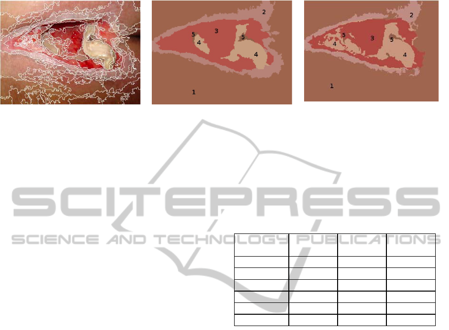
A
B C
Figure 1: Automatic tissue recognition on a PU digital image. Picture A shows the results from the automatic region
segmentation of a PU image using the mean-shift procedure and region growing. For B, a group of expert clinicians have
labeled each one of the regions resulting from the segmented image in A. Picture C shows the automatic labeling done by
the CI system based on neural networks, Bayesian classifiers and heuristics. The tissues shown in B and C have been
labeled with a number that represents: 1: skin; 2: healing tissue; 3: granulation tissue; 4: devitalized tissue; 5: necrotic
tissue. Regions labeled with a same tissue type have been given a same pseudocolor for the sake of clarity. (Figures
included here with permission of (Veredas et al., 2010)).
regions from segmented wound images, obtaining
high precision rates as results.
These CI techniques for automatic tissue
detection have been incorporated into the core of
PULAB tool, which has been designed for the
registering, monitoring, the collaborative expert
evaluation, teleassistance and continuing education
on PUs. At present, PULAB tool is being enriched
with the incorporation of a new e-learning module
designed for education on
PU diagnosis for
undergraduate students as well as for continuing
education for professional clinicians. This e-learning
software will be validated during the next few
months by using it in education and competence
acquisition of Nursing university students.
2 TISSUE RECOGNITION
PULAB tool for management, collaborative
evaluation and teleassisance of PUs is internally
composed of an integrated core based on the CI
strategies and machine learning techniques that have
been developed by the same authors of this current
paper. This software enables automatic segmentation
and precise tissue detection on PU images (Veredas
et al., 2010). Image segmentation on this PU images
is arranged by means of the mean-shift segmentation
method (Comaniciu & Meer, 2002). In figure 1-A, a
typical PU image has been segmented by means of
the mean-shift procedure.
Table 1: Efficiency rates from automatic tissue detection
on 113 PU images. (Data obtained, with permission, from
Table VI in (Veredas et al., 2010)).
Sensitivity Speci-
ficity
Accuracy
Necrotic 86.3 % 98.5 % 98.2 %
Devitalized 67.4 % 95.7 % 93.3 %
Granulation 82.7 % 94.7 % 92.6 %
Healing 59.9 % 91.1 % 85.4 %
Skin 85.2 % 91.0 % 87.9%
GLOBAL 78.7 % 94.7 % 91.5 %
In the table 1, efficiency rates are shown from the
results obtained in the automatic classification of the
tissues present in a set of 113 testing PU images (not
previously “seen” by the machine learning system).
As can be deduced by observing this table, the
automatic classification system based on CI
techniques and used by PULAB for PU evaluation
shows high efficiency rates, not only in the
classification of each particular tissue type, but also
in global terms.
3 THE PULAB TOOL
PULAB is a multiuser teleassistance tool that makes
possible the recording of clinical data and the
collaborative evaluation of PUs. This software tool
has the main purpose of increasing the accuracy of
the diagnosis and the effectiveness of care and
treatment interventions. Moreover, this software
enables the management of each particular wound
case by means of registering digital pictures in the
system and storing contextual clinical data
PULAB - Computational-Intelligence Aided Management, Diagnosis, Teleassistance and e-Learning of Pressure Ulcers
395
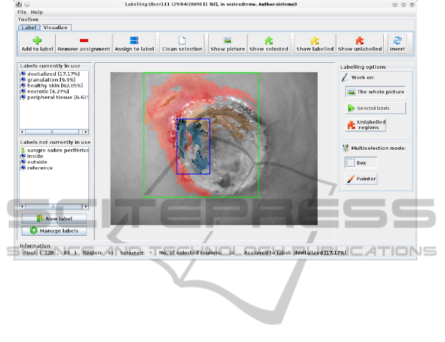
Figure 2: PULAB’s graphical user interface for manual tissue labeling of a registered PU image.
associated with the PU. The system’s users, i.e. the
clinicians and health professionals in general, can
also use PULAB to manage PU series of wounds
grouped by different criteria. Once a PU image has
been uploaded to the system (with its clinical
associated information enclosed), it is immediately
processed by the CI subsystem for region
segmentation and automatic tissue detection. From
that moment on, the user counts on an initial
automatic diagnosis and can look up this automatic
evaluation done by the system to do manual
adjustments on the tissue classification in order to
get a final refined diagnosis. These manual
improvements on the tissue classification are done
with the aid of a friendly graphical user interface
that facilitates the task of region-of-interest selection
and tissue identification. Finally, the system
provides a useful module to efficiently create and
manage collaborative work groups of clinicians who
can share their opinions and reach agreements on
diagnosis of each particular PU case.
PULAB has been designed following a
methodology that is based on a client/server model.
PULAB’s user interface has been developed in Java
and is accessible at the url https://itaca.lcc.uma.es/
ulceras/pulab/launch.jnlp by means of Java Web
Star®.
The main modules of PULAB are the following:
• User-authentication and session-control module:
provides the control of authenticated users,
giving the basis for the management of
collaborative work groups.
• PU-series module: makes possible the creation
and management of series of PUs grouped by
heterogeneous criteria (temporal series of a
same patient, grouping by grade or presence of
different tissue types, grouping by anatomic
location, etc.).
• Collaborative-group module: enables the
management of groups of users to share PU
series, evaluations, diagnosis or decisions on
interventions on the wounds.
• System-record module: provides the
management and controls notification of actions
launched by the systems, such as registrations
and deletions of users, groups, series, etc.;
termination of processes of image segmentation
or tissue detection by the system, etc.
• PU-visualization module: enables the
interaction between the user and the system to
visualize the PU images and navigate on the
segmented regions and classified tissues.
• Labeling module: provides the tools for manual
labeling of the regions resulting from the
automatic segmentation of the images; this
module supplies the user with the necessary
HEALTHINF 2011 - International Conference on Health Informatics
396

tools to manually classify, in a easy and friendly
manner, the segmented regions into the different
tissue types (see the figure 2 for an screenshot
of the user’s interface of the labeling module on
an example of a real PU image).
3 DIAGNOSIS E-LEARNING
An effective strategy to reduce the use of
pharmacological treatments of not-validated benefits
and to homogenize clinical interventions could be to
improve the education of both Health undergraduate
students and professional clinicians. Traditional
education on PU pathology suffers from some
weaknesses that could put the efficacy of the
learning process at risk: on one hand, students
behave usually as merely passive actors during the
learning process, and their interests and motivations
are usually very poor; on the other hand, in clinical
practice, a high variability in the learning procedure
is usually generated. Considering these two issues
above, and as a strategy to improve the learning
process and also guaranty its efficacy and
homogeneity, the current development of PULAB
tool has the main objective of introducing
Information and Communication Technologies
(ICT) to education on PUs for Health undergraduate
students and professionals (i.e. continuing
education).
Very few studies exist on evaluating educational
experiences with ICT-based tools in the specific
field of education on PUs for professional clinicians.
However, some authors conclude that the
development of tools that make e-learning possible
increases the efficacy of the educational processes
since it reduces the time consumed in the learning
process and improves the accessibility of the student
to that learning (Bolwell, 1993). Furthermore, a
recent study by Beeckman et al. (Beeckman et al.,
2008), could be pointed out which deals with
improving the ability of students in the classification
of different PU types and their differentiation from
those other wounds produced as an effect of skin
humidity.
Considering the main goal of minimizing the
user’s system requirements, PULAB’s e-learning
module is being currently developed using web-
based technologies, which will allow using a simple
web browser to have full access to the complete
functionalities offered by this software tool.
Moreover, for the sake of usability, AJAX
technology is being used in the designing of the
graphical user interface with the major objectives of
building complex controls in the forms, minimizing
the data communication and facilitating the
interaction between the user and the system.
PULAB’s e-learning module is being designed as an
adaptive learning interface, which will enable the
students to receive their education in an manner
adapted to his or her particular background-
knowledge level, this way starting from initial
simpler diagnosis cases and progressively going to
more complex examples of PU evaluations and
diagnosis. Experts and professional clinicians will be
included in PULAB with the profile of “teacher” and
will be able to continuously add new evaluated PU
cases in the database, which can be adaptively
included in the sets of PU samples available for their
students. The teachers will be provided with tools
for designing and managing tests for their students,
in order to evaluate their educational progressions.
Both, the teachers and the students, will be supplied
with statistics tools to monitor the learning evolution
and progressions.
Once the PULAB’s e-learning module had been
developed, these authors will proceed to the
validation of this software as an efficient educational
tool, by means of comparing the educational results
obtained from the application of PULAB with those
outcomes coming from the application of traditional
teaching classes. The initial proposed hypothesis for
this validation phase establishes that the PULAB
tutoring system, as an educational software designed
specifically for adaptive education on PU
management, diagnosis and treatment, would
increase the underlying knowledge and improve the
aptitudes for diagnosis, classification, tissue
differentiation and therapeutic decision-making for
undergraduate Health currently students, in
comparison with those results obtained with
traditional teaching methods on PUs management.
4 CONCLUSIONS
PULAB tool has been developed to make possible
the objective evaluation of pressure ulcers. This
software enables teleassitance as well as the
collaborative work of professional clinicians.
PULAB tool consists of an internal core, hosted in
the application-server, that uses computational
intelligence techniques for image segmentation and
tissue detection, which have demonstrated recently
high efficiency rates when applied to real pressure
ulcer images. Finally, the incorporation of an e-
learning module into PULAB tool will make
possible to have an educational tool which will
PULAB - Computational-Intelligence Aided Management, Diagnosis, Teleassistance and e-Learning of Pressure Ulcers
397

increase the efficacy of the learning process on
students or professionals on pressure ulcer
evaluation, diagnosis and care or pharmacological
interventions. The development and validation of
this e-learning module will be concluded in a few
months. Finally, a remarkable issue to be considered
is the possibility of using this same technology
implemented in PULAB for the evaluation and
diagnosis of other sort of skin wounds that require a
teleassistance management similar to the one used
with pressure ulcers and implemented in PULAB.
That could be the case of burn wounds or even
different types of melanomas.
ACKNOWLEDGEMENTS
This project has been supported by the Consejería
de Salud, Servicio Andaluz de Salud, of the Junta de
Andalucía, project id. PI-0502/2009.
REFERENCES
EPUAP, European Pressure Ulcer Advisory Panel., 1999.
Guidelines on treatment of pressure ulcers. EPUAP
Review, 1, 31-33.
Beeckman, D., Schoonhoven, L., Boucque, H., Van
Maele, G., & Defloor, T., 2008. Pressure ulcers: E-
learning to improve classification by nurses and
nursing students. Journal of Clinical Nursing, 17(13),
1697-1707.
Bolwell, C., 1993. Using computers as instructional
technology in the pressure ulcer field. Decubitus, 6(4),
20-25.
Bours, G. J., Halfens, R. J., Lubbers, M., & Haalboom, J.
R., 1999. The development of a national registration
form to measure the prevalence of malnutrition in the
netherlands. Ostomy Wound Manage, 45, 28-40.
Comaniciu, D., & Meer, P., 2002. Mean shift: A robust
approach toward feature space anaylsis. IEEE
Trans.Pattern Anal.Mach.Intell., 24, 603-619.
Edsberg, L. E., 2007. Pressure ulcer tissue histology: An
appraisal of current knowledge. Ostomy/wound
Management, 53(10), 40-49.
Gawlitta, D., Li, W., Oomens, C. W. J., Bader,Frank
P.T.Baaijens and Dan L., & Bouten, C. V. C., 2007.
The relative contributions of compression and hypoxia
to development of muscle tissue damage: An in vitro
study. Annals of Biomedical Engineering, 35(2), 273-
284.
Gunningberg, L., 2004. Risk, prevalence and prevention of
pressure ulcers in three swedish. Journal of Wound
Care, 13(7), 286–-290.
Horn, S. D., Bender, S. A., Bergstrom, N., Cook, A. S.,
Ferguson, M. L., Rimmasch, H. L., et al., 2002.
Description of the national pressure ulcer long-term
care study. Journal of the American Geriatrics
Society, 50(11), 1816-1825.
Melotti, R. M., Fortuna, D., Chiari, P., & Cavicchioli, A.,
2003. Prevalence and prevention and treatment
modalities for pressure sores. Epidemiologia e
Prevenzione, 27(3), 141-146.
Tannen, A., Dassen, T., Bours, G., & Halfens, R., 2004. A
comparison of pressure ulcer prevalence: Concerted
data collection in the netherlands and germany.
International Journal of Nursing Studies, 41(6), 607-
612.
Tsuji, S., Ichioka, S., Sekiya, N., & Nakatsuka, T., 2005.
Analysis of ischemia-reperfusion injury in a
microcirculatory model of pressure ulcers. Wound
Repair and Regeneration, 13(2), 209-215.
Veredas, F., Mesa, H., & Morente, L., 2010. Binary tissue
classification on wound images with neural networks
and bayesian classifiers. Medical Imaging, IEEE
Transactions on, 29(2), 410-427.
Woodbury, M. G., & Houghton, P. E., 2004. Prevalence of
pressure ulcers in canadian healthcare settings. Ostomy
Wound Management, 50(10), 22-38.
HEALTHINF 2011 - International Conference on Health Informatics
398
