
REGISTRATION AND RETRIEVAL OF ELONGATED
STRUCTURES IN MEDICAL IMAGES
Alexei Manso Correa Machado and Christiano Augusto Caldas Teixeira
Pontifical Catholic University of Minas Gerais, Av. Dom Jose Gaspar, 500, Belo Horizonte, MG, Brazil
Keywords:
Medical imaging, morphology, image registration, information retrieval.
Abstract:
This work aims at proposing a set of methods to describe, register and retrieve images of elongated struc-
tures from a database based on their shape content. We propose a registration algorithm that jointly takes
into account the gross shape of the structure and the shape of its boundary, resulting in anatomically consis-
tent deformations. The method determines a medial axis that represents the full extent of the structure with
no branches. Registration follows the linear elasticity model and is implemented through dynamic program-
ming. Discriminative anatomic features are computed from the results of registration and used as variables in a
content-based image retrieval system. A case study on the morphology of the corpus callosum in the chromo-
some 22q11.2 deletion syndrome illustrates the effectiveness of the method and corroborates the hypothesis
that retrieval systems may also act as knowledge discovery tools.
1 INTRODUCTION
Elongated structures such as vessels, bones and brain
ventricles are of interest in many problems and ap-
plications (Toledo et al., 2000; Staal, 2004). Those
structures have in common the fact that their gross
shape can be efficiently represented by centerlines or
medial axes. Contour may present important anatom-
ical features, but the overall shape is, if not more, as
important as the shape of the boundary.
This work aims at proposing a set of methods to
describe, register and ultimately retrieve images of
elongated structures from a database based on their
shape content. Image registration techniques have
been widely used in morphometry, as it provides de-
tailed description of the anatomy, taking a reference
image as a basis for comparison. Registration algo-
rithms are nevertheless computationally costly and,
when applied to the whole image or to the boundary
of elongated structures, may yield unsatisfactory re-
sults. A contribution of this work is a registration al-
gorithm that takes into account both the gross shape
of the structure and the shape of its boundary, with
emphasis to the former aspect.
Figure 1 shows a schematic of a content-based
image retrieval (CBIR) system that follows this ap-
proach. A set of images depicting elongated struc-
tures is segmented and the structures represented by
their boundaries and medial axes. Another image,
taken as a common reference, is deformed through
elastic registration so as to align its anatomy with the
anatomy of the images in the dataset. The result of
registration is a mapping function from each point in
the reference to a point in the target image that en-
able detailed shape description. After the structures
have been described, e.g. based on the curvature of
their boundaries and medial axes, they are stored in
the database for future searching. The querying phase
follows the same steps used to convert the images into
descriptive features. The query image converted to
the corresponding feature vector is compared with the
database, the most similar images are retrieved and
presented to the user. The user may rank the results
according to their relevance, choose one of the re-
trieved images as a new query or redefine a region
of interest that should be given greater priority in the
next retrieving iteration. The query vector is therefore
updated taking into account the user’s feedback.
The characterization of the gross shape is criti-
cal to the registration and retrieval of elongated struc-
tures. We also present a semi-automatic solution to
the extraction of a medial axis that represents the full
extent of the structure with no branches. Finally, dis-
criminative anatomic features are computed from the
results of registration and used as variables in a CBIR
system. A case study on the morphology of the corpus
146
Manso Correa Machado A. and Augusto Caldas Teixeira C. (2008).
REGISTRATION AND RETRIEVAL OF ELONGATED STRUCTURES IN MEDICAL IMAGES.
In Proceedings of the First International Conference on Bio-inspired Systems and Signal Processing, pages 146-153
DOI: 10.5220/0001058901460153
Copyright
c
SciTePress
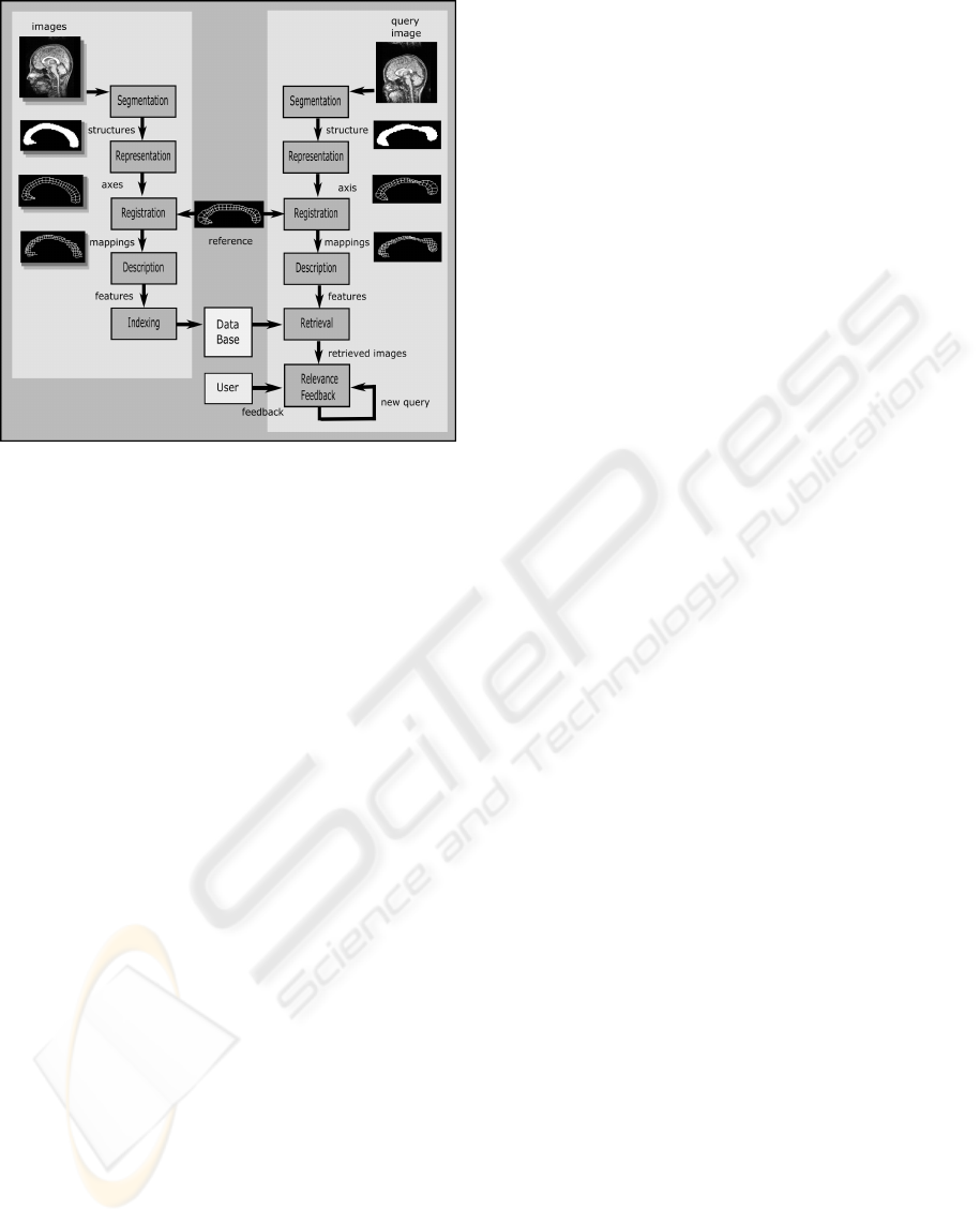
Figure 1: Schematic of a CBIR system based on registra-
tion. The left part of the scheme shows the steps performed
off-line for each image in the database. The on-line part of
the retrieval process is shown in the right. The link between
the on-line and off-line phases is the reference image that
is registered to the query and to the database, establishing a
basis for shape comparison.
callosum in the chromosome 22q11.2 deletion syn-
drome illustrates the effectiveness of the method and
corroborates the hypothesis that CBIR systems may
also act as knowledge discovery tools.
2 RELATED WORKS
The representation of elongated structures through
single sequences of connected points that describe
their intrinsic geometry has been extensively stud-
ied. Pioneered by Blum and Nagel (Blum and Nagel,
1978), the use of medial axes to describe 2D shapes
is based on the removal of points in the boundary un-
til the gross shape is minimally represented. Many
skeleton and thinning algorithms can be found in the
literature, revealing the difficulty on determining a
standard definition for medial axis (Dvies and Plum-
mer, 1981). Other more complex models include the
medial representations (Pizer et al., 2003; Yushkevich
et al., 2003), in which the medial axis and a radial
scalar field are parametrically described such that the
boundary can be further reconstructed, and the medial
profiles (Hamarneh et al., 2004), that provide a shape
representation and deformation operators that can be
used to derive shape distributions.
Registration is considered one of the most im-
portant approaches to provide detailed description of
shape. Automatic registration algorithms (McInerney
and Terzopoulos, 1996; Toga, 1999) may be applied
to the contour (Cootes et al., 1994; Davatzikos and
Prince, 1995) or medial axis (Pizer et al., 1996; Gol-
land et al., 1999) of specific structures. Registration is
also used together with the medial axis transform (Xie
and Heng, 2005) to align the anatomy of structures
based on their skeletons.
Retrieval of images based on their content is still
in its infancy. Smeulders (Smeulders et al., 2000) and
Lew (Lew et al., 2006) present comprehensive dis-
cussions on the main aspects and challenges of im-
age retrieval. Muller (Muller et al., 2004) shows how
CBIR systems can be used to retrieve images in gen-
eral medical databases. In the next section, we dis-
cuss the specific issues related to the registration and
retrieval of images depicting elongated structures and
propose a registration algorithm that jointly considers
the axis and boundaries of such structures.
3 METHODS
The proposed image retrieval method can be divided
into four steps: midline extraction, registration, de-
scription and retrieval.
3.1 Midline Extraction
A midline can be defined as a curve that splits the
structure into dorsal and ventral regions, such that,
at any point, the perpendicular line segments con-
necting the midline to dorsal and ventral parts of the
boundary have roughly the same length (properties
of perpendicularity and congruency). Midline extrac-
tion starts by determining a skeleton based on a vari-
ation of the thinning algorithm described by Gonza-
lez and Woods (Gonzalez and Woods, 2002), for 8-
connected objects. Object points are labeled as 1 and
the background is set to 0. In order for the curve to
fully extend from one extremity to the other, two ob-
ject points are manually chosen and forced to be re-
spectively the starting and ending points of the skele-
ton.Additionally, the thinning algorithm is modified
so as to prune any other branches of the structure’s
skeleton. The final curve is, therefore, a single se-
quence of pixels, each one connected to two neigh-
bors, with the exception of the starting and ending
points.
The following algorithm summarizes skeleton ex-
traction, where p
1
and p
2
are the endpoints; the neigh-
bors of p are denoted as n
i
, numbered counterclock-
wise from 0 (east) to 7 (southeast); function N re-
turns the number of neighbors of p that belong to the
object, i.e., N(p) =
∑
i
n
i
; and function S returns the
REGISTRATION AND RETRIEVAL OF ELONGATED STRUCTURES IN MEDICAL IMAGES
147
number of connected sequences of object points in
the neighborhood of p, i.e., read as an 8-bit string,
the neighbors of p must match the regular expres-
sion 0
+
1
+
0
∗
S
1
+
0
+
1
∗
. It can be shown that only 42
neighborhood configurations satisfy the condition to
mark a point, so that the algorithm can be efficiently
implemented using look-up tables:
Repeat
For each point p of the object, p /∈ {p
1
, p
2
}, do
If N(p) < 7 and S(p) = 1 and n
0
n
6
(n
2
+ n
4
) = 0
Mark p to be removed;
Remove marked points;
For each point p of the object, p /∈ {p
1
, p
2
}, do
If N(p) < 7 and S(p) = 1 and n
2
n
4
(n
0
+ n
6
) = 0
Mark p to be removed;
Remove marked points;
until no more points can be removed.
The linear length of the skeleton is computed con-
sidering the distances between each pair of consecu-
tive pixels: pixels connected by a face with distance
equals to 1 and the ones connected by a vertex with
distance equals to
√
2. The coordinates of the pix-
els are smoothed and interpolated so as to yield an
isotropic rotation-invariant representation of the mid-
line. The derivative of this curve, taken at equidis-
tant points, guides the computation of perpendicu-
lar segments that link the dorsal and ventral bound-
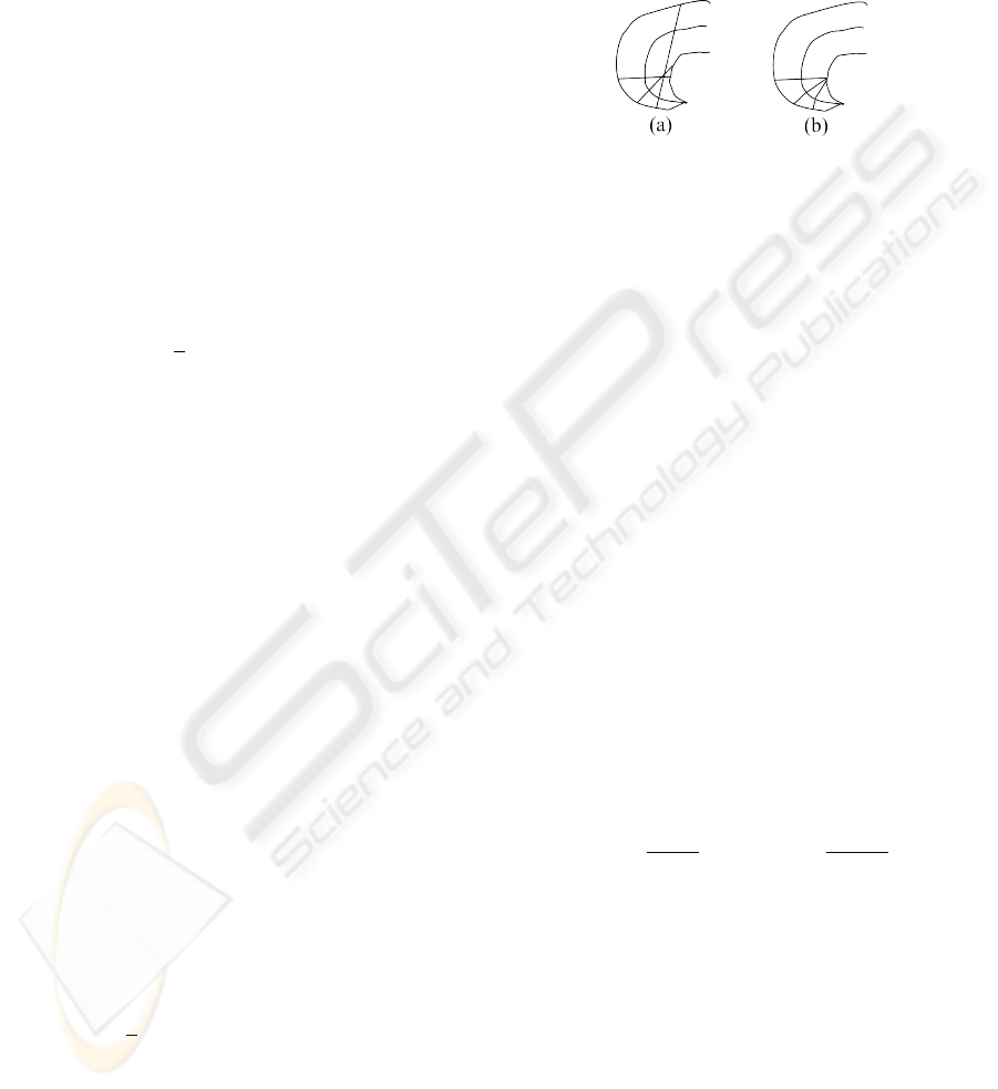
aries of the structure. Problems may occur in regions
where the midline presents increased curvature. In
this case, it may be impossible to satisfy the require-
ments of perpendicularity and congruency for the seg-
ments. Figure 2 shows an example where two con-
secutive segments intersect each other as the result of
increased midline curvature. A solution for this prob-
lem is to violate the property of perpendicularity so
that points with increasing coordinates at the midline
will be connected to points of non-decreasing coordi-
nates at both boundaries. It is however expected that
elongated structures will not frequently incur in this
problem.
The curvature (second derivative) of the midline
can be determined based on the k-curvature metric,
that is defined in each point p
i
= (x
i
,y
i
) as the differ-
ence between the average of the derivatives at the k
next points and the average of the derivatives at the k
previous points (including p
i
):
kcurv(p
i
) =
1
k
(
i+k
∑
j=i+1
d(p
j
) −
i
∑
j=i−k+1
d(p
j
)), (1)
d(p
j
) = tan
−1
(x
j
−x
j−1
,y
j
−y
j−1
).
Parameter k should be empirically chosen so as to pro-
vide enough smoothness. The midline curve should
be extrapolated at the extremities (e.g. based on au-
toregression), so that the curvatures will be computed
over all the midline extension. Analogously, the cur-
vature at the dorsal and ventral boundaries should be
computed at the intersection of the segments. The cur-
vatures at the midline and boundaries will play a fun-
damental role as a measure of similarity during regis-
tration.
Figure 2: Example where consecutive segments intersect
each other as the result of increased midline curvature (a)
and the solution to the problem (b).
3.2 Image Registration
The images in the database should be registered to
a reference in order to establish a common basis
for comparison. Image registration can be stated as
the process of determining a correspondence between
each point p in the midline of the reference image
to a point u(p) in the midline of the subject image.
Let C
M
(p) = kcurv(p) − kcurv(u(p)) be the differ-
ence between the k-curvature taken at point p in the
reference midline and the k-curvature taken at point
u(p) in the subject midline. Analogously, let C
D
and
C
V
be the same difference function computed respec-
tively at the intersection points of the perpendicular
segments emanating from the midline with the dorsal
and ventral boundaries.
The cost function to be minimized is given as
cost = D −S, (2)
where D is the deformation penalty and S is the sim-
ilarity between the curvatures of registered points of
the midline, dorsal and ventral boundaries, given as
D = α
Z
1
0
(
du(p)
d p
)
2
d p + β
Z
1
0
(
d
2
u(p)
d p
2
)
2
d p,
S =
∑
i∈{M,D,V }
γ
i
Z
1
0
C
i
(p)
2
d p (3)
Parameters α and β weight the amount and
smoothness of deformation, respectively. Parameters
γ
M
, γ
D
and γ
V
are negative and weight the importance
of the similarity terms computed respectively for the
midline, dorsal and ventral boundaries.
Registration is performed through dynamic pro-
gramming, in which equidistant points in the refer-
ence midline are mapped to points in the midlines
of the database by minimizing the cost function in
(2). After registering the midlines and corresponding
BIOSIGNALS 2008 - International Conference on Bio-inspired Systems and Signal Processing
148
boundaries, thin plate splines (Barrodale et al., 1993)
are used to interpolate the warping applied to these
curves to the whole structure, so that each pixel in the
reference image is assigned a displacement vector.
An advantage of the proposed registration algo-
rithm is that it will always map a segment perpendic-
ular to the reference midline to a segment perpendic-
ular to the midline of the subject image. This is a
very important constraint to be observed when deal-
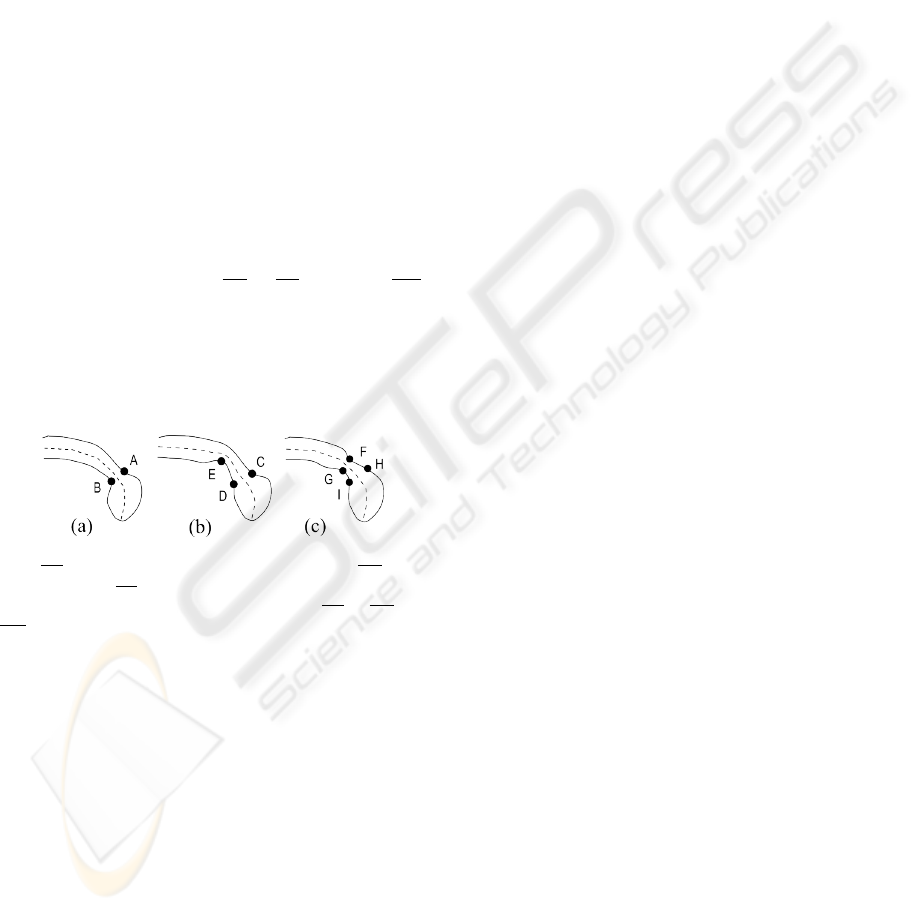
ing with elongated structures. Fig 3 shows two ex-
amples where an image registration algorithm based
only on the boundary or only on the midline would
fail to provide satisfactory deformations. The struc-
ture in (a) is the reference, whose boundary points
A and B must be found correspondence in the other
structures. A registration algorithm that takes into ac-
count only the boundaries would map point A to C
(correctly), but B to E instead of D, since the bound-
ary curvature in B is more similar to the curvature in
E than it is in D. If, on the other hand, the algorithm
is based only on the curvature of the midline, the reg-
istration of the reference to the structure in (c) would
probably map the segment AB to HI instead of FG,
ignoring the similarity between the curvatures at the
boundaries. The similarity function proposed in (3)
avoid both mistakes, since the curvatures at the mid-
line, dorsal and ventral boundaries are jointly taken
into account.
Figure 3: Examples of unsatisfactory registration of the seg-
ment AB in the reference structure (a) to segment CE in (b)
and to segment HI in (c). An algorithm based on both the
boundary and midline would correctly map AB to CD and
FG.
Evaluating the effectiveness of registration meth-
ods is always a difficult task, as ground truth data
is usually inexistent, particularly when the structure
being registered does not present well-defined land-
marks. Alternatively, landmarks may be chosen by
experts, but in this case human subjectivity and lack
of repeatability should be considered in the analysis.
In this work, we designed an interactive interface in
which an expert chooses a set of landmarks in the ref-
erence structure and the corresponding loci in the sub-
jects. The procedure is repeated after 2 weeks, in or-
der to evaluate repeatability. The results achieved by
automatic registration are compared to the mapping
provided by the expert: if the result falls within the
interval of values provided by the expert, it is consid-
ered satisfactory, otherwise the distance in millime-
ters to nearest value is stored and averaged.
3.3 Description
The output of registration is a displacement field that
maps each pixel of the reference image to a point in
the subject. From this set of vectors, it is possible
to obtain diverse measurements that describe the im-
aged objects, such as point-wise area and length varia-
tion, curvature of axes and contours, relationships be-
tween axes of orientation, moments and other shape
descriptors. Feature selection is a fundamental step
in image retrieval systems, as it determines the effec-
tiveness and efficiency of many algorithms. The set
of features that will represent the objects should be
concise and discriminative, as distinguishing features
facilitates the retrieval of relevant images, while non-
relevant characteristics are confounders. Feature se-
lection and information retrieval are synergetic steps:
while the choice of distinguishing features increases
the relevance of retrieval results, retrieval itself act as
a ”mining” tool, selecting the features that discrim-
inate between classes of images. This is the funda-
mental relationship that characterizes image retrieval
as a potential knowledge discovery methodology. In
this work, objects were described as vectors of k-
curvatures (1) taken at each matched point of the sub-
jects, after being registered to the reference.
3.4 Image Retrieval
In a CBIR system, the user presents an image as a
query, which is registered to the reference image. The
features obtained from the resulting mapping func-
tion are compared to the features of the images in the
database, which have been previously processed and
registered to the same reference. Following a measure
of similarity, the most similar images are retrieved and
presented to the user.
The model used to determine the similar-
ity between two images was the Euclidean dis-
tance (Del Bimbo, 1999). If q is the feature vector
representing the query and v
k
is the feature vector rep-
resentation of image k in the database, the similarity
between them can be computed as
sim(v
k
,q) = ((v
k
−q)
T
(v
k
−q))
1/2
The performance of an image retrieval system can
be evaluated by computing two metrics (Del Bimbo,
1999): The recall of the system is the ability to re-
trieve relevant images. It is defined as the ratio be-
tween the number of retrieved images considered rel-
evant and the total number of relevant images in the
database. The precision reflects the ability of the sys-
tem to retrieve only relevant images. It is defined as
REGISTRATION AND RETRIEVAL OF ELONGATED STRUCTURES IN MEDICAL IMAGES
149
the ratio between the number of retrieved images con-
sidered relevant and the total number of retrieved im-
ages. The plot of recall × precision gives an estimate
of the overall effectiveness of a CBIR system, as a
compromise between both performance metrics is ex-
pected.
4 EXPERIMENTS
We illustrate the proposed registration-based retrieval
system with a case study on the morphology of the
corpus callosum in the chromosome 22q11.2 dele-
tion syndrome (DS22q11.2). The DS22q11.2 is an
example of genetic abnormality for which many hy-
potheses on anatomical differences have been re-
cently stated (Machado et al., 2007). This syndrome
is the result of a 1.5 - 3Mb microdeletion on the
long arm of chromosome 22 and is characterized by
a range of medical manifestations that include car-
diac, palatal and immune disorders, as well as par-
ticular problems in cognitive domains associated with
the orienting and executive attention systems and with
numerically related processing. Recent studies have
drawn particular attention to changes in the corpus
callosum — the largest bundle of axons connecting
the two hemispheres of the brain, as differences in the
shape of this structure may indicate changes in brain
connectivity that may be related to the observed cog-
nitive impairments (Simon et al., 2005). We hypothe-
sized that an image retrieval system would be able to
retrieve images of subjects sharing the same diagno-
sis, based on a shape representation of the corpus cal-
losum, if the features used to index the images could
be considered discriminative for the syndrome. In this
sense, the system would reveal the most distinguish-
ing features associated with the disease.
Participants in this study were 18 children with
chromosome 22q11.2 deletion syndrome, ranging in
age from 7.3 to 14.0 years (mean,S.D.=9.9,1.4 years)
and 18 typically developing control children, ranging
in age from 7.5 to 14.2 years (mean,S.D.=10.4,2.0
years) (Simon et al., 2005). Magnetic resonance
imaging was performed on a 1.5 Tesla Siemens MAG-
NETOM Vision scanner (Siemens Medical Solutions,
Erlangen, Germany). For each subject, a high-
resolution three-dimensional structural MRI was ob-
tained using a T1-weighted magnetization prepared
rapid gradient echo (MP-RAGE) sequence with the
following parameters: repetition time (TR) = 9.7 ms,
echo time (TE) = 4 ms, flip angle = 12(, number of
excitations = 1, matrix size = 256x256, slice thick-
ness = 1.0 mm, 160 sagittal slices, in-plane resolution
= 1x1 mm. The midsagittal slice of each brain im-
(a) (b)
(c)
(d)
(e)
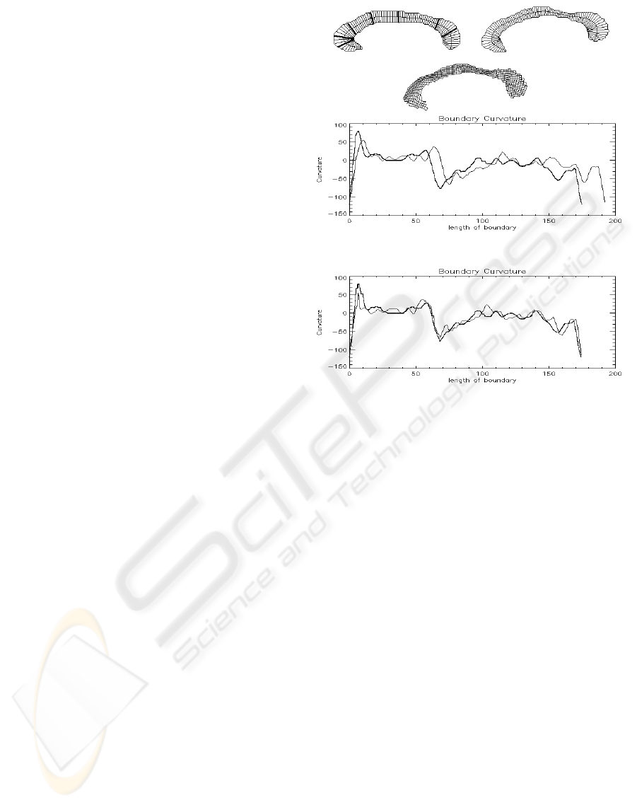
Figure 4: An example of registration. The midline and
boundary of the reference (a) is registered to the subject (b)
and the result interpolated to the whole structure (c). The
original plot of the boundary curvatures (d) and result of
registration (e) are also shown, where the curvatures of the
template and subject are represented by thick and thin lines,
respectively. The 7 landmarks used for registration evalu-
ation, numbered from left to right, are depicted in (a) with
thick lines.
age volume was manually extracted as the best plane
spanning the interhemispheric fissure, and on which
the anterior and posterior commissures and the cere-
bral aqueduct were visible.
The callosa in the midsagittal images were seg-
mented by manual thresholding and delineation. The
boundaries of the callosa were automatically deter-
mined using the Rosenfeld algorithm for 8-connected
contours (Gonzalez and Woods, 2002). The midlines
of the callosa were also extracted based on the algo-
rithm proposed in Section 3.1 and interpolated so as
to yield an isotropic rotation-invariant representation,
in which any two consecutive sampled points were 1
mm apart. The pointwise curvature of the callosum
midline was computed for each subject, using the k-
curvature metric (1), where k was empirically chosen
to be 10% of the length of the midline, so as to pro-
vide enough smoothness.
Shape measurement was performed, by aligning
a reference image of the callosum to subject callosa.
One of the control subjects was arbitrarily chosen as
BIOSIGNALS 2008 - International Conference on Bio-inspired Systems and Signal Processing
150

the reference. The midline of the reference, sampled
at 87 equidistant points, was registered to the sub-
jects’ midlines based on the cost function described in
(2) with parameters α=0.001, β=1000.0 and γ
i
=-1.0
mm
2
/degree
2
for i ∈ {M,D,V }, which were empiri-
cally determined. The midline curves of the subject
callosa were interpolated to provide sub-pixel preci-
sion (0.5 mm). The result of registration was a map-
ping from each of the 87 points in the reference to cor-
responding points in the subjects. Registration took
7.78 seconds to compute. All methods were imple-
mented in IDL language (Research Systems) and run
in a 1.1 GHz Intel Celeron processor computer with
256 MB of RAM, under Windows XP operating sys-
tem.
Figure 4 shows an example of registration where
the reference image described through its midline and
perpendicular segments (a) is deformed to match the
subject (b). The resulting deformation is shown as a
warped grid (c). A plot of the original k-curvatures
(in degrees/mm) at the boundaries of both images
(in mm), taken counterclockwise from the leftmost
endpoint of the midline, is given in (d) and the re-
sulting registration is depicted in (e). The effective-
ness of registration was evaluated based on 2 sets
of landmarks provided by an expert, taken in an in-
terval of 2 weeks. Seven landmarks were defined
at the reference, from anterior to posterior callosum
(Figure 4a), and the expert was asked to determine
their corresponding loci at each of the 36 subjects.
The set of 504 landmarks were compared to the re-
sults of registration. Table 1 summarizes the results,
where it is possible to compare the average error of
the method with the variability of measures provided
by the expert, for each landmark. The average error
of the method for the whole set of landmarks was 1.7
mm, a satisfactory result considering that the aver-
age variability of the expert’s measures was 1.2 mm.
Larger errors were observed at landmarks 3 and 4
(callosal body) where the subjects present larger vari-
ability with respect to curvature. The best results were
achieved at landmarks 5 and 6 (posterior callosum)
where the errors obtained with automatic registration
were smaller than the average variability observed in
manual registration.
The results of image retrieval were evaluated with
the aid of a simple retrieval environment. Initially,
the user browses the database and chooses an image
that will represent the query. The system ranks the
remaining images, showing the n most relevant to the
user appraisal. In this study, we considered as rele-
vant the images that shared the same diagnosis of the
query (with or without the deletion). Following the
recent findings on anatomic differences in the callo-
sum of these populations (Machado et al., 2007)(see
Figure 5), an effective CBIR should be able to re-
trieve images sharing the same diagnosis, unless out-
liers would be present in the database.
(a) (b)
Figure 5: Mean callosal shape for the typically developing
children (a) and children with the deletion (b). Controls
have shorter, more curved anterior callosum (rostrum and
genu) and less curved midbody. Children with the deletion
present more arched callosum (larger height/length ratio).
Table 1: Average error (mm) for each landmark, consider-
ing manual and automatic registration.
Landmark 0 1 2 3 4 5 6
Manual 0.6 0.4 0.7 1.7 1.3 2.6 1.0
Automatic 1.0 0.9 0.8 3.7 2.4 1.6 0.9
An example of the results of image retrieval is
shown in Figure 6. The query image presented by
the user (a) is registered to the same reference used in
the registration of the images stored in the database.
The 10 images that yield greater similarity with re-
spect to the curvature of the midline and boundary are
retrieved and displayed (b). Images of controls are
shown in gray and images of children with the dele-
tion are shown in black. A plot of the recall × pre-
cision computed after the retrieval of each of the 17
relevant images in the database is presented in (d). In
this case, the query is a typical control, yielding high
precision.
An example in which an outlier is retrieved is
given in Figure 7. The third retrieved image is a con-
trol with arched callosum, whereas the query is a child
with the deletion. In this case, the precision is af-
fected. Worse result occurs when the query itself is
an outlier, as exampled in Figure 8. In this case, the
query is a control with longer, less curved rostrum
(left-most end of the midline) that is more common
in children with the deletion. As a consequence, the
precision is drastically affected, staying bellow 50%
from the second retrieved image, a level that would
be expected by pure chance.
5 CONCLUSIONS
We have addressed the problem of registering and re-
trieving images of elongated structures. Traditional
REGISTRATION AND RETRIEVAL OF ELONGATED STRUCTURES IN MEDICAL IMAGES
151
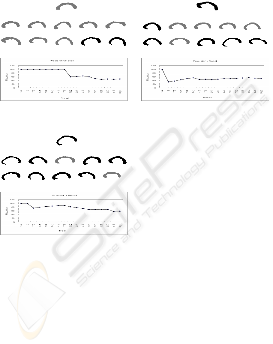
(a)
(b)
(c)
Figure 6: Example of a query image (a) and the result of
retrieval (b). The plot of recall × precision is shown in (c).
(a)
(b)
(c)
Figure 7: Example of a query image (a) and the result of
retrieval (b). In this case, the third best-ranked image is an
outlier. The plot of recall × precision is shown in (c).
registration methods may yield anatomically incon-
sistent results while applying warping models only to
the structure’s contour or medial axis. The method
proposed in this paper jointly registers the medial
axis, dorsal and ventral boundaries, avoiding distor-
tions that may impact substantially in the results of
further morphometric analyses, hypothesis testing or
image retrieval.
The method deserves more systematic evaluation
procedures, as visual inspection is subjective and dif-
ficult to quantify. A case study on the morphology of
the corpus callosum in the 22q11.2 deletion syndrome
was used to illustrate the ability of registration to pro-
vide effective image retrieval. In the experiments, di-
agnosis was considered as the ground truth to evalu-
(a)
(b)
(c)
Figure 8: Example of a query image (a) and the result of
retrieval (b). In this case, the query is an outlier, yielding
poor performance (c).
ate the performance of the retrieval system. Although
evidences of shape differences between controls and
children with the deletion exist, outliers make eval-
uation a difficult task. A deficiency of the method
is the requirement for manual choice of the midline
endpoints, so a fully automated algorithm is already
being designed. Another well-known disadvantage of
registration-driven retrieval methods is its inadequacy
to indexing, limiting the application of these systems
to small datasets. Furthermore, the vector model that
exhibits excellent performance in text retrieval is not
a consensus when dealing with images.
Relevance feedback is an important step that de-
serves attention. Different similarity functions and
query updating models may enhance the effectiveness
of image retrieval, as the user’s preferences are more
rapidly met. Experiments have shown that when the
set of features is restricted to specific regions of in-
terest, the precision is enhanced. In the case of the
study on the corpus callosum morphometry, restrict-
ing the computation of similarity to the anterior-most
part of the structure, where the differences between
groups are more evident, has increased the number of
retrieved images that share the same diagnosis. This
ability to cluster images of the same group may qual-
ify image retrieval as a potential knowledge discovery
tool. It implements new levels of supporting environ-
ments and opens new perspectives to exploratory re-
search in image databases.
BIOSIGNALS 2008 - International Conference on Bio-inspired Systems and Signal Processing
152

ACKNOWLEDGEMENTS
This work was partially supported by FAPEMIG,
PUC Minas and CNPq grant 20043054198. The au-
thors are grateful to the University of Pennsylvania
for sharing the corpus callosum data.
REFERENCES
Barrodale, I., Skea, D., Berkley, M., Kuwahara, R., and
Poeckert, R. (1993). Warping digital images using thin
plate splines. Pattern Recognition, 26(2):375–376.
Blum, H. and Nagel, R. N. (1978). Shape description using
weighted symmetric axis features. Pattern Recogni-
tion, 10(3):167–180.
Cootes, T., Hill, A., Taylor, C. J., and Haslam, J. (1994).
Use of active shape models for locating structures
in medical images. Image and Vision Computing,
12(6):355–365.
Davatzikos, C. and Prince, J. (1995). An active contour
model for mapping the cortex. IEEE Transactions on
Medical Imaging, 14:65–80.
Del Bimbo, A. (1999). Visual Information Retrieval. Mor-
gan Kaufman.
Dvies, E. R. and Plummer, A. P. (1981). Thinning algo-
rithms: a critique and a new methodology. Pattern
Recognition, 14:53–63.
Golland, P., Grimson, W. E. L., and Kikinis, R. (1999). Sta-
tistical shape analysis using fixed topology skeletons:
Corpus callosum study. Lecture Notes in Computer
Science, 1613:382–387.
Gonzalez, R. C. and Woods, R. E. (2002). Digital Image
Processing. Prentice-Hall, Upper Saddle River.
Hamarneh, G., Abu-Gharbieh, R., and McInerney, T.
(2004). Medial profiles for modeling deformation and
statistical analysis of shape and their use in medical
image segmentation. International Journal of Shape
Modeling, 10(2):187–209.
Lew, M., Sebe, N., Djeraba, C., and Jain, R. (2006).
Content-based multimedia information retrieval: State
of the art and challenges. ACM Transactions on Mul-
timedia Computing, Communications, and Applica-
tions, 2(1):1–19.
Machado, A., Simon, T., Nguyen, V., McDonald-McGinn,
D., Zackai, E., and Gee, J. (2007). Corpus cal-
losum morphology and ventricular size in chromo-
some 22q11.2 deletion syndrome. Brain Research,
1131:197–210.
McInerney, T. and Terzopoulos, D. (1996). Deformable
models in medical image analysis: A survey. Medi-
cal Image Analysis, 1(2):91–108.
Muller, H., Michoux, N., Bandon, D., and Geissbuhler, A.
(2004). A review of content-based image retrieval sys-
tems in medical applications-clinical benefits and fu-
ture directions. International Journal of Medical In-
formatics, 73(1):1–23.
Pizer, S. M., Fletcher, P. T., Joshi, S., Thall, A., Chen, J. Z.,
Fritsch, D. S., Gash, A. G., Glotzer, J. M., Jiroutek,
M. R., Lu, C., Muller, K. E., Tracton, G., Yushkevich,
and Chaney, E. (2003). Deformable m-reps for 3D
medical image segmentation. International Journal
of Computer Vision, 55(2):85–106.
Pizer, S. M., Fritsch, D. S., Yushkevich, P., Johnson, V.,
and Chaney, E. (1996). Segmentation, registration
and measurement of shape variation via image ob-
ject shape. IEEE Transactions on Medical Imaging,
18(10):851–865.
Simon, T. J., Ding, L., Bish, J. P., McDonald-McGinn,
D. M., Zackai, E. H., and Gee, J. C. (2005). Volu-
metric, connective, and morphologic changes in the
brains of children with chromosome 22q11.2 deletion
syndrome: an integrative study. Neuroimage, 25:169–
180.
Smeulders, A., Worring, M., Santini, S., Gupta, A., and
Jain, R. (2000). Content-based image retrieval at the
end of the early years. IEEE Transactions on Pat-
tern Analysis and Machine Intelligence, 22(12):1349–
1380.
Staal, J. (2004). Segmentation of elongated structures in
medical imaging. PrintPartners Ipskamp, Enschede.
Toga, A. W. (1999). Brain Warping. Academic Press, New
York.
Toledo, R., Orriols, X., Binefa, X., Radeva, P., Vitria, J., and
Villanueva, J. (2000). Tracking of elongated structures
using statistical snakes. In Proceedings of the CVPR,
pages 157–162, Hilton Head Island.
Xie, J. and Heng, P. A. (2005). Shape modeling using au-
tomatic landmarking. Lecture Notes in Computer Sci-
ence, 3750:709–716.
Yushkevich, P., Fletcher, P. T., Joshi, S., Thall, A., and Pizer,
S. (2003). Continuous medial representations for geo-
metric object modeling in 2d and 3d. Image and Vision
Computing, 21(1):17–28.
REGISTRATION AND RETRIEVAL OF ELONGATED STRUCTURES IN MEDICAL IMAGES
153
